Molecular Biology of the Cell - Volume 1 - Alberts B., Bray D., Lewis J., Raff M., Roberts K., Watson J. 1994
Molecular Organization of Cells
Plasma Membrane
Transport of Macromolecules and Particles Across the Membrane: Exocytosis and Endocytosis
Transport Proteins mediate the passage of many small polar molecules across Cell membranes, but they cannot transport macromolecules such as proteins, polynucleotides, or Polysaccharides. Nevertheless, most Cells can both take up and secrete macromolecules, and some specialized cells can even engulf large particles. The mechanisms by which cells carry out these processes are very different from those mediating the Transport of Small molecules and ions. The transport of macromolecules involves the sequential formation and fusion of membrane-enclosed vesicles. For example, to secrete Insulin, the cells that produce this hormone package it into specialized secretory vesicles. In response to extracellular signals, these vesicles fuse with The Plasma membrane and open to the extracellular space, releasing insulin. This fusion process is called exocytosis. Cells can also take up macromolecules and particles using a similar mechanism, but in reverse. The ingested material is progressively enclosed by a small portion of the plasma membrane, which first invaginates and then pinches off to form an intracellular vesicle containing the engulfed material. This process is called endocytosis. The processes of exocytosis and endocytosis are compared in Fig. 6-68. Both mechanisms involve the fusion of initially separate regions of The Lipid Bilayer and occur in at least two stages: first, the two bilayers adhere (bilayer adhesion), and then they merge (bilayer fusion). Both stages appear to be mediated by specialized proteins, as discussed below (see Section 6.5.16).
Class="center">
Figure 6-68. Bilayer adhesion and fusion during exocytosis and endocytosis. The extracellular space is at the top, separated from the Cytoplasm (bottom) by the plasma membrane. Note that because of the bilayer adhesion stage, exocytosis and endocytosis are not simply the reverse of each other: in exocytosis, the two monolayers of the plasma membrane facing the cytoplasm adhere, whereas in endocytosis, the two outer monolayers of the membrane adhere. In both cases, the asymmetric Nature of the membranes is preserved, and the monolayer facing the cytoplasm always remains in contact with the Cytosol.
An important feature of both exocytosis and endocytosis is that the secreted or ingested macromolecules are localized within vesicles and do not normally mix with other macromolecules or Organelles in The Cell. Vesicles can fuse only with specific membranes, ensuring the directed transport of macromolecules between the extracellular space and the cell interior. A similar process occurs during the transport of newly synthesized macromolecules from The Endoplasmic reticulum to the Golgi apparatus and then to other cell compartments (see Chapter 8). Although it is clear that the rapid and ubiquitous formation and fusion of vesicles is a fundamental feature of all Eukaryotic cells, the molecular mechanisms that drive and direct this transport along specific pathways remain largely to be elucidated.
6.5.1. There are two pathways of exocytosis—constitutive and regulated [38]
In all eukaryotic cells, transport vesicles continually deliver new Components of the plasma membrane from the Golgi apparatus to the plasma membrane via exocytosis. At the same time, cells secrete various types of molecules by The process of exocytosis. Some of these molecules may remain on the cell surface and become part of the cell coat, while others are released into the Extracellular matrix. Some of these then diffuse into the interstitial fluid and/or Blood for Nutrition or signaling to other cells.
As described in Chapter 8, secreted proteins are synthesized on Ribosomes bound to the membranes of the rough endoplasmic reticulum (ER). These proteins enter the ER lumen and are transported to the Golgi apparatus by transport vesicles that bud from the ER. In the Golgi apparatus, the proteins are modified, concentrated, sorted, and then packaged into vesicles that pinch off into the cytosol and eventually fuse with the plasma membrane. Unlike macromolecules, small secreted molecules, such as histamine (see below), are actively transported from the cytosol into preformed vesicles, where they often bind to specific macromolecules (Proteoglycans, in the case of histamine) and can thus accumulate at high concentrations without creating an excessive osmotic gradient.
Some proteins are continuously secreted by the cells that produce them. They are packaged into transport vesicles in the Golgi apparatus and then carried directly to the plasma membrane. This is known as the constitutive secretory pathway. In other cells, specific proteins and/or small molecules are stored in specialized secretory vesicles that fuse with the plasma membrane only after the cell receives an appropriate extracellular signal. This process is called the regulated secretory pathway (Fig. 6-69). The constitutive pathway operates in all cells, whereas the regulated pathway is found mainly in cells specialized for secreting products on demand. These are typically Hormones, Neurotransmitters, or digestive Enzymes. In such specialized secretory cells, the signal for secretion is often a chemical messenger, such as a hormone, that binds to receptors on the cell surface. This receptor activation generates an intracellular signal, which often involves a transient rise in the concentration of free Ca2+ in the cytosol (see Section 12.3.7). By an unknown mechanism, this signal (or signals) triggers exocytosis, causing the secretory vesicles to fuse with the plasma membrane and thereby release their contents into the extracellular space.
During exocytosis, vesicle membranes fuse with the plasma membrane (see Fig. 6-68). At least in the regulated pathway, the protein and Lipid Components of the secretory vesicle membranes are later retrieved in a specific manner by endocytosis to be reused in new secretory vesicles. The total area of secretory vesicle membrane temporarily incorporated into the plasma membrane can be enormous: in a pancreatic acinar cell secreting digestive enzymes, up to 900 μm2 of vesicle membrane is incorporated into the apical plasma membrane (which normally has an area of only 30 μm2) when the cell is stimulated to secrete.

Figure 6-69. Two pathways of protein secretion. Some secreted proteins are packaged into transport vesicles and continuously secreted (constitutive pathway). Others are stored in specialized secretory vesicles and released only in response to stimulation of the cell by extracellular signals (regulated pathway). The constitutive pathway operates in all eukaryotic cells, whereas the regulated pathway occurs only in cells specialized for secretion (secretory cells).
6.5.2. Regulated exocytosis is a localized response of the plasma membrane and the underlying cytoplasm [39]
Mast cells secrete histamine (see Table 12-1) in response to the binding of specific ligands to receptors on their surface. It is the histamine secreted by mast cells that is responsible for many of the unpleasant symptoms, such as itching or sneezing, that accompany allergic reactions. If mast cells are incubated in a medium containing a soluble stimulant, exocytosis is observed over the entire cell surface (Fig. 6-70). If, however, the stimulating Ligand is artificially bound to a solid bead, so that it can interact with only a small area of the mast cell surface, exocytosis is restricted to the site of contact with the bead (Fig. 6-71). Clearly, the mast cell does not respond to stimulation as a whole: receptor activation, the resulting intracellular signals, and subsequent exocytosis evidently occur only in the region of the cell that is stimulated. This demonstrates an important property of the plasma membrane: its individual regions can function independently of the rest of the membrane. As we shall see, this property is equally important for both exocytosis and endocytosis.
6.5.3. There are Two Types of endocytosis: pinocytosis and phagocytosis [40]
Depending on the size of the vesicles formed, two types of endocytosis are distinguished: pinocytosis (from the Greek pino, to drink, + kytos, cell), which involves the ingestion of fluid and solutes via small vesicles (about 150 nm in diameter), and phagocytosis (from the Greek phagein, to devour, + kytos, cell), which involves the ingestion of large particles, such as microorganisms or cell debris. In this case, large vesicles called phagosomes or vacuoles are formed (typically with a diameter > 250 nm).
Fluid and solutes are continuously ingested by most eukaryotic cells via pinocytosis, whereas large particles are engulfed mainly by specialized cells called phagocytes. For this reason, for most cells, the terms "pinocytosis" and "endocytosis" are often used interchangeably.
Most particles and molecules ingested by a cell via phagocytosis or pinocytosis end up in Lysosomes. Large particles are internalized into phagosomes, which then fuse with lysosomes to form phagolysosomes. Fluid and macromolecules taken up by pinocytosis are initially delivered to intermediate membrane-enclosed organelles called endosomes, from which they are eventually either transferred to lysosomes or specifically recycled. Because lysosomes contain A wide variety of hydrolytic enzymes (see Section 8.8.1), most of the material in phagosomes and endosomes that fuse with lysosomes is rapidly degraded; the low-molecular-weight breakdown products, such as Amino Acids, sugars, and NUCLEOTIDES, are transported across the lysosome membrane into the cytosol, where they can be utilized by the cell. Most of the membrane components of endocytic vesicles, however, are retrieved from endosomes and recycled back to the plasma membrane.
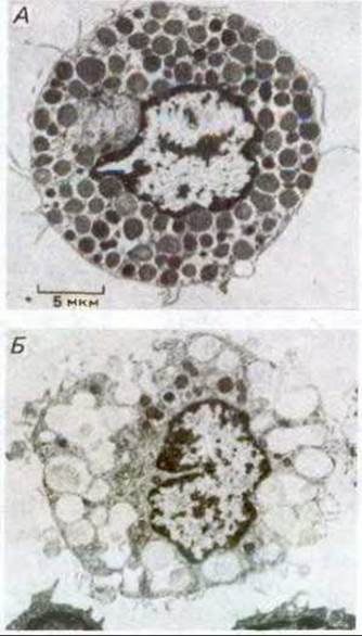
Figure 6-70. Electron micrographs showing exocytosis in rat mast cells. A. An unstimulated cell. B. A cell activated by an extracellular ligand to trigger the secretion of its stored histamine. Vesicles containing histamine appear dark, whereas those that have released their contents appear light. The material remaining in the vesicles after histamine secretion is a network of proteoglycans to which the stored histamine is normally bound. Once a secretory vesicle has fused with the plasma membrane, its own membrane often becomes a target for fusion with other secretory vesicles. Thus, many secretory vesicles in mast cells open to the extracellular space through other already opened vesicles. As a result, the cell (B) contains several large cavities formed by the fused membranes of multiple discharged vesicles, which are now continuous with the plasma membrane. These cavities do not always lie in the same plane of section. (From D. Lawson et al., J. Exp. Med., 142, 391-402, 1975, by permission of the Rockefeller University Press.)
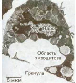
Figure 6-71. Electron micrograph of a mast cell activated to secrete histamine. Activation was induced by a stimulant coupled to a solid bead. Exocytosis occurs only in the region of the cell in contact with the bead. (From D. Lawson et al., J. Cell Biol., 79, 394-400, 1978, by permission of the Rockefeller University Press.)
6.5.4. Pinocytic vesicles form from coated pits in the plasma membrane [41]
Almost all eukaryotic cells continually ingest portions of their plasma membrane in the form of small endocytic (pinocytic) vesicles, which are subsequently returned to the cell surface. This endocytic cycle begins at specialized Regions of the plasma membrane called coated pits. In conventional electron micrographs, these regions appear as invaginations of the plasma membrane, coated with a bristlelike Structure on their cytoplasmic side. In various cells, these structures occupy about 2% of the total surface area of the plasma membrane. The lifetime of coated pits is short: they form within about a minute, then invaginate into the cell, pinch off at the neck, and form coated vesicles (Fig. 6-72). It has been estimated that approximately 2500 coated vesicles pinch off from the plasma membrane of cultured fibroblasts every minute. Their lifetime is even shorter than that of coated pits: they rapidly shed their coats and can then fuse with endosomes. The contents of these endocytic vesicles eventually end up in lysosomes or are recycled.
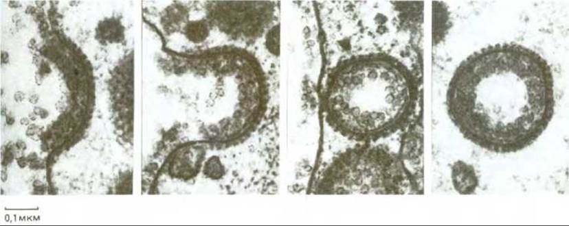
Figure 6-72. Electron micrographs illustrating the probable sequence of events in The formation of a coated vesicle from a coated pit. The coated pit and vesicles shown here are involved in the uptake of lipoprotein particles into a very large hen oocyte during yolk formation; they are incomparably larger here than in cells of normal size. (Courtesy of M. M. Perry and A. B. Gilbert, J. Cell Sci., 39, 257-272, 1979.)
6-30
6.5.5. Coated pits contain clathrin [42]
In Cytology/cytology/93.html">ELECTRON MICROGRAPHS OF samples prepared by rapid freezing and deep etching, The surface of coated pits and vesicles appears as a network of polygons (Fig. 6-73). What is the coat made of, and what are its Functions? Once coated vesicles, which bud from coated pits, were purified, their membranes were found to contain several major proteins. The best characterized of these is clathrin, a protein complex that is highly conserved in evolution. It consists of three large and three small polypeptide chains that form a three-legged protein complex (triskelion). Triskelions assemble on the cytoplasmic surface of the membrane into basketlike lattice structures of hexagons and pentagons (Fig. 6-74). The other proteins, which are more tightly associated with the coated vesicle membrane, are required to bind the clathrin coat to the vesicle and to trap various receptors of the plasma membrane (see below).
It is thought that the invagination of a coated pit is driven by forces generated when clathrin associates with other coat proteins on the cytoplasmic surface of the plasma membrane. Once a coated vesicle is formed, clathrin and its associated proteins dissociate from the vesicle membrane and recycle back to the plasma membrane to form new coated pits. However, it remains unclear how the formation of a coated pit is induced, how a coated pit pinches off to become a coated vesicle, and how this coat is shed from the vesicle. Interestingly, one of the proteins belonging to the hsp 70 (heat Shock protein) family acts in vitro as an ATPase that uncoats clathrin-coated vesicles (see Section 8.8.6). There must be a mechanism to prevent the premature uncoating of a coated pit before it pinches off, since the coat on a pit is much longer-lived.
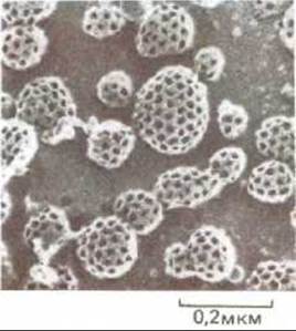
Fig. 6-73. Electron micrograph of numerous coated pits and vesicles on the inner surface of the plasma membrane of cultured fibroblasts. The cells were rapidly frozen in liquid helium, fractured, and then deep-etched to expose the cytoplasmic surface of the plasma membrane. (From J. Heuser, J. Cell Biol., 84, 560-583, 1980, by permission of the Rockefeller University Press.)
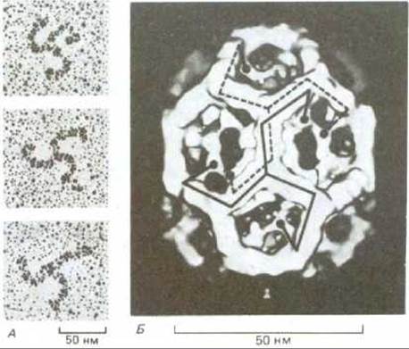
Fig. 6-74. Structure of a clathrin coat. A. Electron micrographs of clathrin triskelions, platinum-shadowed. Each triskelion consists of three heavy and three light clathrin polypeptide chains. Naturally, these details are not visible in the micrographs. B. Three-dimensional model of a clathrin coat. 36 triskelions form a network of 12 pentagons and 8 hexagons. The ends of two triskelions are marked in the photograph. Note that each leg of a triskelion extends along two adjacent polygonal edges and then turns inward, so that their N-terminal domains (black circles) form the inner core of the coat. The overlapping of the flexible ends of the triskelions provides both mechanical strength and flexibility to the entire structure. Clathrin coats of other Sizes and Shapes are constructed in a similar way from 12 pentagons and varying numbers of hexagons. (A - Ungewickell and D. Branton, Nature, 289, 420-422, 1981; B - G.P.A. Vigers et al., EMBO J., 5, 2079-2085, 1986.)
6.5.6. There are at least two types of coated vesicles [43]
In most cells, it is the coated pits and vesicles that mediate the pinocytic uptake of extracellular fluid and membrane-bound ligands. However, other pinocytic pathways using Different types of vesicles are known. Unfortunately, a lack of knowledge about them currently makes it difficult to judge how important they are. Some endothelial cells lining small Blood Vessels apparently transport substances from the bloodstream to the surrounding extracellular fluid using endocytic vesicles that lack a clathrin coat. These vesicles shuttle from one cell surface to the other in a process called transcytosis. However, in most other cells that perform transcytosis, this process is mediated by coated pits and vesicles (see Section 6.5.11).
Not all coated vesicles in the cell originate from the plasma membrane. In Chapter 8, we will discuss how many vesicles are continuously formed from the Endoplasmic reticulum and the Golgi apparatus. They carry out large-scale Vesicular Transport between these and other organelles. There are at least two types of coated vesicles: 1) clathrin-coated vesicles, which are involved in both endocytosis and vesicular Transport from the trans-Golgi network to endolysosomes (see below) and secretory vesicles (see Section 8.9); 2) non-clathrin-coated vesicles, which carry out vesicular transport from the endoplasmic reticulum to the Golgi apparatus, from one Golgi cisterna to another, and from the Golgi apparatus to the plasma membrane. The molecules forming the coat on these vesicles are not yet characterized. Coated (clathrin) vesicles and pits may be much more complex than non-clathrin ones because they are able to recognize specific macromolecules for internalization, whereas non-clathrin vesicles cannot.
6-35
6.5.7. Receptor-mediated endocytosis serves as a concentrating device for the uptake of specific extracellular macromolecules
In most animal cells, clathrin-coated pits and vesicles provide An Efficient Pathway for taking up specific macromolecules from the extracellular fluid in a process called receptor-mediated endocytosis. Macromolecules bind to their receptors on the cell surface, accumulate in coated pits, and enter The Cell as macromolecular complexes enclosed in endocytic vesicles. Because extracellular fluid is trapped in the coated pit and internalized in coated vesicles, solutes dissolved in it are also taken up, but at a much lower rate—a process called fluid-phase endocytosis. Receptor-mediated endocytosis provides a selective concentrating mechanism that increases the efficiency of uptake of specific ligands more than 1000-fold, so that even minor components of the extracellular fluid can be specifically internalized in large amounts (without taking up large volumes of extracellular fluid).
6-33
6-34
6.5.8. Cells import Cholesterol via low-density Lipoproteins (LDL) by receptor-mediated endocytosis [44]
An important process that occurs in many animal cells via receptor-mediated endocytosis is the uptake of cholesterol from the extracellular medium. This provides most of the cell's requirement for cholesterol, which is needed to synthesize new membranes. If cholesterol uptake into cells is blocked, cholesterol accumulates in the blood and can contribute to the formation of atherosclerotic plaques on blood vessel walls. Most cholesterol is transported in the blood as Protein Complexes. These complexes are called low-density lipoproteins, or LDL, and are large spherical particles (22 nm in diameter), each having a core filled with 1500 cholesterol molecules esterified to long-chain Fatty acids. The LDL core is surrounded by a lipid monolayer containing a single protein molecule that organizes The structure of the particle (Fig. 6-75).
When a cell needs cholesterol for membrane synthesis, it produces LDL receptor proteins and inserts them into the plasma membrane. Once in the membrane, the LDL receptor diffuses until it associates with a forming coated pit and becomes incorporated into it (Fig. 6-76, A). Because coated pits are constantly pinching off to form coated vesicles, any LDL particles bound to LDL receptors in a coated pit are rapidly internalized. After shedding their clathrin coats, the vesicles deliver their contents to endosomes. In endosomes, the LDL particles and their receptors dissociate: the receptors are subsequently recycled back to the membrane, while the LDL is delivered to lysosomes (Fig. 6-77). In lysosomes, the cholesterol esters in the LDL particles are hydrolyzed to free cholesterol, which can then be used by the cell for new membrane synthesis. If too much cholesterol accumulates in a cell, both its synthesis and the synthesis of LDL receptor proteins are shut down, so that less cholesterol is both produced and imported.
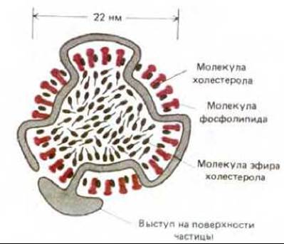
Fig. 6-75. Schematic cross-section of a low-density lipoprotein (LDL) particle. Each spherical particle with a mass of 3 x 10⁶ Da contains about 1500 cholesterol ester molecules surrounded by a lipid monolayer consisting of ~800 phospholipid and 50 cholesterol (unesterified) molecules. The STRUCTURE OF THE particle is organized by a single protein molecule (500,000 Da) responsible for the specific binding of LDL to the cell-surface receptor protein.
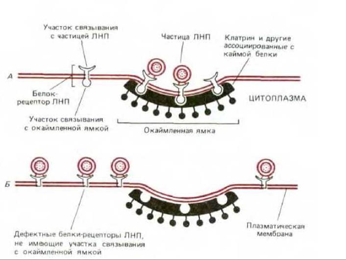
Fig. 6-76. LDL receptor proteins bind to coated pit regions in the plasma membrane of normal cells (A). Human LDL receptors are transmembrane Glycoproteins that span the bilayer once. They consist of 840 residues, of which only 50 are on the cytoplasmic side. B. Mutant cell: LDL receptor proteins are defective because they lack the regions in their cytoplasmic domain that enable them to bind to coated pits. Such cells bind LDL but cannot internalize them. In humans, about one in 500 individuals has one defective LDL receptor Gene; such individuals are at high risk of dying at an early age from Heart disease.
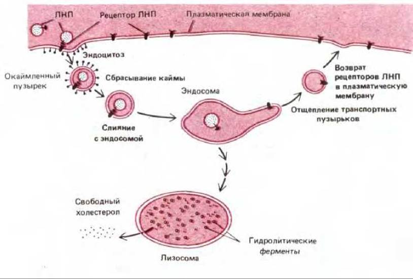
Fig. 6-77. Receptor-mediated endocytosis of LDL. Note that LDL dissociates from its receptor in the acidic environment of the endosome. The transport mechanism is unknown. LDL enters the lysosome and is degraded, releasing cholesterol. LDL receptors are recycled back to the plasma membrane via transport vesicles that bud off from the tubular region of the endosome. For simplicity, only one LDL receptor is shown internalizing and recycling back to the plasma membrane. Whether or not it binds LDL, the receptor undergoes an internalization-recycling cycle every 10 min, making several hundred round trips during its lifetime (about 20 hours).
This pathway is disrupted in certain individuals who inherit defective genes for LDL receptor proteins: their cells are unable to take up LDL from the blood. The resulting high blood cholesterol levels in these individuals predispose them to premature atherosclerosis, so that most of them die at an early age from CORONARY HEART DISEASE. The abnormality may also be associated with the loss of the receptor's binding site for either LDL or the coated pit (see Fig. 6-76, B). In the latter case, There is a sufficient number of LDL receptor proteins, but they do not concentrate in the coated regions of the plasma membrane. LDL binds to the surface of these mutant cells but is not internalized. This directly demonstrates The Importance of coated pits in receptor-mediated endocytosis of cholesterol.
More than 25 different receptors for various molecules involved in endocytosis have already been identified. All of them apparently use the same pathway through coated pits. Many of these receptors associate with coated pits regardless of whether they are bound to specific ligands or not. Not all plasma Membrane Proteins are found in coated pits. This means that the pits function as molecular filters, concentrating certain plasma membrane proteins on their surface while excluding others. Electron microscopic studies of cells grown in media with different ligands (labeled so they can be distinguished under Electron Microscopy) have shown that a single coated pit contains many types of receptors. A coated pit region of the plasma membrane can probably accommodate about 1000 receptors of various types. All receptor-ligand complexes internalized via clathrin-coated vesicles evidently end up in the same endosome. However, the subsequent fate of these molecules is determined by the type of receptor.
6.5.9. Endosomal contents reach lysosomes unless specifically recycled [45]
In electron micrographs, the endosomal compartment can be made easily visible if cells are briefly incubated in a medium containing a labeled ligand. The internalized ligand reveals the endosomal compartment as a complex set of heterogeneous, membrane-enclosed tubules and vesicles. They extend from the cell periphery to its perinuclear region, often found near the Golgi apparatus, but not within it (Fig. 6-78). In experiments with labeled ligands, two sets of endosomes are observed: peripheral endosomes, which appear immediately near the plasma membrane within 1 minute, and perinuclear (late) endosomes, which appear after 5-15 min. The internal environment of endosomes is acidic (pH 5-6) due to the pumping of H+ ions from the cytosol into the interior by an ATP-dependent H+ pump located in the endosomal membrane. The contents of late endosomes are more acidic than those of peripheral endosomes. As we will see, the acidic environment of internalized molecules plays an exceptional role in the functioning of these organelles. Apparently, the same (or several similar) endosomal H+-ATPase acidifies all endocytic and exocytic organelles, including phagosomes, lysosomes, specific compartments of the Golgi apparatus, and many transport or secretory vesicles.
Most of the contents of perinuclear endosomes end up in lysosomes. However, many molecules escape this fate and are recycled from peripheral (and possibly perinuclear) endosomes back to the plasma membrane. This occurs through the budding of transport vesicles from endosomes. As a result, only those internalized molecules that are not specifically destined for recycling undergo degradation.
It is now known that the pathway from endosomes to lysosomes is much more complex than previously thought. Hydrolytic enzymes destined for lysosomes initially arrive from the Golgi apparatus (in transport vesicles) at a special prelysosomal compartment, which is located alongside but separate from the perinuclear endosomes. Although these membrane-enclosed organelles might be mistaken for endosomes in the final stages of maturation, the process of hydrolytic degradation, which will be completed in lysosomes, is actually just beginning. Therefore, we would call these organelles endolysosomes (Fig. 6-79).
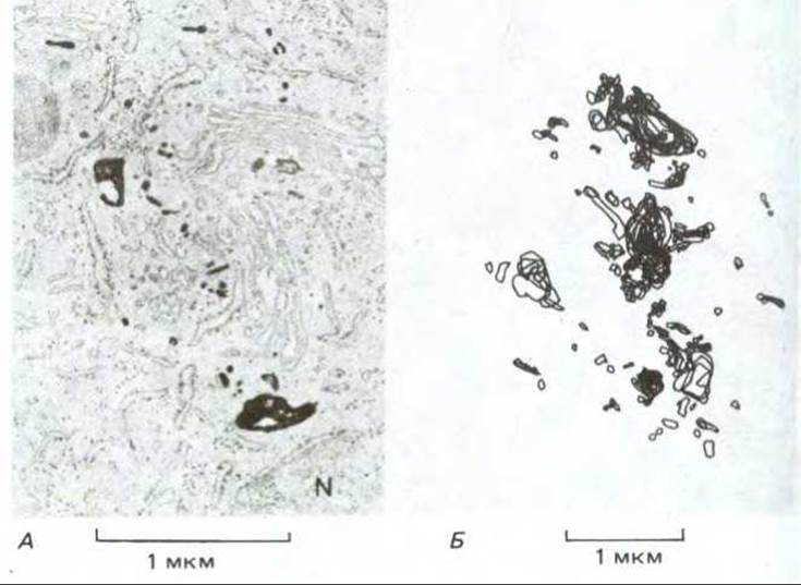
Fig. 6-78. Electron micrograph (A) and schematic representation (B) of perinuclear endosomes from cultured baby hamster Kidney cells. Cells were incubated in a medium containing peroxidase for 15 min at 37 °C, which is sufficient for peroxidase uptake by fluid-phase endocytosis and transport to endosomes (and endolysosomes, see text), but insufficient for delivery to lysosomes. After cell fixation and incubation with the peroxidase substrate (diaminobenzidine), the enzymatic reaction products were fixed with osmium tetroxide to increase electron density. (B) shows an average view reconstructed from 18 thin sections. The Cell Nucleus is labeled N. (A). (Marsh et al., Proc. Natl. Acad. Sci. USA, 83, 2899-2903, 1983.)
It remains unclear how molecules internalized by endocytosis move from one endosomal compartment to another to eventually end up in lysosomes. One hypothesis is that peripheral endosomes slowly move inward, maturing into perinuclear endosomes, which then transform into endolysosomes and subsequently lysosomes through fusion with transport vesicles from the Golgi apparatus, continuous membrane recycling, and increasing acidity. Another hypothesis suggests that each endosomal and lysosomal compartment may be a permanent structure, similar to a stack of cisternae in the Golgi apparatus (see Section 8.7.6). Transport between them would then be mediated by transport vesicles, just as between adjacent cisternae of the Golgi apparatus. We will return to the question of how lysosomes are formed in Chapter 8.
6.5.10. Ligand-receptor complexes are sorted within endosomes [46]
The endosomal compartment functions as the main sorting station in the endocytic pathway, much like the Golgi apparatus performs a similar function in the biosynthetic secretory pathway (see Section 8.7.6). The acidic environment inside endosomes plays a key role in the sorting process by affecting receptor-ligand complexes and thus determining their subsequent fate. As the pH drops, the receptors within the endosome change their conformation and dissociate from their ligands. Once released inside the endosome, these ligands are typically destined for degradation. Other ligands that remain bound to their receptors ultimately share The Fate of their receptors.
The fate of specific receptors depends on their function: 1) they can be recycled to the same regions of the plasma membrane from which they were internalized (this is the most common pathway for receptors), 2) they can be delivered to lysosomes, or 3) they can be transported to a different region of the plasma membrane via transcytosis (Fig. 6-80).
The LDL receptor described in Section 6.5.8 follows the first pathway. It dissociates from its ligand (LDL) in the endosome and returns to the plasma membrane for reuse, while the released LDL is transported to lysosomes (see Fig. 6-77). A similar but more complex cycle occurs during the endocytosis of transferrin, a blood iron-transport protein. The transferrin receptor on the cell surface delivers transferrin bound to iron ions into peripheral endosomes via endocytosis. The low pH in the endosome induces the release of iron from transferrin, but the transferrin (now called apotransferrin) remains bound to the receptor and is recycled to the plasma membrane as a receptor-apotransferrin complex. Upon reaching the extracellular fluid, which has a neutral pH, apotransferrin dissociates from the receptor, enabling it to bind iron ions again and enter a new transport cycle. Thus, transferrin shuttles between the extracellular fluid and the endosomal compartment without entering lysosomes, delivering the iron necessary for cell growth.
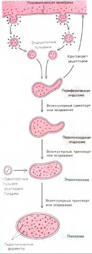
Fig. 6-79. Molecules internalized in coated pits enter peripheral endosomes and then sequentially appear in perinuclear endosomes, endolysosomes, and lysosomes. However, some of them can be specifically recycled from endosomes or endolysosomes. Lysosomal hydrolytic enzymes are initially transported from the Golgi apparatus to endosomes. It remains unclear how internalized molecules that are not recycled move from one compartment to another, eventually ending up in lysosomes where they degrade.
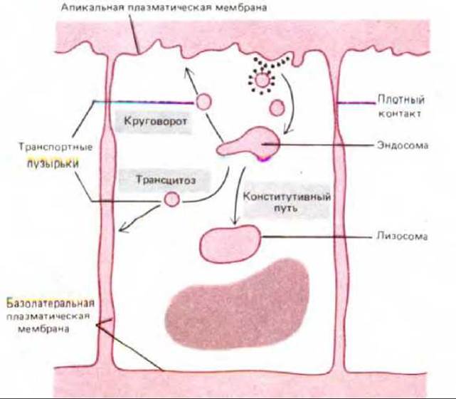
Fig. 6-80. Three pathways from the endosomal compartment in epithelial cells. Most molecules that are not recycled from endosomes (or endolysosomes, not shown) back to the membrane proceed along the constitutive pathway from the endosomal compartment to lysosomes. Recycled molecules return either to the same region of the plasma membrane from which they were internalized (recycling) or to a different domain of the plasma membrane (transcytosis).
An example of a receptor that follows the second endocytic pathway is the receptor that binds a small protein, epidermal growth factor (EGF). EGF receptors are somewhat unusual because they accumulate in coated pits only after binding to EGF. Apparently, ligand-induced Conformational Changes in the receptor are required for its localization to the pits. Moreover, EGF dissociates from its receptor at a lower pH than is required for the dissociation of many other ligand-receptor complexes. Perhaps for this reason, many internalized EGF receptors end up in lysosomes, where they degrade along with EGF. Thus, the binding of epidermal growth factor to its receptor leads to a decrease in the concentration of EGF receptors on the cell surface. As a result, the concentration of the signaling ligand in the extracellular fluid regulates the number of complementary receptor molecules On the surface of the target cell. This regulatory mechanism is fundamentally different from that of LDL receptors, whose number depends on the intracellular concentration of cholesterol (see Section 6.5.8).
6.5.11. Macromolecules can be transported across epithelial cell sheets by transcytosis [47]
Some receptors on the surface of polarized epithelial cells transfer specific macromolecules from one extracellular space to another via transcytosis. These receptors follow the third pathway through the endosomal compartment. For example, newborn rats acquire Antibodies from maternal milk (which helps protect them from infection) by transporting them across the intestinal epithelium. The environment in the intestinal lumen is slightly acidic; at this low pH, antibodies from the milk bind to specific receptors on the apical (absorptive) side of the intestinal epithelial cells and are internalized via coated vesicles. Once in the endosome, the receptor-antibody complexes remain intact. Transport vesicles then fuse with the basolateral domain of the plasma membrane, exposing the complexes to the neutral pH of the extracellular fluid. At this pH, the antibodies dissociate from the receptors and eventually enter the newborn's bloodstream. In turn, antibody secretion into milk by the mother is also mediated by transcytosis, but in the opposite direction—from the blood into the milk (see Fig. 18-20).
The Diversity of pathways followed by different receptors from endosomes implies that many of them possess sorting signals In addition to their ligand-binding and coated-pit-association domains. These signals direct the receptors into the appropriate transport vesicles budding from endosomes, thereby delivering them to the correct Location in The cell membrane. The Nature of these signals remains unclear. Since much more is known about a similar process in the Golgi apparatus, where the trans-Golgi network sorts different proteins into distinct transport vesicles, we will defer further Discussion of intracellular sorting and return to this topic in Chapter 8.
6-31
6-32
6.5.12. Coated pits and vesicles provide the main pathway for fluid-phase endocytosis in many cells [48]
The rate of internalization of the cell's own plasma membrane by endocytosis can be calculated by briefly adding a tracer to the extracellular fluid to monitor and measure its rate of uptake by endocytic vesicles. Two types of molecules are used for this purpose: either those dissolved in the extracellular fluid and taken up by fluid-phase endocytosis (see Fig. 678), or those that bind to cell-surface receptors and are internalized via receptor-mediated endocytosis. Typically, both Methods of measuring rates yield similar results. This indicates that the coated pit-coated vesicle pathway is the major route in many cells not only for receptor-mediated endocytosis but also for fluid-phase endocytosis.
The rate of plasma membrane internalization depends on the cell type, but it is usually surprisingly high. Macrophages, for example, ingest an amount of fluid equal to 25% of their volume every hour. This means they must internalize 3% of their own membrane every minute, or 100% of the membrane in about half an hour. The rate of endocytosis in fibroblasts is slightly lower, whereas in some amoebae, membrane internalization is much faster. Since both cell surface area and volume remain constant throughout this process, it is clear that an equivalent amount of membrane must be returned to the cell surface by exocytosis.
6-8
6.5.13. The endocytic cycle may be related to cell movement and The phenomenon of "capping" [49]
Coated pits are distributed more or less randomly over the cell surface, so that internalization of the plasma membrane (Lipids together with specific receptor proteins) occurs all over the cell surface. In unpolarized cells, the internalized membrane patches are also returned randomly to the cell surface. In polarized cells, however, such as fibroblasts crawling on a substrate, the centrosome and its associated Golgi apparatus are displaced toward the front of the cell, and internalized membrane pieces are returned preferentially to the membrane of the leading edge. This spatial Asymmetry of the endocytic-exocytic cycle may help the cell extend its leading edge forward during movement (see Section 11.6.6). Moreover, because the sites of Endocytosis and Exocytosis do not coincide in such cells, there will be a continuous flow of lipids and receptors along the plasma membrane from the leading edge to the rear of the cell (Fig. 6-81). This membrane flow may explain why objects like charcoal particles, when placed on the surface of cultured fibroblasts, move from the front of the cell to the rear as the cell migrates forward.
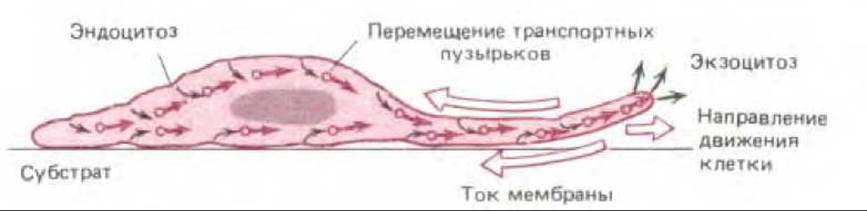
Fig. 6-81. Plasma membrane flow in motile cells as a result of the asymmetry of the endocytic cycle in these cells. A fibroblast (shown in cross-section) moves from left to right. Endocytosis of receptor-containing membrane occurs in coated pits distributed randomly over the cell surface. Internalized membrane is returned from the endosomal compartment (not shown) as exocytic vesicles that fuse with the membrane at the leading edge of the cell (relative to its movement). Thus, endocytosis over the entire surface and directed exocytosis cause membrane components to flow in the direction opposite to cell movement (indicated by large colored arrows). (After M. S. Bretscher, Science, 224, 681-686, 1984.)
When multivalent ligands (such as antibodies and Lectins) bind to specific membrane proteins, they form clusters that aggregate to form patches. This process is called patching. These patches move rapidly over the cell surface (e.g., of a lymphocyte), gathering into a so-called "cap" (Fig. 6-82). The process of cap formation, capping, takes only a few minutes. It is ATP-dependent and culminates (in lymphocytes) in the appearance of this unusual structure at the rear of the cell. Interestingly, only cells capable of crawling on a substrate can form caps, although cell movement itself is not required for this process. This suggests that The Mechanism of capping may share similarities with the mechanisms the cell uses for locomotion. One hypothesis is that the membrane flow described above, which arises during cyclic endocytosis, carries large clusters of bound molecules to the rear end of the cell, whereas free (un-crosslinked) proteins diffuse rapidly enough that their distribution remains random despite the flow. Alternative hypothesis is that clusters of membrane proteins interact (directly or indirectly) with a motor system of cellular Actin filaments (located in the cortex, directly beneath the plasma membrane) that moves the clusters to the rear end of the cell. So far, there are no clear results that rule out either hypothesis.
6.5.14. Specialized phagocytic cells ingest particles that bind to specific receptors on their surface [50]
Phagocytosis is a specialized form of endocytosis in which large particles, such as microorganisms or cell debris, are ingested. This occurs through the formation of large endocytic vesicles called phagosomes. In Protozoa, phagocytosis is a form of feeding: large particles are captured by phagosomes and then delivered to lysosomes. The Digestion products pass into the cytosol and are used as food. In Multicellular Organisms, most cells are unable to ingest large particles efficiently. Therefore, in the gut, large food particles are broken down extracellularly before absorption, and throughout the body, phagocytosis is carried out by "professional" phagocytes. In mammals, there are two classes of white Blood Cells that mediate phagocytosis: macrophages (widely distributed in both Tissues and blood) and neutrophils. These two cell types derive from a common progenitor cell (see Section 17.5.8) and defend us against infection by ingesting invading microorganisms. Macrophages also play an important role in disposing of senescent or damaged cells and cellular debris. Quantitatively, this latter function is particularly important: in each of us, macrophages daily ingest more than 1011 senescent red blood cells by phagocytosis.
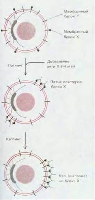
Fig. 6-82. Antibody-induced patching and capping of cell surface proteins on lymphocytes. Antibodies cross-link the protein molecules to which they are bound, forming large clusters. Over time, these clusters accumulate in one area, forming a "cap" at the rear pole of the cell. Note that the front pole is defined by THE POSITION OF the centrosome, and the cross-linked proteins form a "cap" at the opposite pole, even when the cells are in suspension and not migrating.
Endocytic vesicles budding from coated pits are relatively small (~150 nm in diameter). Phagosomes, however, have a diameter determined by the size of the particle being ingested. Sometimes they are almost as large as the phagocytic cells themselves (Fig. 6-83). Phagosomes fuse with lysosomes to form phagolysosomes. This is where the degradation of the ingested material takes place. Indigestible substances remain in the phagolysosomes, forming residual bodies. As in endocytosis, some of the internalized components of the cell's own plasma membrane are recycled back to the plasma membrane. In some macrophages, Peptides derived from the degradation of ingested proteins are returned to the cell surface bound to glycoproteins of the Major Histocompatibility Complex (see Section 18.6.10). The surface of these macrophages is then scrutinized by T lymphocytes of The Immune System. If the peptides are derived from a foreign agent, they activate T lymphocytes to mount an Immune Response. Thus, in this case, macrophages act as antigen-presenting cells (see Section 18.6.10).
For phagocytosis to occur, the particles to be ingested must first bind to the surface of the phagocyte. However, not all bound particles are ingested. There is a set of specialized surface receptors functionally linked to the cell's phagocytic machinery. Unlike pinocytosis, which is a constitutive process that occurs continuously, phagocytosis is an inducible phenomenon in which activated receptors transmit signals into the cell to initiate a response. The best-characterized triggers of phagocytosis are antibodies. Antibodies protect us from microbial infection by binding to their surface to form a coat in which the Fc regions of each antibody molecule are exposed. This coat is then recognized by specific Fc receptors on the surface of macrophages and neutrophils. The binding of antibody-coated particles to these receptors triggers the formation of pseudopodia in the cell's plasma membrane, which envelop the particle and fuse at their tips to form a phagosome (Fig. 6-84).
Two other classes of receptors are known only to promote phagocytosis. Receptors of one class recognize Complement components, while those of the other recognize Oligosaccharides on the surface of certain microorganisms. In addition, macrophages can recognize and ingest senescent and damaged cells, but virtually nothing is known about the receptors involved in these processes.
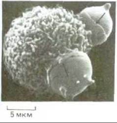
Fig. 6-83. Scanning electron micrograph of a mouse macrophage ingesting two chemically damaged red blood cells by phagocytosis. The arrows point to the cell margins involved in this delicate process (pseudopodia), where the macrophage appears to drape itself over THE RED BLOOD cell to engulf it. (Courtesy of Jean Paul Revel.)
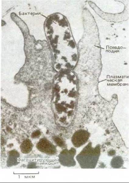
Fig. 6-84. Electron micrograph of a neutrophil ingesting a dividing bacterium by phagocytosis. (Courtesy of Dorothy F. Bainton.)
6.5.15. Phagocytosis is a localized response that proceeds by a membrane "zippering" mechanism [51]
Red blood cells can be treated so that they bind to the surface of macrophages but are not phagocytosed. If these macrophages are then allowed to phagocytose antibody-coated Bacteria, only the bacteria are ingested; the red blood cells, even those in the immediate vicinity of active phagocytosis, are ignored. This indicates that phagocytosis, like the induced exocytosis of mast cells (see Section 6.5.2), is a localized response of a region of the plasma membrane and its underlying cytoplasmic structures.
If a macrophage binds to target cells that are uniformly coated with antibodies, it engulfs them. However, if the antibody molecules are concentrated by capping at one pole of the cell (see Section 6.5.13), the macrophage plasma membrane closely apposes the target cell surface only at the cap region, and phagocytosis does not occur (Fig. 6-85). This indicates that the initial interaction of antibody-coated cells with Fc receptors on the macrophage surface is not sufficient to trigger engulfment. The binding of the target cell to the macrophage merely induces a progressively spreading process of membrane attachment that requires continuous receptor-antibody contact; only under this condition is the ingested cell completely enclosed within a phagosome. This demonstrates that phagocytosis occurs by a membrane "zippering" mechanism.

Fig. 6-85. Schematic diagram of an experiment showing that phagocytosis occurs by a membrane "zippering" mechanism. (After F. M. Griffin et al., J. Exp. Med., 144, 788-809, 1976.)
Neither the mechanism that initiates engulfment upon Antibody Binding to Fc receptors nor the nature of the driving forces that extend pseudopodia are known. However, pseudopodia accumulate actin and actin-binding proteins, and cytochalasin (a drug that prevents actin polymerization) inhibits phagocytosis. This suggests that the mechanism of pseudopod extension is actin-dependent. Since clathrin is sometimes present on the cytoplasmic surface of phagosomes forming in macrophages, it is likely that it plays a role not only in pinocytosis but also in phagocytosis. These two forms of endocytosis, however, are clearly distinct, as cytochalasin does not inhibit pinocytosis.
How do the pseudopodia that have engulfed particles fuse at their tips to form a phagosome? This question brings us back to the fundamental problems we briefly touched upon at the beginning of our discussion of exocytosis and endocytosis.
6.5.16. Membrane fusion during exocytosis and endocytosis is likely catalyzed by specialized fusion proteins [52]
Bilayer adherence and bilayer joining are successive steps in membrane fusion. These are fundamental cellular membrane processes occurring not only during exocytosis and endocytosis but also during Cell Division or Cell Fusion (Fig. 6-86). In none of these cases is the mechanism of membrane fusion yet understood; however, several interesting insights can be drawn from analyzing the fusion of certain enveloped Viruses with cells during infection. Cell membranes never fuse spontaneously. For membranes to fuse, Water molecules must be displaced from between the interacting lipid bilayers, which must come within 1.5 nm of each other. This process is energetically highly unfavorable. Therefore, it is highly likely that all membrane fusion in cells is catalyzed by specialized fusion proteins. Although such cellular proteins have not yet been directly identified, viral fusion proteins are known to play a key role in The entry of enveloped viruses (i.e., those having a lipid bilayer-based membrane envelope) into the host cell (see Section 8.9.5). Viruses such as the Influenza virus, for example, enter the cell via receptor-mediated endocytosis and end up in endosomes. At low pH within the endosome, a fusion protein (fusogen) in the viral envelope is activated. It catalyzes the fusion of the viral membrane with the endosomal membrane, releasing the viral nucleic acid into the cytosol (Fig. 6-87).
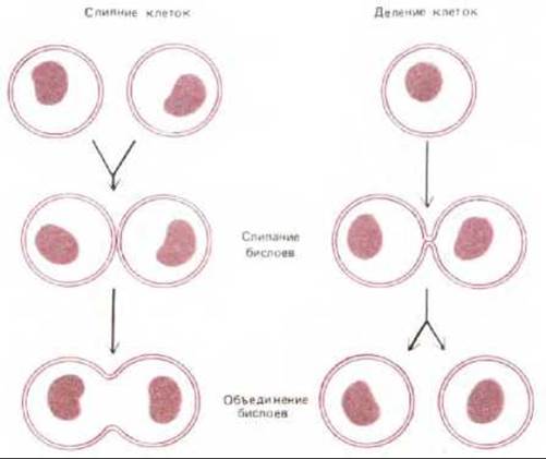
Fig. 6-86. Membrane fusion (comprising two steps: bilayer adherence and bilayer joining) occurring during cell division and cell fusion. In nature, cell fusion is observed during Fertilization (fusion of sperm and egg) and during the formation of multinucleated Skeletal Muscle cells (myoblast fusion).
The genes encoding several viral fusion proteins have been cloned and used to transfect eukaryotic cells in culture. The transfected cells expressed the viral proteins on their membrane surface. Upon brief incubation at low pH, these cells fused with one another to form a Giant multinucleated cell. For the best-studied fusion protein, from the influenza virus, the three-dimensional structure has been determined by X-ray crystallography (see Section 8.6.12). It was shown that low pH induces major conformational Changes in the fusion protein, exposing a previously hidden hydrophobic region on the protein surface. This enables its interaction with the lipid bilayer of the target membrane. Apparently, a cluster of such hydrophobic regions located close to one another in the fusion protein molecule brings the two lipid bilayers into tight contact and destabilizes them so that the bilayers fuse (Fig. 6-87).
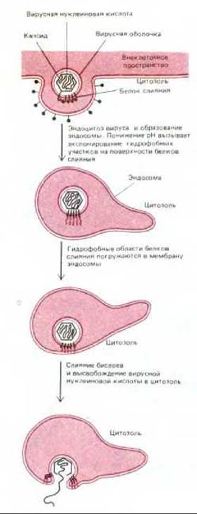
Fig. 6-87. Schematic diagram showing how fusion proteins on the surface of many enveloped viruses catalyze the fusion of viral and endosomal membranes. Viruses enter the cell via receptor-mediated endocytosis and end up in endosomes; the low pH inside the endosomes activates the protein that catalyzes membrane fusion. This allows the viral capsid to be released into the cytosol, where the viral nucleic acid can replicate.
Summary
Most cells secrete and ingest macromolecules by the processes of exocytosis and endocytosis, respectively. In exocytosis, the contents of transport or secretory vesicles are released into the extracellular space when they fuse with the plasma membrane. In endocytosis, the process is reversed: localized regions of the plasma membrane invaginate and pinch off to form an endocytic vesicle. Most ingested particles are then delivered to lysosomes, where they are degraded. Both exocytosis and endocytosis can be either constitutive or regulated in response to extracellular signals.
Most cells continuously endocytose parts of their plasma membrane and then return them to the cell surface in an endocytosis-exocytosis cycle, mediated mainly by clathrin-coated pits and vesicles. Many cell-surface receptors that bind specific extracellular macromolecules localize to clathrin-coated pits and are consequently internalized in coated vesicles. This process is called receptor-mediated endocytosis. Coated endocytic vesicles rapidly shed their clathrin coats and fuse with endosomes, where receptors and ligands are sorted. Most ligands dissociate from their receptors within the endosome and are eventually delivered to lysosomes, whereas most receptors are recycled back to the cell surface via transport vesicles. Some receptor-ligand complexes follow other pathways from the endosomal compartment. Sometimes both receptor and ligand are delivered to lysosomes and degraded. In other cases, the receptor and ligand are transported across the cell, and the ligand is released at another cell surface by exocytosis, a process called transcytosis.
General
Bretscher M. S. The molecules of the cell membrane. .Sci. Am., 253(4), 100-109, 1985.
Datta D. B. A Comprehensive Introduction to Membrane Biochemistry. Madison. Floral., 1987.
Finean J. B. R., Coleman R., Michell R. H. Membranes and Their Cellular Functions, 3rd rd. Oxford, Blackwell, 1984.
Singer S. J., Nicolson G. I. The Fluid Mosaic Model of the Structure of Cell membranes. Science, 175, 720-731, 1972.
Veagle P. The Membranes of Cells. Orlando, Academic, 1987.
Cited
1. Branton D. Fracture faces of frozen membranes. Proc. Natl. Acad. Sci. USA, 55, 1048-1056, 1966.
Ceve G., Marsh D. Phospholipid Bilayers: Physical Principles and Models. New York., Wiley, 1987.
Gorter E., Grendel F. On bimolecular layers of lipoids on the chromocytes of the blood. J. Exp. Med., 41, 439-443, 1925.
2. Hawthorne J. N., Ansell G. B. Phospholipids. New Comprehensive Biochemistry, Vol. 4. Amsterdam, Elsevier, 1982.
Storch J., Kleinfeld A. M. The lipid structure of Biological Membranes. Trends Biochem. Sci., 10., 418-421, 1982.
3. Bangham A. D. Models of cell membranes. In: Cell Membranes: Biochemistry, Cell Biology and Pathology (G. Weissmann, R. Claiborne eds.), pp. 24-34. New York. Hospital Practice, 1975.
Edidin M. Rotational and lateral diffusion of membrane proteins and lipids: phenomena and function. Curr. Top. Memb. Transp., 29, 91-127, 1987.
Kornberg R. D., McConnell H. M. Lateral diffusion of phospholipids in a vesicle membrane. Proc. Natl. Acad. Sci. USA, 68, 2564-2568, 1971.
4. Chapman D. Lipid dynamics in cell membranes. In: Cell Membranes: Biochemistry, Cell Biology and Pathology (G. Weissmann, R. Claiborne eds.), pp. 13-22. New York, Hospital Practice, 1975.
Chapman D., Benga G. Biomembrane fluidity - studies of model and natural membranes. In: Biological Membranes (D. Chapman ed.), Vol. 5, pp. 1-56. London, Academic, 1984.
Kates M., Manson L. A. eds. Membrane Fluidity. Biomembranes, Vol 12. New York, Plenum, 1984.
Kimelberg H. K. METABOLISM/18.html">The Influence of membrane fluidity on The activity of membrane-bound enzymes. In: Dynamic Aspects of Cell Surface Organization. Cell Surface Reviews (G. Poste, G. L. Nicolson eds.), Vol. 3, pp. 205-293. Amsterdam, Elsevier,
5. Carruthers A., Melchior D. I. How bilayer lipids affect membrane protein activity. Trends Biochem. Sci., 11, 331-335, 1986. de Kruiff B. et al. Lipid polymorphism and membrane function. In: The ENZYMES OF BIOLOGICAL Membranes, Vol. 1, Membrane Structure and Dynamics, 2nd ed. (A.N. Martonosi ed.), pp. 131-204. New York, Plenum, 1985.
Kleinfeld A. Current views of membrane structure. Curr. Top. Memb. Transp., 29, 1-27, 1987.
6. Bretscher M. Membrane structure: some General Principles. Science, 181, 622-629, 1973.
Rothman J., Lenard J. Membrane asymmetry. Science, 195, 743-753, 1977.
7. Hakomori S. Glycosphingolipids. Sci. Am., 254(5), 44-53, 1986.
Weigandt H. The gangliosides. Adv. Neurochem., 4, 149-223, 1982.
8. Eisenberg D. Three-dimensional structure of membrane and surface proteins. Annu. Rev. Biochem., 53, 595-623, 1984.
Engelman D. M., Steitz T. A., Goldman A. Identifying nonpolar transbilayer helices in Amino acid sequences of membrane proteins. Annu. Rev. Biophys. Biophys. Chem., 15, 321-353, 1986.
Henderson R. The structure of Bacteriorhodopsin and its relevance to other membrane proteins. In: Membrane Transduction Mechanisms (R. A. Cone, J.E. Dowling eds.), pp. 3-15. New York, Raven, 1979.
Sefton B. M., Buss J. E. The Covalent Modification of eukaryotic proteins with lipid. J. Cell Biol., 104, 1449-1453, 1987.
Unwin N., Henderson R. The structure of proteins in biological membranes. Sci. Am., 250(2), 78-94, 1984.
9. Helenius A., Simons K. Solubilization of membranes by detergents. Biochim. Biophys. Acta, 415, 29-79, 1975.
Montal M. Functional reconstruction of membrane proteins in planar lipid bilayer membranes. In: Techniques of the Analysis of Membrane Proteins (C. J. Regan, R.J. Cherry eds.), pp. 97-128. London, Chapman and Hall, 1986.
Racker E. Reconstruction of Transporters, Receptors and Pathological States. Orlando, Academic, 1985.
10. Bretscher M. Membrane structure: some general principles. Science, 181, 622-629, 1973.
Steck T. L. The organization of proteins in the human red blood cell membrane. J. Cell Biol., 62, 1-19, 1974.
11. Bennett V. The membrane Skeleton of human erythrocytes and its implications for more complex cells. Annu. Rev. Biochem., 54, 273-304, 1985.
Branton D., Cohen C.M., Tyler J. Interaction of cytoskeletal proteins on the human erythrocyte membrane. Cell, 24, 24-32, 1981.
Byers T. J., Branton D. Visualization of the protein associations in The erythrocyte membrane skeleton. Proc. Natl. Acad. Sci. USA, 82, 6153-6157, 1985.
Marchesi V. T. Stabilizing infrastructure of cell membranes. Annu. Rev. Cell Biol., 1, 531-561, 1985.
Shen B. W., Josephs R., Steck T. L. ULTRASTRUCTURE OF THE intact Skeleton of the human erythrocyte membrane. J. Cell Biol., 102, 997-1006, 1986.
12. Marchesi V. Т., Furthmayr H., Tomita M. The red cell membrane. Annu. Rev. Biochem., 45, 667-698, 1976.
13. Jay D., Cantley L. Structural aspects of the red cell anion exchange protein. Annu. Rev. Biochem., 55, 511-538, 1986.
Kopito R. R., Lodish H. F. Primary Structure and transmembrane orientation of the murine anion exchange protein. Nature, 316, 234-238, 1985.
14. Henderson R., Unwin P. N. T. Three-dimensional model of purple membrane obtained by electron microscopy. Nature, 257, 28-32, 1975.
Stoeckenius W., Bogomolni R. A. Bacteriorhodopsin and related pigments of Halobacteria. Annu. Rev. Biochem., 51, 587-616, 1982.
15. Deisenhofer J., Epp. O., Miki K., Huber R., Michel H. The structure of the protein subunits in the photosynthetic reaction centre of Rhodopseudomonas viridis at 3 A resolution. Nature, 318, 618-624, 1985.
16. de Petris S., Rqff M.C. Normal distribution patching and capping of lymphocyte surface immunoglobulin studied by electron microscopy. Nature New Biol., 241, 257-259, 1973.
Edidin M. Rotational and lateral diffusion of membrane proteins and lipids: phenomena and function. Curr. Top. Memb. Transp., 29, 91-127, 1987.
Frye L. D., Edidin M. The rapid intermixing of cell surface Antigens after formation of mouse-human heterokaryons. J. Cell Sci., 7, 319-335, 1970.
MvCloskey M., Poo M.-M. Protein diffusion in cell membranes: some biological implications. Int. Rev. Cytpl., 87, 19-81, 1984.
Poo M., Cone R.A. Lateral diffusion of rhodopsin in the photoreceptor membrane. Nature, 247, 438-441, 1974.
Wier M., Edidin M. Constraint of the translational diffusion of a membrane glycoprotein by its' external domains. Science, 242, 412-414, 1988.
17. Gumbiner В., Louvard D. Localized barriers in the plasma membrane: a common way to form domains. Trends Biochem. Sci., 10, 435-438, 1985.
Myles D. G., Primakoff P. Sperm surface domains. In: Hybridoma Technology in the Biosciences and Medicine. (T. A. Springer ed.), pp. 239250. New York, Plenum, 1985.
Simons K., Fuller S. D. Cell Surface Polarity in Epithelia. Annu. Rev. Cell Biol., 1, 243-288, 1985.
18. Hirano H., Parkhouse В., Nicolson G.L., Lennox E.S., Singer SJ. Distribution of saccharide residues on membrane fragments from a myeloma cell homogenate: its implications for membrane biogenesis. Proc. Natl. Acad Sci. USA, 69, 2945-2949,1972.
Kornfeld R., Kornfeld S. Structure of glycoproteins and their oligosaccharide units. In: The Biochemistry of Glycoproteins and Proteoglycans (W.J. Lennarz ed.), pp. 1-84. New York, Plenum, 1980.
Lis H., Sharon N. Lectins as molecules and tools. Annu. Rev. Biochem., 55, 35-67, 1986.
Olden K., Bernard B. A., Humphries M. J., Yeo T.-K., Yeo K.T., White S.L., Newton S. A., Bauer H. C., Parent J. B. Function of glycoprotein glycans. Trends Biochem. Sci., 10, 78-82, 1985.
19. Hille B. Ionic Channels of Excitable Membranes, Sunderland, MA, Sinauer, 1984.
Martonosi A. N. ed. The Enzymes of Biological Membranes, Vol. 3, Membrane Transport, 2nd ed. New York, Plenum, 1985.
West I. S. The Biochemistry of Membrane Transport. London, Chapman and Hall, 1983.
Stein W.D. Transport and Diffusion Across Cell Membranes Orlando, Academic, 1986.
20. Andersen 0. S. Permeability properties of unmodified lipid bilayer membranes. In: Membrane Transport in Biology. Vol. 1 (G. Giebisch, D. C.
Tosteson, H. H. Ussing eds.), pp. 369-446. New York, Springer-Verlag, 1978.
Finkelstein A. Water movement through membrane channels. Curr. Top. Memb. Trans., 21, 295-308, 1984.
Walter A., Gutknecht J. Permeability of small nonelectrolytes through lipid bilayer membranes. J. Membr. Biol., 90, 207-217, 1986.
21. Hobbs A.S., Alberts R. W. The structure of proteins involved in active membrane transport. Annu. Biophys. Bioeng., 9, 259-291, 1980.
Kyte J. Molecular considerations relevant to the mechanism of Active Transport. Nature, 292, 201-204, 1981.
Stein W. D. ed. Ion pumps: Structure, function and regulation. New York, Alan Liss, 1988.
Tanford C. Mechanism of the energy coupling in active transport. Annu. Rev. Biochem., 52, 379-409, 1983.
Wilson D.B. Cellular transport mechanisms. Annu. Rev. Biochem., 47, 933-965, 1978.
22. Cantley L. C. Structure and mechanisms of the (Na, K) ATPase. Curr. Topics Bioenerget., 11, 201-237, 1981.
Glynn I. M., Ellory C. eds. The Sodium Pump. Cambridge, U. K. The Company of Biologists, 1985.
Shull G. E., Schwartz A., Lingrel J. B. Amino Acid Sequence of the catalytic subunit of the (Na+-K+) ATPase deduced from a complementary DNA. Nature, 316, 691-695, 1985.
23. Swecdner K. J., Goldin S. M. Active transport of sodium and potassium ions: mechanism, function and regulation. N. Engl. J. Med., 302, 777783, 1980.
Glynn I.M. The Na+-K+ transporting adenosine triphosphatase. In: The Enzymes of Biological Membranes, 2nd ed. (A. Martonosi ed.), Vol. 3, pp. 34-114. New York, Plenum, 1985.
24. Hassebach W., Oetliker H. Energetics and electrogenicity of the sarcoplasmic retiulum calcium pump. Annu. Rev. Physiol., 45, 325-339,
1983.
MacLennan D. H., Brandl C. J., Korezak В., Green N. M. Amino acid sequence of a Ca2+, Mg2+-dependent ATPase from rabbit muscle sarcoplasmic reticulum, deduced from its complementary DNA sequence. Nature, 316, 696-700, 1985.
Schatzman H.J. The red cell calcium pump. Annu. Rev. Physiol., 45, 303-312, 1983.
25. Hinkle P.C., McCarty R.E. How cell make ATP. Sci. Am., 238(3), 104-123, 1978.
Nicholls D. G. An Introduction to the Chemiosmotic Theory, 2nd ed. New York, Academic, 1987.
26. Scott D. M. Sodium cotransport systems: cellular, molecular and regulatory aspects. Bioessays, 7, 71-78, 1987.
Wright J. K., Seckler R., Overath P. Molecular aspects of sugar: ion transport. Annu. Rev. Biochem., 55, 225-248, 1986.
27. Grinstein S., Rotnstein A. Mechanisms of Regulation of the Na+/H+ exchanger. J. Membrane Biol., 90, 1-12, 1986.
Olsnes S., Tonnessen T. I., Sandvig K. pH-regulation anion antiport in nucleated mammalian cells. J. Cell Biol., 102, 967-971, 1986. Pouyssegur J., Franchi A., Kohno M., LAllemain G., Paris S. Na+-H+ exchange and growth control in fibroblasts: a genetic approach. Curr. Top. Memb. Transp., 26, 201-220, 1986.
Rozengunt E., Mendoza S. Early stimulation of Na+-H+ antiport, Na+-K+ pump activity and Ca2+ fluxes in fibroblast mitogenesis. Curr. Top. Membr. Transp., 27, 163-191, 1986.
28. Aimers W., Stirling C. Distribution of transport proteins over animal cell membranes. J. Membrane Biol. 77, 169-186, 1984.
Semenza G. Anchoring and Biosynthesis of stalked brush border membrane proteins: Glycosidases and peptidases of Enterocytes and renal tubuli. Annu. Rev. Cell Biol., 2, 255-313, 1986.
29. Postma P. W., Lengeler J. W. Phosphoenolpyruvate: carbohydrate phosphotransferase system of bacteria. Microbiol. Rev., 49, 232-269, 1985.
30. Ames G. F. L. Bacterial periplasmic transport systems: structure, mechanism and evolution. Annu. Rev. Biochem., 55, 397-425, 1986.
31. Hille B. Ionic Channels of Excitable Membranes, Sunderland, MA, Sinauer, 1984.
32. Baker P. F., Hodgkin A. L., Shaw T. L. The effects of changes in internal ionic concentration on the electrical properties of perfused giant axons. J. Physiol., 164, 355-374, 1962.
Hodgkin A. L., Keynes R. D. Active transport of cations in giant axons from Sepia and Loligo. J. Physiol., 128, 26-60, 1955.
Kuffler S.W., Nicholls J. G., Martin A. R. From Neuron to Brain, pp. 111-125, Sunderland MA, Sinauer, 1984.
33. Hodgkin A. L., Huxley A. F. A quantitative description of membrane current and its application to conduction and excitation in nerve. J. Physiol., 117, 500-544, 1952. Kuffler S.W., Nicholls J. G., Martin A. R. From Neuron to Brain, pp. 125-152. Sunderland MA, Sinauer, 1984.
34. Catterall W.A. Molecular properties of voltage-sensitive sodium channels. Annu. Rev. Biochem., 55, 953-985, 1986.
Noda M. et al. Primary structure of Electrophorus electricus sodium channel deduced from cDNA sequence. Nature, 312, 121-127, 1984. Sigworth F. J., Neher E. Single Na+ channel currents observed in cultured rat muscle cells. Nature, 287, 447-449, 1980.
Stevens C.F. Biophysical studies of Ion Channels. Science, 225, 1346-1350, 1984.
Tanabe T. et al. Primary structure of the receptor for calcium channel blockers from skeletal muscle. Nature, 328, 313-318, 1987.
35. Grenningloh G. et al. The strychnine-binding subunit of the Glycine receptor shows Homology with nicotinic acetylcholine receptors. Nature, 328, 215-220, 1987.
Guy H. R., Hucho F. The ion channel of the nicotinic Acetylcholine Receptor Trends Neurosci., 10, 318-321, 1987.
Hille B. Ionic Channels of Excitable Membranes, pp. 117-138 and 335-360. Sunderland MA, Sinauer, 1984.
Noda M. et al. Structural homology of Torpedo californica acetylcholine receptor subunits. Nature, 302, 528-532, 1983.
Schoefield P. R. et al. Sequence and functional expression of the GABAa receptor shows a ligand-gated receptor superfamily. Nature, 328, 221-227, 1987.
36. Stevens C. F. The neuron. Sci. Am., 241(3), 54-65, 1979.
37. Gomperts B. D. The Plasma Membrane: Models for Its Structure and function, pp. 109-212. New York, Academic Press, 1976.
Pressman B. C. Biological Applications of ionophores. Annu. Rev. Biochem., 45, 501-530, 1976.
38. Burgess T. L., Kelly R. B. Constitutive and regulated secretion of proteins. Annu. Rev. Cell Biol., 3, 243-294, 1987.
39. Lawson D., Fewtrell C., Raff M. Localized mast cell degranulation induced by concanavalin A-sepharose beads: implications for the Ca2+ hypothesis of stimulus-secretion coupling. J. Cell Biol., 79, 394-400, 1978.
40. Steinman R. M., Mellman I. S., Muller W. A., Cohn Z. Endocytosis and recycling of plasma membrane. J. Cell Biol., 96, 1-27, 1983.
41. Goldstein J.L., Anderson R.G.W., Brown M.S. Coated pits, coated vesicles, and receptor-mediated endocytosis. Nature, 279, 679-685, 1979.
Roth T. F., Porter K. R. Yolk protein uptake in the oocyte of the mosquito, Aedes aegypti L. J. Cell Biol., 30, 313-332, 1964.
42. Pearse B. M. F., Bretscher M. S. Membrane recycling by coated vesicles. Annu. Rev. Biochem., 50, 85-101, 1981.
Pearse B.M.F., Growther R.A. Structure and assembly of coated vesicles. Ann. Rev. Biophys. Biophys. Chem., 16, 49-68, 1987.
Schmid S. L., Rothman J. E. Enzymatic dissociation of clathrin in a two-stage process. J. Biol., 260, 10044-10049, 1985.
43. Orci L., Click B. S., Rothman J. E. A new type of coated vesicular carrier that appears not to contain clathrin: its possible role in protein transport within the Golgi stack. Cell, 46, 171-184, 1986.
Simionescu N., Simionescu M., Palade G.E. Premeability of muscle capillaries to small heme-peptides: Evidence for the existence of patent transendothelial channels. J. Cell Biol., 64, 586-607, 1975.
44. Brown M. S., Goldstein J. L. How LDL receptors influence cholesterol and atherosclerosis. Sci., Am., 251(5), 58-66, 1984.
Brown M. S., Goldstein J. L. A receptor-mediated pathway for cholesterol Homeostasis. Science, 232, 34-48, 1986.
45. Brown W.J., Goodhouse J., Farquhar M,G. Mannose-6-phosphate receptors for lysosomal enzymes cycle between the Golgi complex and endosomes. J. Cell Biol., 103, 1235-1247, 1986.
Griffiths G., Hoflack В., Simons K., Mellman I., Kornfleld S. The mannose-6-phosphate receptor and the biogenesis of lysosomes. Cell, 52, 329-341, 1988.
Helenius A., Mellman I., Wall D., Hubbard A. Endosomes. Trends Biochem. Sci., 8, 245-250, 1983.
Mellman L, Howe C., Helenius A. The control of membrane traffic on the endocytis pathway. Curr. Top. Memb. Transp., 29, 255-288, 1987.
46. Carpenter G. Receptors for epidermal growth factor and other polypeptide mitogens. Annu. Rev. Biochem., 56, 881-914, 1987.
Dautry-Varsat A., Ciechanover A., Lodish H.F. pH and the recycling of transferrin during receptor-mediated endocytosis. Proc. Natl. Acad. Sci. USA, 80, 2258-2262, 1983.
Goldstein J.L., Brown M.S., Anderson R.G. W., Russell D. W., Schneider WJ. Receptor-mediated endocytosis. Annu. Rev. Cell Biol., 1, 1-39, 1985.
Mellman L, Fuchs R., Helenius A. Acidification of the endocytic and exocytic pathways. Annu. Rev. Biochem., 55, 663-700, 1986.
47. Mostov K.E., Simister N.E. Transcytosis. Cell, 43, 389-390, 1985.
Rodewald R., Abrahamsom D. R. Receptor-mediated transport of IgG across the intestinal epithelium of the neonatal rat. In: Membrane Recycling (Ciba Foundation Symposium 92), pp. 209-232. London, Pitman, 1982.
48. Helenius A., Marsh M. Endocytosis of enveloped animal viruses. In: Membrane Recycling (Cida, Foundation Symposium 92), pp. 59-76. London, Pitman, 1982.
Thilo I. Quantification of endocytosis-derived membrane traffic. Biochim. Biophys. Acta, 822, 243-266, 1985.
49. Abercrombie M., Haysman J. E. M., Pegrum S. M. The locomotion of fibroblasts in culture. III. Movements of particles on the dorsal surface of the leading lamella. Exp. Cell Res., 62, 389-398, 1970.
Bretscher M. S. Endocytosis: relation to capping and cell locomotion. Science, 224, 681-686, 1984.
Taylor R.B., Duffus W.P.H., Raff M. C., de Petris S. Redistribution and pinocytosis of limphocyte surface immunoglobulin molecules induced by anti-immunoglobulin intobody. Nature New Biol., 233, 225-229, 1971.
50. Wright S. D., Silverstein S. C. Overview: the function of receptors in phagocytosis. In: Handbook of Experimental Immunology, 4th ed. (D. M. Weir, L. Herzenberg eds.), pp. 41-1-41-14. Oxford UK, Blackwell, 1983.
51. Aggeler J., Werb Z. Initial events during phagocytosis by macrophages viewed from outside and inside the cell: Membrane-particle interaction and clathrin. J. Cell Biol., 94, 613-623, 1982.
Griffin F. M., Jr., Griffin J. A., Silverstein S. C. Studies on the mechanism of phagocytosis. II. The interaction of macrophages with antiimmunoglobulin IgG-coated Bone Marrow-derived lymphocytes. J. Exp. Med., 144, 788-809, 1976.
Griffin F. M., Jr., Silverstein S. C. Segmental response of the macrophage plasma membrane to a phagocytic stimulus. J. Exp. Med., 139, 323336, 1974.
52. Blumenthal R. Membrane Fusion. Curr. Top. Memb. Transp., 29, 203-254, 1987.
Gething M. J., Doms R., York D., White J. Studies on the mechanism of membrane fusion: site-specific mutagenesis of the hemagglutinin of influenza virus. J. Cell Biol., 102, 11-23, 1986.
White J., Kielian M., Helenius A. Membrane fusion proteins of enveloped animal viruses. Quart. Rev. Biophys., 16, 151-195, 1983.
Wiley D. C., Skehel J. J. The structure and function of the hemagglutinin membrane glycoprotein of influenza virus. Annu. Rev. Biochem., 56, 365-394, 1987.
Last update: 12/08/2026
Editorial and Educational Adaptation: This material has been compiled based on the primary/original source text. The project team performed an editorial review, corrected technical inaccuracies, structured sections, and adapted the content for an educational format.
What was processed:
- elimination of formatting defects (OCR errors, structural breaks, corrupted characters);
- editorial organization of content;
- standardization of terminology in accordance with academic sources;
- verification of factual statements against the original source text.
All mentions of the author, publication year, and origin of the primary text have been preserved in accordance with the source.