Biochemistry - The Chemical Reactions of Living Cells, Volume 2 - D. Metzler 1980
Organization of Metabolism: Catabolic Pathways
The Tricarboxylic Acid Cycle
An Efficient Pathway for Cleaving Strong Bonds
To complete Fatty acid oxidation, the acetyl residues of the acetyl-CoA molecules generated via ß-Oxidation must be fully oxidized to carbon dioxide and Water [14]. The Tricarboxylic Acid Cycle, through which this oxidation takes place, is a vital metabolic pathway in nearly all aerobic organisms. The Central Role of this cycle in METABOLISM is further reinforced by the fact that acetyl-CoA is also produced during the Catabolism of CARBOHYDRATES and Certain Amino Acids.
Oxidizing a chemically stable two-carbon acetyl group presents a significant chemical challenge. As we already know, C—C bond Cleavage most commonly occurs between the carbon atoms at the $\alpha$ and $\beta$ positions relative to a carbonyl group. Such $\beta$-cleavage (Chap. 7, Sec. I) is naturally impossible for an acetyl group. The only standard route available is thiamine-dependent C—C bond cleavage adjacent to a carbonyl group ($\alpha$-cleavage, Chap. 8, Sec. D). However, $\alpha$-cleavage requires prior oxidation (hydroxylation) of the acetate methyl group. Although numerous Examples of biological hydroxylation reactions are known (Chap. 10, Sec. G), they are very rarely utilized in major Catabolic pathways1).
For the efficient oxidation of acetyl groups, nature has devised another widespread solution: a catalytic cycle. Although direct breakdown is unfeasible, the aldol Condensation of the two-carbon acetyl group from acetyl-CoA with another carbonyl compound is entirely possible. The resulting condensation product contains more than two carbon atoms, thereby enabling $\beta$-cleavage with the release of CO2. Because the cycle Functions to oxidize acetyl groups, we will consider acetyl-CoA as its primary substrate. The second carbonyl compound participating in the condensation can be referred to as the regenerating substrate. To complete the catalytic cycle, two carbons must be removed from the product of the condensation between both substrates, and the remaining molecular framework must be converted back into the original regenerating substrate. The reader might find it an engaging exercise to attempt to devise such a cyclic pathway for acetyl group oxidation using the simplest possible regenerating substrate. Let us ask ourselves, however, could nature have found anything simpler for this purpose than oxaloacetate—the very compound actually employed in the tricarboxylic acid cycle?
1) We might note here that hydroxylation is a metabolically "difficult" reaction, far too slow for primary catabolic pathways. On the other hand, there is no apparent reason why these processes could not proceed with the aid of fast and efficient hydroxylases. Hydroxylation is presumably not utilized because it ultimately yields less energy compared to dehydrogenation reactions followed by the Electron Transport Chain processes.
Class="center">
FIG. 9-2. Reactions of the tricarboxylic acid cycle. Asterisks indicate THE POSITION OF the label introduced into the cycle via carboxyl-labeled acetate. Note that the two carbon atoms leaving the cycle as CO2 originate not from the entering acetyl-CoA molecule, but from oxaloacetate. Only after several turns of the cycle are the carbon atoms of acetyl-CoA fully released as CO2. Nevertheless, the cycle can be legitimately viewed as a mechanism for The oxidation of acetyl groups to CO2. The plus sign denotes the position of 2H incorporated into malate from a medium containing 2H+. FAD designates covalently bound 8-histidyl-FAD (Chap. 8, Sec. I, 3).
The reactions of the tricarboxylic acid cycle are outlined in Fig. 9-2. The First stage (a) is the condensation of acetyl-CoA with oxaloacetate. Note that the enzyme citrate synthase, which catalyzes this condensation, also removes the CoA molecule via Hydrolysis once it has fulfilled its role as an activator of the methyl hydrogens (Eq. 7-67). However, before the citrate produced by this condensation can undergo $\beta$-cleavage, its tertiary hydroxyl group must be shifted to the adjacent carbon, where, as a secondary alcohol group, it can be oxidized to a carbonyl group. This is accomplished in steps b and c, catalyzed by the enzyme aconitase [Eq. (7-47)]. Isocitrate is oxidized to the $\beta$-keto acid oxalosuccinate, and the latter readily decarboxylates while remaining bound to the enzyme surface [steps c and d; see also Eq. (7-75)].
The second carbon cleaved from citrate is also released as CO2 via the oxidative decarcase of the $\alpha$-keto acid $\alpha$-ketoglutarate ($\alpha$-oxoglutarate, Chap. 8, Sec. K, 2). To complete the cycle, it remains to convert the four-carbon succinyl group of succinyl-CoA back into oxaloacetate. This is achieved through two oxidation stages. First, succinyl-CoA is converted to free succinate (step e), followed by $\beta$-oxidation reactions (steps f through i in Fig. 9-2; see also Fig. 9-1). Substrate-level phosphorylation takes place during steps d and e (reaction sequence S7B, Fig. 8-19) [15]. Succinyl-CoA is a "high-energy," unstable thioester; if step e were to consist merely of simple thioester hydrolysis, it would represent a wasteful dissipation of energy. Consequently, thioester cleavage is coupled to the synthesis of ATP (in E. coli and higher plants) or GTP (in mammals). A portion of the succinyl-CoA generated in Cell/35.html">Mitochondria is utilized through alternative pathways, such as the one illustrated in Eq. (9-8).
Box 9-B
The Discovery of the Tricarboxylic Acid Cycle
One of the earliest investigators to study the oxidation of Organic compounds in animal Tissues was Thunberg, who between 1911 and 1920 discovered roughly 40 organic compounds capable of being oxidized by animal tissues. Succinate, fumarate, malate, and citrate were oxidized the most rapidly. Well-acquainted with Knoop's theory of $\beta$-oxidation, Thunberg proposed a cyclic mechanism for acetate oxidation. He hypothesized that two molecules of this two-carbon compound condense (with reduction) to form succinate, which is subsequently oxidized to oxaloacetate via the same pathway as in the tricarboxylic acid cycle. Oxaloacetate is then decarboxylated to Pyruvate, and the latter is converted to acetate through oxidative decarboxylation, thereby completing the cycle. Only one of the reactions in this proposed cycle could not be experimentally verified (let the reader determine for themselves which reaction this refers to).
In 1935, Szent-Györgyi established that all those carboxylic acids now known to participate in the tricarboxylic acid cycle are capable of stimulating Respiration in animal tissues under conditions where another substrate, such as glucose, is simultaneously being oxidized in those tissues. Taking all these findings into account, Krebs1) and Johnson proposed the tricarboxylic acid cycle in 1937. In 1940, Krebs obtained further confirmation for the proposed scheme by demonstrating that malonate—a structural analog of succinate and a competitive inhibitor of its conversion—blocks tissue respiration even at concentrations as low as 0.01 M by preventing the oxidation of succinate to fumarate.
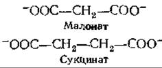
In Muscle tissue, respiration was inhibited by 90%, accompanied by an accumulation of succinate—providing yet another compelling piece of Evidence for the crucial role of the tricarboxylic acid cycle in animal tissue respiration.
Box 9-G
Poisons: Fluoroacetate and "Lethal synthesis"
Among simple compounds, sodium fluoroacetate is one of the most lethal poisons known. The LD50 (the dose lethal to 50% of test animals) for rats is a mere 0.2 mg/kg—nearly 10 times lower than the lethal dose of the nerve agent diisopropyl phosphorofluoridate (Chap. 7, Sec. D, 1a,b). Widely used (though controversial in its application) as a rodenticide known as "1080," fluoroacetate has also been identified in the leaves of certain poisonous plants found in Africa, Australia, and South America. Notably, difluoroacetate HCF2—COO- is entirely non-toxic.
Biochemical research has established that fluoroacetate itself is not toxic to Cells. Toxicity manifests only after fluoroacetate is metabolically converted into fluorocitrate, which acts as a highly specific inhibitor of aconitase (Chap. 7, Sec. I, 6b,c). This fact is quite remarkable because the isomeric fluorocitrate formed in the reaction of fluoroxaloacetate with acetyl-CoA exerts only a weak inhibitory effect on the same enzyme, even though it is precisely this isomer that positions the fluorine atom at the site attacked by aconitase.
The fluorine atom has a small Structure/103.html">Van der Waals radius (0.135 nm), comparable to that of hydrogen (0.12 nm); this property is frequently cited when explaining the ability of fluorine-containing compounds to "trick" Enzymes. However, it is more likely that the high electronegativity of fluorine and its capacity to participate in Hydrogen Bonds make it metabolically more comparable to a —OH group. In the case of fluorocitrate, it is hypothesized that the inhibitory isomer binds to aconitase in an "incorrect mode," wherein the fluorine atom becomes coordinationally bonded to the iron atom in the Active Site of aconitasec.
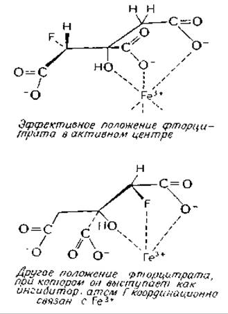
A fair number of naturally occurring fluorine-containing compounds are knownc. The biochemical properties of these compounds and fluorine-containing therapeutic drugs warrant further investigation.
1) Nobel Laureate in Physiology or Medicine, 1953.
a Gibble G. W., J. Chem. Educ., 50, 460-462 (1973).
b Elliot K., Birch I. eds., Ciba Foundation, Carbon-Fluorine Compounds, Elsevier, Amsterdam, 1972.
c Glusker J. P. In: The Enzymes (P. D. Boyer, ed.), 3rd ed., Vol. 5, pp. 413—439, Academic Press, New York, 1971.
Last update: 06/08/2026
Editorial and Educational Adaptation: This material has been compiled based on the primary/original source text. The project team performed an editorial review, corrected technical inaccuracies, structured sections, and adapted the content for an educational format.
What was processed:
- elimination of formatting defects (OCR errors, structural breaks, corrupted characters);
- editorial organization of content;
- standardization of terminology in accordance with academic sources;
- verification of factual statements against the original source text.
All mentions of the author, publication year, and origin of the primary text have been preserved in accordance with the source.