Molecular Biology of the Cell - Volume 3 - Alberts B., Bray D., Lewis J., Raff M., Roberts K., Watson J. 1994
From Cells to Multicellular Organisms
Plant Cell Characteristics
The Central Role of the Cell Wall
The Cell wall in plant Tissues is a complex Extracellular matrix that surrounds every cell. In contrast to animal Cells—most of which also feature an extracellular surface matrix (see Section 14.2)—The plant cell wall is typically much thicker, stronger, and, above all, more rigid. Most differences between plants and animals regarding Nutrition, Digestion, osmoregulation, growth, reproduction, Intercellular Communication, defense mechanisms, and Morphology are directly related to The properties of The Cell wall. For instance, the acquisition of rigidity by the plant cell wall led to the loss of locomotive ability. This sessile lifestyle has been retained in multicellular plants. It was precisely these thick cell walls, clearly visible under a Microscope, that enabled Robert Hooke in 1663 to first observe cells and give them the name we still use today.
Class="center">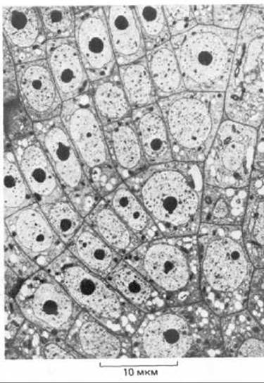
Fig. 20.1. Electron micrograph of cells from a reed ROOT tip. The regular Structure resulting from the strict sequence of Cell Division with rigid walls is clearly visible. (Courtesy of B. Gunning.)
The plant cell wall primarily protects the living Contents of the cell. Each such wall serves as a link between its own cell and neighboring cells, ensuring the unity and integrity of the entire plant Organism (Fig. 20-1). Although every plant cell is thus encased in its own "wooden box," the possibility of direct intercellular contact is not lost and is maintained through plasmodesmata. Thousands of these cytoplasmic channels, lined with Plasma Membrane, pierce the cell wall to connect adjacent cells and facilitate the movement of small molecules from Cell to Cell. In addition, fluids circulate along and through the cell wall. Thus, in plants, the cell wall performs not only protective and supportive Functions, but also transport roles.
During specialization, plant cells form walls that are particularly well adapted to perform specific functions. In this section, we examine the role that the surrounding cell wall plays in the life of a plant cell. We begin with a Description of the wall's structure.
20.1.1. The cell wall is formed of Cellulose fibers embedded in a polysaccharide-protein matrix [1]
Most newly formed cells in a multicellular plant arise in specialized regions called Meristems (see Section 20.5.1). These new cells are usually small compared to already differentiated ones. Their enlargement is possible because the walls of such cells (primary cell walls, Fig. 20-2) are very thin and represent semi-rigid structures. Once cell growth ceases, the wall no longer needs to expand. Mature (non-growing) cells may retain their primary cell wall, but much more frequently the cell builds up an additional secondary cell wall. This occurs either by thickening the primary wall or by depositing new, strong layers of varying composition on its inner side (see Section 20.1.7).
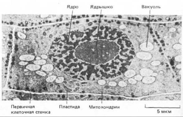
Fig. 20-2. Electron micrograph of a cell from a young onion root tip. The main Organelles and the thin primary cell wall are visible. (Courtesy of B. Wells.)
Although the primary cell walls of higher plants vary greatly in composition and fine Organization, they are all built on a single unified principle common to extracellular matrices: long fibers providing tensile strength are held together by a network of Proteins and Polysaccharides that confers resistance to compression. This same engineering principle (strong tensile-stress fibers surrounded by an amorphous, compression-resistant matrix) is used in the construction of animal bones (see Section 17.8) and is characteristic of widely used building Materials such as fiberglass and reinforced concrete. The fibrils making up Plant Cell Walls typically consist of cellulose polysaccharide—the most abundant organic macromolecule on Earth. The matrix, in turn, consists mainly of two Other types of polysaccharides: hemicelluloses and Pectins, as well as structural Glycoproteins (Fig. 20-3). The fibril molecules and the cross-linking matrix are bound together by covalent bonds and non-covalent interactions, forming an unusually complex structure whose composition is typically specific to each cell type (Fig. 20-4). While The structure of the main molecules forming cell walls in many cell types is known, it remains unclear whether all these molecular species are present in the walls of different cell types, nor has the exact manner in which these molecules assemble into a three-dimensional structure been fully established.

Fig. 20-3. Diagram of the putative linkage between the two Main Components of the primary cell wall—cellulose microfibrils and the matrix. Hemicellulose molecules (such as xyloglucans) are attached to The surface of cellulose microfibrils by Hydrogen Bonds. Some of these molecules are cross-linked by short molecules of neutral pectins (such as arabinogalactans) and acidic pectins (such as rhamnogalacturonans). Glycoproteins are tightly interwoven into the cell wall fabric.

Fig. 20-4. A. Electron micrograph of a primary cell wall in carrot (prepared by freeze-fracture and deep-etching, see Section 4.1.11). Cellulose microfibrils are interconnected by a complex network of matrix molecules. Compare this micrograph with the diagram in Fig. 20-3. B. Thin section of a typical primary cell wall. (A—courtesy of B. Wells and K. Roberts; B—courtesy of J. Burgess.)
20.1.2. Cellulose microfibrils are cross-linked with molecules of hemicellulose, pectin, and glycoprotein, resulting in The formation of a complex network [1,2]
A cellulose molecule is an unbranched chain containing at least 500 glucose residues covalently linked by ß1 → 4 glycosidic bonds, giving the entire molecule a ribbon-like structure stabilized by intramolecular hydrogen bonds. Intermolecular hydrogen bonds link adjacent cellulose molecules, helping to bind them into parallel strands that overlap to form a bundle of 60–70 chains of identical polarity. Such ordered crystalline aggregates, measuring many micrometers in length, are called cellulose microfibrils (Fig. 20-5).
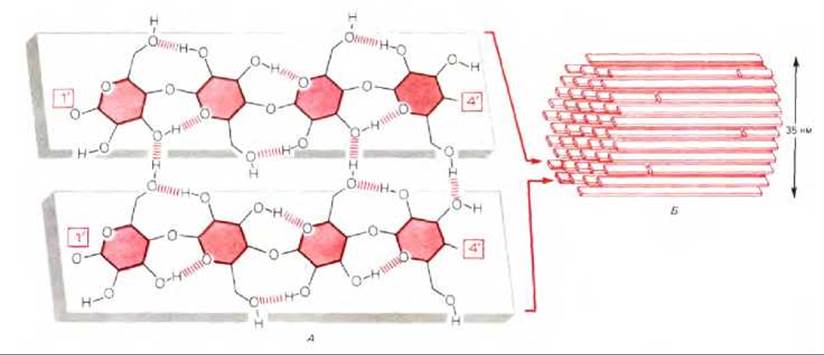
Fig. 20-5. Structure of cellulose. A. A small fragment of two cellulose molecules, each consisting of a long, flat chain of glucose residues linked by ß1 → 4 bonds; these chains reach many micrometers in length. Intramolecular hydrogen bonds stabilize each chain, while intermolecular hydrogen bonds tightly cross-link neighboring chains. Hydrogen bonds not shown in the figure connect each chain to those lying above and below it. B. Cellulose microfibrils consisting of numerous parallel cellulose molecules held together by hydrogen bonds. In most higher plants, the microfibril diameter is approximately 3.5 nm, although in some Algae it can be up to 10 times larger. Each cellulose molecule is polar (having 1'- and 4'-ends), and all molecules within a given microfibril share the same polarity.
Hemicelluloses are a heterogeneous group of branched polysaccharides that bind tightly to the surface of each cellulose microfibril and to one another, thereby coating the microfibrils and facilitating their assembly into a complex network via hydrogen bonds (see Fig. 20-3). Many different hemicelluloses exist, but all share a basic long linear backbone composed of molecules of a single monosaccharide linked by ß1 → 4 bonds, with short side chains of different sugar residues branching off this backbone (Fig. 20-6). The Monosaccharides of both the backbone and side chains are specific to the plant species and its developmental stage. Another important polysaccharide component of the cell wall is pectin. Pectins comprise a heterogeneous group of branched polymers containing numerous negatively charged galacturonic acid residues (Fig. 20-7). Due to their negative charge, pectins are heavily hydrated and actively bind cations. When Ca2+ ions are added to a pectin solution, cross-linking occurs to form a semi-solid gel (which is why pectin is added to fruit juices to make jelly). Such Ca2+-mediated cross-linking is believed to play a role in integrating cell wall components. The middle lamella—a specialized central region that cements together the cell walls of adjacent cells—is particularly rich in pectins (see Fig. 20-17). This layer is interrupted in places to form intercellular air spaces characteristic of many plant tissues (Fig. 20-8).

Fig. 20-6. Diagram of the structure of a hemicellulose molecule from the cell wall of a typical flowering plant. The cellulose-like backbone of the molecule consists of glucose residues and is attached by hydrogen bonds to the surface of a cellulose microfibril within the cell wall. Depicted here is a xyloglucan, in which xylose residues are attached to the glucose units of the backbone chain; other sugars, such as galactose and fucose, may also be Components of the side oligosaccharide chains.

Fig. 20-7. Diagram of the structure of an acidic pectin (rhamnogalacturonan) from a Higher Plant Cell wall. Kinks in the linear backbone chain of negatively charged galacturonic acid residues are shown. These kinks are caused by the presence of rhamnose residues, which serve as attachment sites for neutral pectins that link acidic pectins to hemicellulose molecules (see Fig. 20-3).

Fig. 20-8. Scanning electron micrograph of a freeze-fractured bean leaf. The upper and lower epidermis are clearly visible. Between them lie numerous leaf mesophyll cells. As these photosynthetic cells grow, their walls separate from one another at specific points along the middle lamella, forming an open cellular network where each cell has unrestricted access to carbon dioxide resources within the relatively large surrounding air spaces. (C. E. Jeffree, N. D. Read, V. A. Smith, J. E. Dale, Planta, 172, 20-37, 1987.)
In addition to the Three types of polysaccharides described above, the primary cell wall contains glycoproteins, which can account for up to 10% of its total mass. Researchers have successfully cloned the DNA encoding these major glycoproteins and determined its nucleotide sequence. Cell wall glycoproteins are unusual in that they contain many repeating Amino acid sequences. Up to 30% of their amino acid residues consist of hydroxyproline, which is formed through the post-translational hydroxylation of Proline (much like in Collagen, see Section 14.2.7). Short oligosaccharide chains are attached to the side chains of hydroxyproline and Serine, meaning that more than half of each glycoprotein's mass is made up of its carbohydrate moiety. Isolating these glycoproteins without disrupting the overall STRUCTURE OF THE cell wall is extremely difficult, indicating that they are firmly embedded within the complex three-dimensional network of polysaccharides that comprises the wall. It is believed that glycoproteins act much like glue, enhancing the tensile strength of the cell envelope. Notably, the levels of certain mRNAs encoding these glycoproteins increase dramatically in response to infection or wounding.
For a plant cell to increase in size or change its shape, its cell wall must stretch or deform. Due to their crystal-like structure, individual cellulose microfibrils are incapable of stretching; therefore, such changes require the microfibrils to slide relative to one another and/or the adjacent microfibrils to separate. As discussed below, the direction of elongation in a growing cell depends on the orientation of the stretch-resistant cellulose microfibrils within the primary cell wall (see Section 20.4.7).
20.1.3. Small pore size in the plant cell wall restricts the exchange of macromolecules between the cell and its environment [3]
All cells take up nutrients and excrete Metabolic waste products through The Plasma Membrane. In plant cells, these molecules must also pass through the cell wall. Because the cell wall matrix is a highly hydrated polysaccharide gel (the primary wall contains up to 60% Water by mass), water, gases, and small water-soluble molecules diffuse through it rapidly. (Even when the cell wall thickness reaches 15 µm, it accounts for only about 10% of the total resistance experienced by water molecules circulating between the Cytoplasm and the external environment; the remaining 90% is attributable to the plasma membrane.) The average diameter of the pores between the cross-linked macromolecules that form most cell walls is about 5 nm. This is sufficient to severely retard the movement of any globular macromolecule with a molecular weight exceeding 20,000. Consequently, plants are forced to utilize small molecules as nutrients. Molecules that mediate intercellular signals must also be small and water-soluble. Indeed, in plants, most known signaling molecules—such as growth regulators like Auxins, Cytokinins, and Gibberellins (see Section 20.5.8)—have molecular weights of less than 500.
20.1.4. The high tensile strength of the cell wall allows cells to maintain excess internal hydrostatic pressure, known as turgor [4]
The mechanical strength of the cell wall enables plant cells to survive in an environment that is hypotonic relative to the cell's interior. The extracellular fluid of higher plants includes the aqueous phase within all cell walls, as well as the liquid contained in the long tubes formed by the cell walls of dead xylem cells (see Section 20.2.5). These tubes transport water (the Transpiration stream) from the roots to evaporation sites located primarily in the leaves. Although extracellular fluid contains more dissolved solutes than the less concentrated solution in the plant's surrounding environment (such as soil), it nonetheless remains hypotonic with respect to the intracellular fluid. This can be easily demonstrated by degrading the cell wall with cellulases and Other Enzymes and observing The behavior of such wall-less cells, known as protoplasts (see Fig. 20-7). If this spherical protoplast is kept under hypotonic conditions for a time, it takes up water osmotically and bursts (Fig. 20-9). A cell that has retained its wall will only swell slightly in the same situation. This is because the cell generates its own internal hydrostatic pressure, which Supports the cell wall much like an inflated inner tube supports a bicycle tire casing. This hydrostatic pressure leads to osmotic equilibrium and prevents any further influx of water (for a detailed illustration of osmosis, see Panel 6-1).
The excess hydrostatic pressure inside a plant cell—known as turgor pressure (or simply turgor)—resulting from the osmotic imbalance between these two environments is vital to the plant's survival. Turgor is the primary force driving cell expansion during growth; it is also largely responsible for the rigidity of living plant tissues (compare the wilted leaf of a dehydrated plant with the turgid leaves of a well-watered one).
20.1.5. Plant cell growth is determined by both turgor pressure and controlled cell wall synthesis [5]
Turgor pressure is important not only for maintaining plant tissue turgidity. Whenever the cell wall yields to internal turgor pressure and stretches, an irreversible increase in cell volume occurs—in other words, the cell grows. Cell growth takes place only when the internal turgor pressure exceeds the local yield strength of the wall. In principle, a plant can employ two strategies for growth: either increase turgor pressure or weaken the cell wall in specific regions. There is solid evidence that plant cells adopt the latter strategy, weakening the cell wall through various mechanisms (for example, by pumping H+ ions into the wall via a plasma membrane-localized H+-ATPase that acts as a proton pump). Although the details of this mechanism remain incompletely understood, it is believed that a local decrease in pH leads to a reduction in the weak bonds holding the wall components together, allowing the wall macromolecules to slide past one another under METABOLISM/18.html">The Influence of turgor pressure. To further facilitate wall expansion, other more complex modifications take place, including the activation of enzymes that catalyze the Hydrolysis of glycosidic and other covalent bonds.
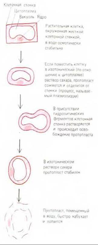
Fig. 20-9. A plant cell lacking a wall—that is, a protoplast—is osmotically unstable and, when placed in water or the hypotonic extracellular fluid that bathes plant cells, swells and bursts. By contrast, if the cell is surrounded by a rigid wall, it can swell only to a limited extent. The pressure exerted by the cell against its cell wall makes it turgid and establishes osmotic equilibrium, halting any further net entry of water into the cell (see Panel 6-1).

Fig. 20-10. A substantial increase in cell size can be achieved without a proportional increase in cytoplasmic volume. Wall loosening allows the cell to expand in a specific direction driven by turgor pressure, accompanied by water uptake into the Swelling vacuole. As a result, the cytoplasm becomes a thin peripheral layer connected to the nuclear region by strands that traverse the vacuole and contain bundles of Actin filaments (see Fig. 20-53).
In most cases, the increase in cell volume occurs unevenly, driven primarily by vacuolar expansion rather than cytoplasmic growth. Maintaining a nearly constant amount of nitrogen-rich cytoplasm reduces the metabolic cost of producing large cells (Fig. 20-10). This provides an additional advantage for plants growing in environments where nitrogen is a limiting nutrient. To sustain the turgor pressure required for continuous cell enlargement, the growing vacuole must actively take up and accumulate solutes to maintain its osmotic pressure.
However, plant cell growth is a far more complex process than simply inflating a balloon, as the cell must also acquire a specific shape. Cell growth is actually governed by two intricate processes. One of these, operating via a feedback loop, involves the interaction between two events: cell wall stretching and turgor recovery. The other entails the cell assuming a defined shape as certain Regions of the wall remain rigid while other areas soften and stretch. Such Regulation of Cell shape is mediated by events occurring within the cytoplasm, as described below (see Section 20.4.8).
20.1.6. Turgor is regulated via a feedback mechanism through changes in intracellular solute concentrations [6]
Given the crucial role of turgor pressure in plant life, it is hardly surprising that plant cells have evolved sophisticated mechanisms to regulate its magnitude. This value varies widely depending on the plant species and cell type, ranging from as little as half an atmosphere in certain large-celled algae to as much as 50 atmospheres in stomatal guard cells. Cells can increase turgor pressure by raising the concentration of osmotically active molecules in the Cytosol—either by pumping them inward from the extracellular fluid across the plasma membrane or by breaking down osmotically inactive polymers typically stored in vacuoles. In both cases, changes in turgor pressure are regulated by feedback control.
How do such feedback systems operate? Experiments indicate that a "turgor pressure detector" in the plasma membrane triggers ion transport (most commonly the active uptake of K+ ions into the cell) in response to a sudden drop in turgor pressure, whereas a sharp increase in turgor leads to the efflux of K+ ions. These processes occur very rapidly and appear to involve alterations in specific transport proteins within the plasma membrane.
Membrane-associated detectors likely also modulate the rate at which osmotically active substances are synthesized in the cytoplasm and vacuole, though these changes occur much more slowly. Systems that regulate cellular turgor are particularly critical for plants inhabiting environments with extreme or fluctuating osmotic properties. For instance, plants growing in saline soils must accumulate very high concentrations of solutes in their fluids to maintain turgor. Because accumulating such massive quantities of ions like K+ would likely disrupt The activity of vital enzymes, these cells instead accumulate specialized organic molecules—polyhydroxylated compounds such as glycerol or mannitol, Amino Acids like proline, or N-methylated Amino Acid Derivatives such as Glycine betaine. The concentration of these substances in the cytosol can reach very high levels (0.5 M) without interfering with cellular metabolism. The vacuole and its contents play a direct role in regulating turgor pressure in response to environmental shifts (see Section 20.4.2).
Regulated changes in turgor also underlie the limited movements observed in plants. For example, stomatal guard cells control The rate of gas exchange between leaves and the surrounding air by opening and closing the stomatal pores (Fig. 20-11). During the day, when Stomata are open, light activates a K+ ion pump in the plasma membrane of the guard cells; as a result of K+ influx, turgor pressure rises, causing the guard cells to swell and open the stomatal pore. Extremely rapid turgor changes in strategically positioned cells drive more conspicuous movements, such as the snapping shut of traps in carnivorous plants and the rapid motion of certain flower parts during pollination. The turgor shifts responsible for these movements result from a sudden surge in membrane permeability in specialized key cells. These cells act as turgor-regulated "hinges." Exactly how a light Touch to a sensitive surface triggers a turgor-driven burst in these cells remains unclear, but it likely involves Ion Channels coupled with action potentials.

Fig. 20-11. Scanning electron micrographs at different magnifications showing stomata in the leaf epidermis of a tropical plant. Stomata are pores formed on the leaf surface by a pair of guard cells. Their turgor-regulated movements determine the size of the stomatal aperture and, consequently, the rate of gas exchange between the leaf and the environment. In most plants, stomata are open during the day to admit carbon dioxide and release photorespiratory byproducts. At night, stomata generally close. The epidermal cells are coated on the outside by a waterproof waxy cuticle (see also Fig. 20-18). (Courtesy of N.W. Woolhouse and G.J. Hills.)
20.1.7. The formation of specialized cells involves the Modification of the cell wall [7]
Land plants vary widely in their structure and reproductive strategies (Fig. 20-12). Nevertheless, they are all built upon common principles from a small set of cell and tissue types. The body plan of nearly all plants is based on longitudinal modular units, where a typical module consists of a stem, a leaf, and a bud (see Fig. 20-58). Furthermore, they all contain the same specialized cell types, which are invariably organized into three major tissue systems: dermal (providing protective coverage), ground (providing support and nutrition), and vascular (providing fluid transport) (Fig. 20-13).
The principal cell types are illustrated in Panel 20-1. All of them originate from cells with a primary cell wall through growth and subsequent differentiation. As cells differentiate, the primary wall becomes elaborated to form a secondary cell wall. In some cases, this process simply involves The addition of successive cellulose layers, but it frequently entails the deposition of new layers with a distinct chemical composition. Cellulose molecules deposited in the secondary wall are typically much longer (~15,000 glucose residues) than those found in the primary wall (500 to 5,000 glucose residues). Moreover, the highly hydrated pectin components characteristic of the primary cell wall are largely replaced by other polymers, making the secondary wall denser and significantly less hydrated than the primary wall.
The secondary cell wall bears the brunt of the mechanical load experienced by the plant. It also serves as a vital nutritional component for many animals and forms The basis of materials such as wood and paper. The shape and COMPOSITION OF THE resulting cell wall are closely tied to the function of specific specialized cell types: each cell type differs from another in morphology—for instance, in the architecture of the mature pollen grain wall, which is unique to each specific plant species (Fig. 20-14).

Fig. 20-12. The evolution of land plants. All plants (except algae) can be divided into vascular plants, in which transport occurs through specialized conducting tissues (xylem and phloem), and non-vascular plants (such as mosses), which are small in size and relatively simple in organization. As noted previously, vascular plants appeared on Earth approximately 350 million years ago with the advent of seeds. The seed provides protection for the developing embryo, which can remain dormant until conditions necessary for its further development are met. Because seed-bearing vascular plants are the most widespread on Earth, this chapter focuses primarily on them.

Fig. 20-13. Three tissue systems can be distinguished in various Organs of higher plants (leaves, stems, and roots): the vascular, ground (supporting), and dermal systems. As seen in the schematic cross-section of a root tip, Vascular Tissues are embedded in the ground tissue, which in turn is surrounded by the dermal tissue. With some variations in arrangement, these three systems make up all parts of a higher plant. Each system consists of a relatively small number of principal cell types, some of which are shown in Panel 20-1.
All significant changes in both the Composition and Structure of the Primary and secondary cell walls reflect processes occurring within the cytoplasm; this is best illustrated by The Development of xylem vessels. During Cell Differentiation in young growing tissues, thickenings rich in cellulose are deposited on the walls of xylem vessel elements. Their arrangement is guided by bundles of Cytoplasmic microtubules lying in the cortical layer directly beneath the plasma membrane. These microtubule bundles typically form spirals or rings (Fig. 20-15) and arise from the reorganization of cortical microtubules, which are normally distributed rather evenly (see Section 20.4.8). This is a specific example of a more general rule discussed below (in the same section), which states that extracellular cellulose microfibrils are laid down parallel to the microtubules located in the cortical cytoplasm.
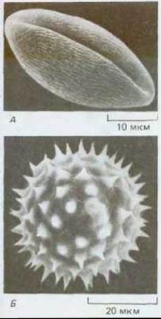
Fig. 20-14. Scanning electron micrograph of pollen grains from petunia (A) and sunflower (B). The wall is composed of sporopollenin, a complex and exceptionally durable hydrocarbon polymer that determines the characteristic shape of the pollen grains. Pores in the wall allow the pollen tube to emerge during germination. (Courtesy of S. MacFarlane and C. Jeffree.)
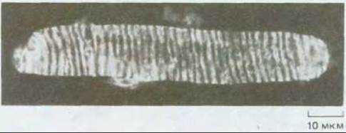
Fig. 20-15. Cortical microtubules in a cell from a developing pea SHOOT xylem. Staining with fluorescent Antibodies reveals a helical arrangement of microtubules that define the region of the cell wall to be thickened, first by cellulose deposition and subsequently by Lignin (see Panel 20-1). Because the cell is large, the depth of focus allows only one side of the helical pattern to be seen. (Courtesy of I. Roberts.)
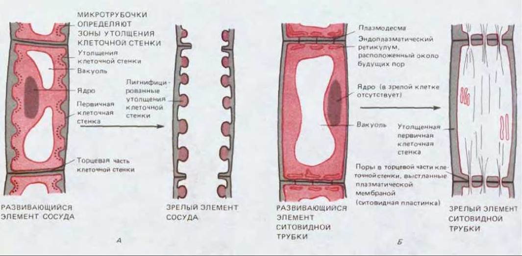
Fig. 20-16. Two Examples of potential cell wall modifications during the formation of specialized cells. A. Schematic Cytology/practical/54.html">Longitudinal section of a developing small xylem vessel element. Annular thickenings form in this cell, along with other types of deposits. Eventually, the protoplast and the end cell wall disappear, creating a continuous open tube. The mature element dies after losing its protoplast. B. Schematic longitudinal section through a developing phloem sieve tube element. The primary cell wall thickens, and pores appear in its end walls to form a sieve plate connecting adjacent tube elements. Mature cells retain their plasma membrane, but lose The Nucleus and most of the cytoplasm.
The thickened cellulosic regions of the developing xylem cell walls are subsequently reinforced by the deposition of lignin, an insoluble polymer composed of aromatic phenolic units that form a cross-linked, branched network within the cell wall, providing the structural basis of wood. Through the localized removal of material from the end walls, rigid, low-resistance vessels are formed for efficient water transport in the xylem (Fig. 20-16). A similarly profound remodeling of the primary cell wall occurs during the development of phloem sieve tubes in the plant's vascular tissues (see Fig. 20-16 and Panel 20-1).

Fig. 20-17. Formation of a secondary fiber cell wall (schematic cross-section). In this case, three new layers of cell wall material have been deposited inside the primary wall. Because the orientation of cellulose microfibrils differs in each layer, the cell wall structurally resembles plywood. In many fibers, the completion of secondary Cell wall formation culminates in cell death.
The secondary cell wall is typically deposited between the plasma membrane and the primary cell wall, sometimes with layers laid down sequentially one after another (Fig. 20-17). However, in certain instances, specialized macromolecules are deposited either within the primary wall (such as lignin in xylem cells) or on its outer surface. For example, epidermal cells covering the outer surface of a plant generally possess a thickened primary cell wall, the outer portion of which is covered by a thick, waterproof cuticle that protects the plant against infection, mechanical damage, water loss, and harmful ultraviolet radiation (see Panel 20-1). The cuticle is secreted as the epidermal cells differentiate. It consists predominantly of cutin (or suberin, a chemically related substance in bark), which is a long-chain fatty acid polymer forming an extensive cross-linked network on the plant surface. The cutin layer is frequently impregnated and layered with Waxes, which are esters of long-chain alcohols and Fatty acids (Fig. 20-18). The plant cell cuticle differs fundamentally in composition from the cuticles of insects and crustaceans, which are constructed of proteins and polysaccharides.
20.1.8. Even the mature cell wall remains a dynamic structure [8]
The composition and structure of an adult plant's cell wall are not static: constituents can be added and removed, and the linkages between components can change. The localized removal of wall material during the development of xylem vessel end walls and phloem sieve tubes striking examples of such modifications (Fig. 20-16).
Cell wall modification in already mature cells is also exemplified by processes occurring during the loss of a plant part, such as a fallen leaf. As a leaf dies, its cell polymers break down, and sugars, amino acids, and ions are reclaimed by the plant. In addition, the Aging leaf releases small amounts of Ethylene gas. In the region located between the Base of the leaf petiole and the stem (the abscission zone), cells respond to complex and still poorly understood combinations of ethylene and other endogenous plant growth regulators (see Fig. 20-67) by synthesizing and secreting cell wall-degrading enzymes (such as pectinase and cellulase). These enzymes act locally on a specific target area, partially dissolving the cell walls within the abscission zone (Fig. 20-19). Concurrently, water-resistant suberin is deposited in the layer of stem-side cells, protecting the "wound" created after leaf detachment caused by enzymatic digestion.
A similar localized cellular response occurs during fruit ripening. In this case, low concentrations of ethylene (one part per million) stimulate the secretion of Proteolytic Enzymes in target Cells of the fruit (such as oranges or bananas), which weaken the adhesion between neighboring cells. This leads to the softening—that is, the "ripening"—of the fruit.

Panel 20-1. Cell types in higher plants.

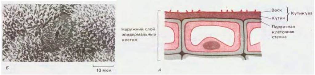
Fig. 20-18. A. Typical mature cell from the leaf epidermis (schematic section). Waterproof layers of cutin and wax are deposited on the outer surface of the thick primary cell wall, together forming the cuticle. Cuticular wax deposits frequently create complex surface patterns. B. Scanning electron micrograph of the lower epidermis of a pea leaf, clearly showing the characteristic wax deposition around the stomatal pore. (Courtesy of P. Linstead.)
Higher plants consist of a vast number of cells bound together in specific ways by their surrounding cell walls. Many Characteristic Properties of plants are directly or indirectly linked to the presence of these cell walls. The composition and appearance of cell walls are directly determined by the cell type and its specific functions. At the same time, the fundamental structural principles of all cell walls are remarkably similar: rigid cellulose microfibrils are embedded in a cross-linked matrix consisting of polysaccharides, such as pectins and hemicelluloses, as well as glycoproteins. Due to this architecture, the primary cell wall possesses a high tensile strength and is permeable only to relatively small molecules. If a plant cell lacking a cell wall (a protoplast) is placed in water, it will take up water by osmosis, swell, and burst. Conversely, when the living contents of a cell are enclosed within a wall, they swell and exert pressure against it, generating what is known as turgor pressure. Turgor is strictly regulated and is essential both for cell expansion and for providing mechanical rigidity to young plants.
The species diversity of higher plants is shaped by a relatively small number of specialized cell types. During their formation—such as the vascular elements of the two conducting tissues, xylem and phloem—the cell wall undergoes significant modifications. Certain regions of the cell wall may be reinforced, often through the addition of one or more layers that form the secondary cell wall. Other areas may be selectively degraded, as occurs with the end walls when a continuous vessel is formed from a long file of cylindrical cells. These structural alterations of the cell wall are governed by temporal and spatial changes within the cytoplasm of developing cells. The cell wall is a dynamic structure whose composition and morphology can undergo marked changes not only during cell growth and differentiation, but also after maturation.
Last update: 12/08/2026
Editorial and Educational Adaptation: This material has been compiled based on the primary/original source text. The project team performed an editorial review, corrected technical inaccuracies, structured sections, and adapted the content for an educational format.
What was processed:
- elimination of formatting defects (OCR errors, structural breaks, corrupted characters);
- editorial organization of content;
- standardization of terminology in accordance with academic sources;
- verification of factual statements against the original source text.
All mentions of the author, publication year, and origin of the primary text have been preserved in accordance with the source.