LEHNINGER PRINCIPLES OF BIOCHEMISTRY - VOLUME 1. THE FOUNDATIONS OF BIOCHEMISTRY: STRUCTURE AND CATALYSIS - 2011
PART I. STRUCTURE AND CATALYSIS
Class="center">Good fences make good neighbours.
Robert Frost, "Mending Wall" in *North of Boston*, 1914
11. BIOLOGICAL MEMBRANES AND TRANSPORT
The first Cell likely arose when a membrane formed, separating a small volume of aqueous solution from the outside world. Membranes form the outer boundaries of Cells and regulate the movement of molecules across these boundaries (Fig. 11-1). In Eukaryotic cells, they compartmentalize the intracellular space to isolate specific processes and components. They organize complex reaction sequences and play a central role in both biological energy storage and The formation of intercellular contacts. The BIOLOGICAL FUNCTIONS OF membranes stem from their remarkable physical properties. Membranes are plastic, capable of self-sealing, and selectively permeable to polar solutes. Because of their plasticity, cell growth and movement (such as amoeboid movement) can alter cell shape. Possessing The ability to rupture and reseal, two membranes can fuse, as in exocytosis; a single membrane-bounded compartment can divide into two separate compartments without major leakage of its contents, as in endocytosis and Cell Division. Because Membranes are selectively permeable, they retain certain substances and ions within cells and within individual cellular compartments while excluding others.
Figure 11-1. Introduction/36.html">Biological Membranes. In cross-section, all cellular membranes share a characteristic trilaminar appearance. After staining an erythrocyte with osmium tetroxide, its Plasma Membrane appears in the Electron microscope as a trilaminar Structure 5 to 8 nm thick (50–80 Å). The trilaminar image consists of two electron-dense layers separated by a less dense central region (osmium binds to the inner and outer surfaces of the membrane).

Membranes are not merely passive barriers. They contain a multitude of Proteins designed to facilitate or catalyze various cellular processes. At The Cell surface, transporters move specific Organic compounds and inorganic ions across the membrane; receptors perceive extracellular
signals and trigger molecular changes within the cell; adhesion molecules hold neighboring cells together. Within the cell, membranes organize cellular processes such as the synthesis of Lipids and certain proteins, as well as energy Transduction in Mitochondria and METABOLISM/14.html">Chloroplasts. Because membranes consist of just two molecular layers, they are extremely thin—practically two-dimensional. Intermolecular collisions in this two-dimensional space are far more probable, vastly increasing the efficiency of enzymatic processes within the membrane.
In this chapter, we will first describe the composition of cell membranes and their chemical architecture—the molecular structures that underpin their biological functions. Next, we will examine the remarkable dynamic properties of membranes, in which lipids and proteins undergo lateral and rotational diffusion. We will discuss the dynamic role of Membrane Proteins in Cell Adhesion, endocytosis, and the membrane fusion that accompanies neurotransmitter secretion. We then turn to protein-mediated Transport of substances across membranes via carriers and Ion Channels. In subsequent chapters, we will discuss The Role of membranes in signal transduction (Chapters 12 and 23), energy transduction (Chapter 19), lipid synthesis (Chapter 21), and Protein Synthesis (Chapter 27).
11.1. Composition and Architecture of Membranes
Understanding membrane function requires studying their composition—which molecules are found in all membranes and which are unique to membranes with specialized functions. Therefore, before delving into membrane Structure and function, let us examine the molecular makeup of biological membranes: proteins and polar lipids, which make up the bulk of the membrane, and CARBOHYDRATES (Glycoproteins and Glycolipids).
Each membrane type contains characteristic lipids and proteins
The relative proportions of proteins and lipids vary depending on the membrane type (Table 11-1), reflecting differences in biological function. For example, some Neurons possess a myelin sheath—an extensive plasma membrane that wraps around the cell multiple times and acts as a passive electrical insulator. The myelin sheath consists predominantly of lipids, whereas bacterial Plasma Membranes, as well as the Mitochondrial and Chloroplast membranes where many enzymatic processes are concentrated, contain a higher proportion of protein to lipid by weight.
Table 11-1. Composition of plasma membranes in various organisms
Components (wt %) Sterol Other lipids Proteins |
Sterols |
||
Human nerve myelin |
30 30 19 |
galactolipids, plasmalogens |
|
Mouse Liver |
45 27 25 |
cholesterol |
- |
Corn leaves |
47 26 7 |
sitosterol |
galactolipids |
52 7 4 |
ergosterol |
triacylglycerols, sterol esters |
|
Paramecium |
56 40 4 |
stigmasterol |
- |
E. coli |
75 25 0 |
- |
- |
Note: The total composition does not add up to 100% because, In addition to proteins, phospholipids, and sterols, other substances are present. For instance, plants contain large amounts of glycolipids.
The first step in studying membrane composition is isolation. When cells are mechanically disrupted, their plasma membranes rupture and fragment, releasing cytoplasmic components and membrane-bound Organelles—mitochondria, chloroplasts, Lysosomes, and nuclei. Plasma membrane fragments and intact organelles can be isolated by centrifugation (Figs. 1-8 and Box 2-1, p. 87).
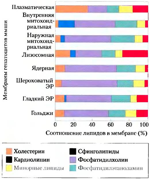
Cells undoubtedly possess mechanisms to regulate the types and amounts of Membrane Lipids, from their synthesis to the delivery of specific lipids to designated organelles. Every kingdom, species, tissue, cell type, and organelle type possesses a characteristic set of membrane lipids. For example, plasma membranes are rich in cholesterol and contain no appreciable amounts of cardiolipin (Fig. 11-2); in The inner mitochondrial membranes of hepatocytes, the distribution is reversed: very little cholesterol and abundant cardiolipin. With few exceptions, we can attribute an adaptive role to these various combinations of membrane lipids; in other cases, the Functional Significance of these combinations remains unknown.
Figure 11-2. Lipid Composition of plasma membranes and organelle membranes in rat hepatocytes. The functional specialization of each membrane type is reflected in its unique lipid composition. Cholesterol is present in substantial quantities in plasma membranes, but is found only in trace amounts in mitochondrial membranes. Cardiolipin is an essential component of the inner mitochondrial membrane, but not of The Plasma Membrane. In most membranes, phosphatidylserine, phosphatidylinositol, and phosphatidylglycerol are minor components (yellow), yet they perform critical functions; for instance, phosphatidylinositol and its derivatives participate in hormone signaling. Sphingolipids, phosphatidylcholine, and phosphatidylethanolamine are present in most membranes, but in varying ratios. Glycolipids are abundant in plant chloroplast membranes while being practically absent in animal Tissues.
The protein composition of membranes varies even more widely than their lipid composition, reflecting their functional specialization. Furthermore, some membrane proteins are covalently linked to Oligosaccharides. For example, 60% (by weight) of Glycophorin—an erythrocyte membrane glycoprotein—consists of complex oligosaccharide units covalently attached to specific amino acid residues. Sugars are most frequently attached via Ser, Thr, and Asn residues (Fig. 7-29). Sugar residues on surface glycoproteins influence protein folding, stability, and intracellular localization. They play an essential role in the specific binding of ligands to cell-surface glycoprotein receptors (Fig. 7-35).
Some membrane proteins are covalently attached to one or more lipids, which serve as hydrophobic anchors that, as we will see, help tether the proteins to the membrane.
All biological membranes share a set of fundamental properties
Membranes are impermeable to most polar or charged species, but permeable to nonpolar compounds; membrane thickness ranges from 5 to 8 nm (50–80 Å); in cross-section under an electron microscope, they appear trilaminar (Fig. 11-1). Based on Electron Microscopy, chemical composition analysis, and physical Methods investigating membrane permeability and the movement of individual lipid and protein molecules within the membrane, the Fluid Mosaic Model of biological membranes was developed (Fig. 11-3). Phospholipids form a bilayer, with the nonpolar portions of the molecules oriented toward the interior of the bilayer, while their polar “heads” face outward and interact with the aqueous phase on both sides of the membrane. Proteins are embedded in this bilayer matrix, anchored between membrane lipids and the hydrophobic domains of the proteins via hydrophobic interactions. Some proteins protrude from only one side of the membrane, whereas others have domains exposed on both sides. The orientation of proteins within the bilayer is asymmetric, giving the membrane a “sidedness”: Protein domains on one side of the bilayer differ from those on the other side, which also reflects functional Asymmetry. Individual lipid and protein units in the membrane form a constantly changing fluid mosaic structure (unlike a mosaic of ceramic tiles laid on cement). The membrane mosaic is fluid because most interactions between its components are noncovalent; lipids and proteins are capable of free lateral movement along the plane of the membrane.
Fig. 11-3. The fluid mosaic model of Membrane Structure. Fatty acyl chains within the membrane form a Hydrophobic core. Integral proteins float in this lipid “sea” owing to their hydrophobic properties, conferred by nonpolar amino acid side chains. Both proteins and lipids can move freely along the plane of the bilayer, whereas movement from one leaflet of the bilayer to the other is restricted. Carbohydrate moieties attached to certain proteins and lipids of the plasma membrane are located on the outer surface of the membrane.

We will now examine some Features of the fluid-crystal model in greater detail and provide experimental evidence supporting the core Concept of the proposed model, along with necessary refinements.
The Lipid Bilayer Is the Structural Foundation of Biomembranes
Glycerophospholipids, sphingolipids, and sterols are virtually insoluble in Water. When mixed with water, they spontaneously form microscopic lipid aggregates (clusters) as a separate phase, with their hydrophobic moieties in contact with one another while their hydrophilic groups interact with the surrounding aqueous medium. Recall that the aggregation of lipids into clusters decreases the Hydrophobic surface area exposed to water, thereby minimizing the number of ordered water molecules at the lipid-water interface (Fig. 2-7) and leading to an increase in Entropy. Hydrophobic interactions between lipid molecules provide the thermodynamic driving force for the formation and maintenance of these clusters.
When lipids are mixed with water, depending on conditions and The Nature of the lipids, Three types of lipid clusters can form (Fig. 11-4). Micelles are spherical structures containing nothing other than amphiphilic molecules (ranging from several dozen to several thousand). These molecules are arranged such that their hydrophobic regions aggregate in the interior, excluding water, whereas their hydrophilic “heads” face the surface and contact the aqueous medium. Micelle formation is favored when the cross-sectional area of the HEAD group is larger than that of the acyl side chain(s), as is the case for free Fatty acids, lysophospholipids (phospholipids lacking one fatty acid), and detergents such as sodium dodecyl sulfate (SDS; p. 136).
The second type of lipid aggregate in water is the bilayer, in which two lipid monolayers form a two-dimensional sheet. Bilayer formation occurs most readily when the cross-sectional areas of the head group and the acyl chain(s) are well-matched, as in glycerophospholipids and sphingolipids. The hydrophobic regions of each monolayer interact with one another, avoiding contact with water. The hydrophilic heads interact with water at both surfaces of the bilayer. Because the hydrophobic regions at the edges (Fig. 11-4b) have transient contact with water, the bilayer sheet is relatively unstable and spontaneously folds back on itself to form a hollow sphere, a vesicle (Fig. 11-4c). By forming vesicles, bilayers eliminate their exposed edge hydrophobic regions, achieving maximal stability in an aqueous environment. Water is entrapped within these bilayer vesicles as a separate aqueous compartment. The precursors of the earliest living cells likely resembled such lipid vesicles, with their aqueous contents segregated from the rest of the world by a hydrophobic shell.
Fig. 11-4. Aggregates formed by amphiphilic lipids in water, a) In micelles, the hydrophobic fatty acyl chains are packed at the center of the sphere. There is virtually no water within the hydrophobic core. b) In an open bilayer, all acyl side chains except those at the edges of the sheet are protected from interaction with water, c) When the two-dimensional bilayer folds, it forms a closed bilayer, a three-dimensional hollow vesicle enclosing water in its interior.

Biological membranes consist of lipid bilayers 3 nm (30 Å) thick. The hydrocarbon core of the membrane, composed of the —СН2- and -СН3 groups of fatty acids, is nonpolar and resembles decane in this respect; vesicles formed in the laboratory from pure lipids (Liposomes) are nearly as impermeable to polar molecules as biological membranes (although the latter are permeable to specific solutes via transporters).
The lipids of the plasma membrane are distributed asymmetrically between the two monolayers, although, unlike membrane proteins, this asymmetry is not absolute. In the human erythrocyte plasma membrane, for example, Choline-containing lipids (phosphatidylcholine and sphingomyelin) are predominantly located in the outer (exoplasmic) monolayer (Fig. 11-5), whereas phosphatidylserine, phosphatidylethanolamine, and phosphatidylinositols are much more prevalent in the inner (cytoplasmic) monolayer. Alterations in lipid distribution between plasma membrane monolayers can have profound consequences. For instance, only when phosphatidylserine is translocated to the outer monolayer of the plasma membrane can a platelet fulfill its role in Blood clotting. For many other cell types, the exposure of phosphatidylserine on the outer membrane surface serves as an “eat-me” signal for cell destruction via programmed cell death.
Fig. 11-5. Asymmetric distribution of phospholipids between the inner and outer monolayers of the erythrocyte plasma membrane. The Distribution of a specific phospholipid is determined by treating intact cells with phospholipase C, which cannot act on lipids in the inner monolayer but removes the polar head groups of lipids in the outer monolayer. The amount of released head groups serves as a measure of the proportion of each lipid in the outer monolayer.

Three Types of Membrane Proteins Differ in Their Membrane Association
Integral membrane proteins are firmly embedded in the membrane and can be removed only by agents that disrupt hydrophobic interactions, such as detergents, organic Solvents, or denaturing Reagents (Fig. 11-6). Peripheral membrane proteins are associated with the membrane through Electrostatic Interactions and hydrogen bonding with the hydrophilic domains of integral proteins and the polar head groups of membrane lipids. They can be released by relatively mild treatments that disrupt electrostatic interactions or break Hydrogen Bonds, such as high-pH carbonate extraction. Amphitropic proteins can exist in both cytosolic and membrane-bound states. Their affinity for membranes is driven in some cases by noncovalent interactions with membrane proteins or lipids, and in other cases by the covalent attachment of one or more lipids to the amphitropic protein (see Fig. 11-14). Typically, the Reversible Binding of amphitropic proteins to membranes is regulated; for example, phosphorylation or Ligand binding can induce Conformational Changes in the protein that expose a previously inaccessible membrane-binding site.
Fig. 11-6. Peripheral, integral, and amphitropic proteins. Membrane Proteins can be classified According to the conditions required for their release from the membrane. Most peripheral proteins are released by changes in pH or Ionic strength, chelation of Ca2+, or The addition of urea or carbonate. Integral proteins are extracted by detergents that disrupt hydrophobic interactions with the lipid bilayer and form micelle-like clusters around individual protein molecules. Integral proteins covalently linked to membrane lipids, such as glycosylphosphatidylinositol (GPI, Fig. 11-14), can be released by Treatment with phospholipase C. Amphitropic proteins alternate between membrane-bound and cytosolic states; this binding process is regulated, for example, via reversible palmitoylation.

Many Membrane Proteins Span the Lipid Bilayer
The topology of a membrane protein (its orientation relative to the lipid bilayer) can be determined using reagents that react with protein side chains but cannot cross membranes. Examples include polar chemical reagents that react with primary amines of Lysine residues, or Enzymes like Trypsin that cleave proteins but cannot penetrate the membrane. Human erythrocytes are a convenient model for such studies because they lack membrane-bound organelles, leaving the plasma membrane as their only membrane. If a membrane protein in an intact erythrocyte reacts with an impermeable reagent, that protein must have at least one domain exposed on the outer (extracellular) surface of the membrane. Trypsin digests extracellular domains without affecting domains hidden within the bilayer or located exclusively on the inner surface, unless the plasma membrane is intentionally disrupted to make those domains accessible to the enzyme.
Experiments with such topologically specific reagents reveal that the erythrocyte glycoprotein glycophorin spans the plasma membrane. Its N-terminal domain (bearing carbohydrate chains) is located on the outer surface and is cleaved by trypsin. The carboxyl terminus protrudes into the intracellular space, where it is inaccessible to membrane-impermeable reagents. Both the N- and C-terminal domains contain numerous polar or charged amino acid residues and are thus fully hydrophilic. However, a segment in the middle of the protein (residues 75–93) is rich in hydrophobic Amino Acids, leading to the Conclusion that glycophorin possesses a transmembrane segment positioned as shown in Figure 11-7.
Fig. 11-7. Glycophorin spans the erythrocyte bilayer. One hydrophilic domain containing all the sugar residues is located on the outer surface, and another hydrophilic domain protrudes from the inner surface of the membrane. Each red hexagon represents a tetrasaccharide containing two molecules of Neu5Ac (sialic acid), Gal, and GalNAc linked via oxygen to a Serine (Ser) or Threonine (Thr) residue; the blue hexagon represents an N-linked oligosaccharide chain attached to an Asn residue. In reality, the oligosaccharide units are relatively larger. A segment of 19 hydrophobic residues (75–93) forms an α-helix that traverses the membrane bilayer (Fig. 11-11a). A segment comprising amino acid residues 64–74 contains several hydrophobic residues and is likely embedded in the membrane from the extracellular side, as illustrated.
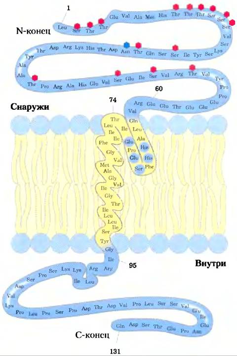
Experiments have demonstrated that glycophorin is arranged asymmetrically in the membrane: its N-terminus is always directed outward. Similarly, other membrane proteins have been shown to possess a specific orientation within the bilayer, such that one domain of a transmembrane protein always faces the exterior while the other faces the interior. Furthermore, plasma membrane glycoproteins are always oriented such that their carbohydrate residues reside on the outer surface of the cell. As we will see, the asymmetric arrangement of membrane proteins dictates their functional asymmetry. For instance, The structure of an ion pump reveals that all of its molecules share the same orientation within the membrane and therefore operate in a uniform direction.
Integral Proteins Interact with Lipids via Their Hydrophobic Domains, Thereby Anchoring Themselves in the Membrane
A strong connection between integral proteins and membranes is achieved through hydrophobic interactions between membrane lipids and the hydrophobic domains of the protein. Some proteins contain a single hydrophobic sequence in the middle (such as glycophorin), at the N- or C-terminus. Other proteins have multiple hydrophobic sequences, each of which, if it has an α-helical conformation, is long enough to span the lipid bilayer (Fig. 11-8).
Fig. 11-8. Integral membrane proteins. Known plasma membrane proteins can be classified into six types based on the spatial relationships of their Protein Domains and the lipid bilayer. Types I and II contain only a single transmembrane α-helix; the N-terminal domain is located extracellularly in Type I and intracellularly in Type II. Type III proteins feature multiple transmembrane helices within a single polypeptide. In Type IV proteins, transmembrane domains from several different Polypeptides associate to form a channel across the membrane. Type V proteins are anchored to the bilayer primarily via covalently attached lipids (Fig. 11-14), whereas Type VI proteins contain both transmembrane helices and lipid (GPI) anchors.
In this figure and throughout the book, we depict transmembrane protein segments in their most probable Conformations—as α-helices of six to seven turns. Sometimes these helices are simply shown as cylinders. Because relatively few membrane protein structures have been resolved by X-ray crystallography, the illustration of extramembrane domains is schematic and does not necessarily correspond to true scale.

One of the most extensively studied membrane-spanning proteins, Bacteriorhodopsin, contains seven highly hydrophobic internal sequences and traverses the lipid bilayer seven times. Bacteriorhodopsin is a light-driven proton pump densely packed into ordered arrays within the purple membrane of the bacterium Halobacterium salinarium. X-ray crystallography revealed a structure with seven α-helical segments (each spanning the lipid bilayer), connected by non-helical loops on the extracellular and cytoplasmic sides of the membrane (Fig. 11-9). The Amino Acid Sequence of bacteriorhodopsin features seven segments, each consisting of approximately 20 hydrophobic residues—meaning the length of each segment is just sufficient to form a bilayer-spanning α-helix. The seven helices cluster together and are oriented almost perpendicularly to the plane of the bilayer, providing a transmembrane pathway for proton translocation. As we will see in Chapter 12, this set of seven hydrophobic membrane-spanning helices is a common motif for membrane proteins involved in signal transduction. Hydrophobic interactions between nonpolar Amino Acids and the fatty acyl groups of membrane lipids firmly anchor the protein in the membrane.
Fig. 11-9. Bacteriorhodopsin, a membrane-spanning protein (PDB ID 2AT9). A single polypeptide chain folds into seven hydrophobic α-helices, each crossing the lipid bilayer almost perpendicular to the plane of the membrane. The seven transmembrane helices cluster together, and the space around and between them is filled with the acyl chains of membrane lipids. The light-absorbing pigment retinal (Fig. 10-21) is embedded deep within the membrane and contacts several helical segments (not shown). The color intensity of the helices corresponds to their Hydrophobicity (see Fig. 11-11, b).

Membrane proteins investigated via crystallographic methods (thereby resolving their molecular structure) frequently contain phospholipid molecules; these are believed to appear in crystals because they originate from the native membranes. Many of these phospholipid molecules lie on the protein surface, with their polar head groups interacting with polar amino acid residues at the membrane-water interface on both the inner and outer sides, while their acyl side chains associate with nonpolar residues. These annular lipids form a bilayer-like shell (annulus) around the protein, oriented approximately as phospholipids would be in a bilayer (Fig. 11-10). Other phospholipids have been found at the interfaces between monomers in multi-subunit membrane proteins, where they form a "grease seal." Additionally, phospholipids can be deeply embedded within membrane proteins, typically with their head groups positioned below the membrane surface level. For example, succinate dehydrogenase (Complex II, found in mitochondria; see Fig. 19-10) contains several deeply sequestered phospholipid molecules.
Fig. 11-10. Annular lipids associated with two integral membrane proteins. (a) Crystal structure of sheep aquaporin (a transmembrane water channel; PDB ID 2B60), consisting of a phospholipid shell in which the head groups (blue) are positioned on the outer and inner membrane surfaces, while the hydrophobic tails (beige) closely associate with the protein surface. The lipid forms a lipid layer around the protein, rendered as a green surface. (b) Crystal STRUCTURE OF THE Type V integral protein complex Na+-ATPase from Enterococcus hirae (PDB ID 2BL2), composed of 10 identical subunits, each containing four transmembrane helices surrounding a central cavity that houses phosphatidylglycerol (PG). In this view, five subunits have been removed to reveal the phosphatidylglycerol molecules bound to each subunit.

The three-dimensional structure of a membrane protein can be predicted from its sequence
Determining the three-dimensional structure (topology) of a membrane protein is typically a much more formidable task than identifying its amino acid sequence via protein or Gene sequencing. Although thousands of membrane protein Amino acid sequences are known, far fewer three-dimensional structures have been resolved using X-ray crystallography or NMR spectroscopy. The presence of continuous sequences of more than 20 hydrophobic residues in a membrane protein is generally taken as evidence that these sequences are immersed in the lipid bilayer, functioning as hydrophobic anchors or forming transmembrane channels. Virtually every integral protein features at least one such sequence. Applying this reasoning to genomic sequences reveals that in many species, 20% to 30% of all proteins are integral membrane proteins.
What predictions can be made regarding the Secondary structure of integral proteins that span the membrane? An α-helical sequence of 20–25 residues is just long enough to traverse the entire thickness (30 Å) of the lipid bilayer (recall that the rise of an α-helix is 1.5 Å [0.15 nm] per amino acid residue). A polypeptide chain surrounded by lipids cannot form hydrogen bonds with water molecules and therefore tends to adopt an α-helical or β-sheet structure, where hydrogen bonding is maximized. If the side chains of all amino acids within the helix are nonpolar, hydrophobic interactions with the surrounding lipids will further stabilize the helix.
Several straightforward methods for analyzing amino acid sequences allow for fairly accurate prediction of the secondary Structure of Transmembrane proteins. The relative polarity of each amino acid is determined experimentally by measuring the Free energy change accompanying The transfer of an amino acid side chain from a hydrophobic solvent to water. The energetics of this process, which can be expressed via the hydrophobicity index (hydropathic index) for a given amino acid sequence (Table 3-1), ranges from highly exergonic for charged or polar residues to highly endergonic for amino acids with aromatic or aliphatic hydrocarbon side chains. The overall hydrophobicity of an amino acid sequence is estimated by summing the free energies of transfer for the constituent residues. To screen a polypeptide sequence for membrane-spanning segments, researchers calculate the hydrophobicity index of a sequence window of a specific size—typically 7 to 20 residues. For windows of seven residues (e.g., residues 1–7, 2–8, 3–9, etc.), the hydrophobicity indices are plotted as shown in Fig. 11-11, with the midpoint plotted at the center of each window (e.g., residue 4 for the 1–7 interval). Any region containing more than 20 residues with a high hydrophobicity index is considered a transmembrane segment. When the sequences of membrane proteins of known 3D structure are scanned in this manner, There is a remarkably good correlation between predicted and experimentally determined transmembrane segments. Hydropathy analysis successfully predicts a single hydrophobic helix in glycophorin (Fig. 11-11, a) and seven transmembrane segments in bacteriorhodopsin (Fig. 11-11, b), consistent with experimental findings.
Fig. 11-11. Hydropathy profiles. The hydrophobicity index (Table 3-1) is plotted against residue numbers for two integral proteins. The hydrophobicity index for each amino acid residue within a sequence of a defined length (window) is used to calculate the average hydrophobicity of the residues in that window. The abscissa represents the residue number at the center of the window. (a) Human erythrocyte glycophorin contains a single hydrophobic sequence between residues 75 and 93 (yellow); cf. Fig. 11-7. (b) Based on structural studies using various methods, bacteriorhodopsin possesses seven transmembrane helices (Fig. 11-9) and seven hydrophobic regions. Note, however, that the hydropathy profile in the region of segments 6 and 7 is somewhat ambiguous. Subsequent studies confirmed the presence of two transmembrane segments in this region.

Based on amino acid sequences and hydropathy plots, many of the transport proteins described in this chapter are predicted to contain multiple membrane-embedded helical segments—in other words, they belong to Type III or Type IV integral proteins (Fig. 11-8). When these predictions align with chemical studies of protein localization (as described for glycophorin and bacteriorhodopsin), the probability that these hydrophobic regions represent genuine membrane-spanning domains increases significantly.
Another remarkable feature of many transmembrane proteins whose structures have been resolved is the presence of Tyr and Trp residues clustered at the lipid-water interface (Fig. 11-12). The side chains of these residues appear to act as interfacial anchors capable of interacting simultaneously with the central hydrophobic lipid phase and the aqueous phases on either side of the membrane. Another general observation regarding the distribution of amino acid residues relative to the lipid bilayer is that positively charged Lys, His, and Arg residues in membrane proteins are predominantly found on the cytoplasmic side of the membrane (the "positive-inside rule").
Fig. 11-12. Clustering of Tyr and Trp residues of membrane proteins at the lipid-water interface. The structures of these five integral membrane proteins were determined crystallographically: the K+ channel (PDB ID 1BL8) from Streptomyces lividans (Fig. 11-48); maltoporin (PDB ID 1AF6), outer membrane phospholipase A (PDB ID 1QD5), OmpX (PDB ID 1QJ9), and OmpE (PDB ID 1PHO)—outer membrane proteins of E. coli. Tyr (orange) and Trp (red) residues are located predominantly where the nonpolar region of the acyl chains meets the polar head group region. Charged residues (Lys, Arg, Glu, Asp) are colored blue and are found almost exclusively in the aqueous phases.

Not all integral membrane proteins are composed of transmembrane α-helices. Another structural motif common to membrane proteins is the β-barrel (Fig. 4-17, d), in which 20 or more transmembrane segments form β-sheets that roll up into a cylinder (Fig. 11-13). The same thermodynamic factors that favor α-helix formation in the hydrophobic environment of the lipid bilayer also stabilize β-barrels. In the absence of water molecules to hydrogen-bond with peptide carbonyl oxygens or nitrogens, the most stable conformation is achieved by maximizing interchain hydrogen bonds. In flat β-sheets, not all potentially capable atoms are hydrogen-bonded, and consequently, flat β-sheets are generally not found in membrane proteins; β-barrels allow for maximal hydrogen bonding and are characteristic of membrane proteins. Porins—proteins that allow certain polar solutes to cross the outer membrane of Gram-negative Bacteria such as E. coli—contain multi-stranded β-barrels that form a polar transmembrane channel.
Fig. 11-13. Membrane proteins with a β-barrel structure. Three outer membrane proteins of E. coli are shown in the plane of the membrane. The iron-uptake protein FepA (PDB ID 1FEP) contains 22 membrane-spanning β-strands. Outer membrane phospholipase A, OmpLA (from PDB ID 1QD5), features a 12-stranded β-barrel (existing as a dimer in the membrane). Maltoporin (from PDB ID 1MAL), a maltose transporter, is a trimer in which each monomer consists of a 16-stranded β-barrel.

Polypeptides can adopt a β conformation more readily than an α Helix; as few as seven to nine residues are sufficient to span a membrane in a β conformation. Recall that in the β conformation, the side chains project alternately above and below the plane of the sheet (Fig. 4-6). In the β structures of membrane proteins, every second residue in the membrane-spanning segment is hydrophobic and interacts with the lipid bilayer, while the aromatic side chains typically reside at the lipid-protein interface. Other residues may or may not be hydrophilic. For proteins with β-barrel motifs, standard hydropathy plots are generally unreliable for predicting transmembrane segments. However, as Databases containing such Structural motifs expand, sequence-based predictions of transmembrane β conformations are becoming increasingly feasible. For instance, sequence analysis has successfully predicted that A number of Gram-negative bacterial membrane proteins (Fig. 11-13) contain β-barrels.
Covalently linked lipids anchor certain membrane proteins
Some membrane proteins are covalently attached to one or more lipids of several types: long-chain fatty acids, Isoprenoids, sterols, or glycosylated derivatives of phosphatidylinositol (GPI; Fig. 11-14). These attached lipids serve as hydrophobic anchors embedded in the lipid bilayer, which hold the protein on the membrane surface. The strength of the hydrophobic interaction between the bilayer and a single hydrocarbon chain linked to the protein may be insufficient to anchor the protein securely, but many proteins possess more than one lipid-binding site. Other interactions, such as ionic attraction between positively charged Lys residues in the protein and negatively charged lipid head groups, likely also contribute to stabilizing the attachment. The association of these lipid-linked proteins with the membrane is naturally weaker than that of integral membrane proteins and, in some cases at least, is readily disrupted. However, treatment with alkali metal carbonates does not release GPI-linked proteins, and they can therefore be considered integral.
Beyond simply anchoring a protein to the membrane, the attached lipid may play a specific functional role. In the plasma membrane, GPI-anchored proteins are found exclusively on the outer surface and form clusters in specific regions, as we will see below (pp. 543-546), whereas other lipid-linked proteins (with attached farnesyl or geranylgeranyl groups; Fig. 11-14) are present solely on the inner surface. In polarized epithelial cells (such as intestinal epithelial cells; see Fig. 11-44), where the apical and basal surfaces perform different functions, GPI-anchored proteins are targeted specifically to the apical surface. The attachment of a specific lipid to a newly synthesized membrane protein thus directs its localization.
Fig. 11-14. Lipid-linked membrane proteins. Covalently attached lipids anchor membrane proteins in the lipid bilayer. A palmitoyl group is linked via a thioester bond to a Cys residue; an N-myristoyl group is typically attached to an N-terminal Gly; farnesyl and geranylgeranyl groups, linked to C-terminal Cys residues, are isoprenoids containing 15 and 20 carbon atoms, respectively. These three lipid-Protein Assemblies are found exclusively on the inner face of the plasma membrane. Glycosylphosphatidylinositol (GPI) anchors are derivatives of phosphatidylinositol in which the Inositol carries a short oligosaccharide covalently attached to the C-terminal residue of the protein via phosphoethanolamine. GPI-linked proteins are always located on the extracellular surface of the plasma membrane.

Summary of Section 11.1 Composition and Architecture of Membranes
■ Biological membranes define cell boundaries, divide cells into discrete compartments, organize complex reaction sequences, and function in signal transduction and energy transformations.
■ Membranes are composed of lipids and proteins in varying combinations characteristic of each species, cell type, and organelle. The fluid-mosaic model describes the properties common to all biological membranes. The fundamental structural unit of membranes is the lipid bilayer.
■ Peripheral membrane proteins are loosely attached to the membrane via electrostatic interactions and hydrogen bonds or through covalently attached lipid anchor molecules. Integral proteins are firmly bound to the membrane by hydrophobic interactions between the lipid bilayer and nonpolar amino acid side chains exposed On the surface of the protein molecule. Amphiphilic proteins associate with the membrane in a reversible manner.
■ Many membrane proteins span the lipid bilayer multiple times, with hydrophobic sequences of about 20 amino acid residues forming transmembrane α helices. Multi-stranded β barrels are also characteristic of integral membrane proteins. Tyr and Trp residues of transmembrane proteins are typically located at the lipid-water interface.
■ The membrane-associated lipids and proteins exhibit a specific sidedness in their distribution across the membrane. Thus, membranes are structurally and functionally asymmetric. Many membrane proteins contain covalently attached oligosaccharides. Plasma membrane glycoproteins are always oriented such that the carbohydrate-bearing domain resides on the extracellular surface of the membrane.
Last update: 06/08/2026
Editorial and Educational Adaptation: This material has been compiled based on the primary/original source text. The project team performed an editorial review, corrected technical inaccuracies, structured sections, and adapted the content for an educational format.
What was processed:
- elimination of formatting defects (OCR errors, structural breaks, corrupted characters);
- editorial organization of content;
- standardization of terminology in accordance with academic sources;
- verification of factual statements against the original source text.
All mentions of the author, publication year, and origin of the primary text have been preserved in accordance with the source.