Biological Membranes - A. N. Ogurtsov 2012
Structure and Functions of Biomembranes
Cellular Membrane Structures
Fluid-Mosaic Model of Biomembrane Structure
The realization that Cell/30.html">The Plasma Membrane is a well-defined Structure was achieved in the mid-19th century. In the late 19th century, Overton noted a correlation between the rate at which small molecules penetrate PLANT Cells AND their oil-Water partition coefficient, which led him to METABOLISM/2.html">THE CONCEPT OF the lipid nature of membranes.
In 1925, Gorter and Grendel hypothesized that the Lipids in The erythrocyte membrane form a bimolecular layer (lipid bilayer) (Figure 3(a)).
Class="center">
Figure 3 - Capacitor model of The Lipid Bilayer: a - bimolecular lipid layer, b - membrane as a capacitor
The Gorter and Grendel model emerged from the results of a simple experiment. Erythrocyte lipids were extracted with acetone and then spread into a thin film on a water surface using a Langmuir trough. The layer of lipid molecules at the water-air interface was compressed using a float until this layer began to exert
resistance to further compression; this phenomenon was explained by The formation of a tightly packed monomolecular lipid film.
Measuring the area occupied by the lipids and comparing it with the surface area of the erythrocytes from which these lipids were extracted gave a ratio of 2:1. From this, it was concluded that the erythrocyte membrane consists of lipid molecules arranged in two layers. Historically, this work was of great importance, since since then the Concept of the lipid bilayer as the structural basis of Introduction/36.html">Biological Membranes has become dominant and, in fact, proved to be correct. This hypothesis was confirmed in 1935 by Cole and Curtis, who determined the electrical parameters of biological membranes: high electrical resistance R ≈ 107 Ω∙m2 and large specific capacitance Cs ≈ 0.5 ∙ 10-2 F/m2.
A biological membrane can be considered as an electrical capacitor (Figure 3(b)), in which the plates are the electrolytes of the external and internal solutions (extracellular and Cytoplasm) with the polar heads of the lipid molecules immersed in them. The conductors are separated by a dielectric layer formed by the non-polar part of the lipid molecules—the double layer of their tails. The dielectric permittivity of the lipid layer ε = 2. The capacitance of a parallel-plate capacitor

where ε0 = 8.85 ∙ 10-12 F/m is the vacuum permittivity; d is the distance between the capacitor plates; S is the plate area.
The specific capacitance (capacitance per unit area) is defined as
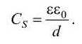
Therefore, we can calculate the distance between the capacitor plates, which in our case corresponds to the thickness of the lipid part of the membrane

This corresponds in order of magnitude to the observed membrane thickness.
In addition to lipids, protein molecules are also part of the membrane. Thus, the surface tension coefficient of cell membranes is much closer to the surface tension coefficient at the protein-water interface (~10-4 N/m) than at the lipid-water interface (~10-2 N/m).
The concept of the bilayer lipid membrane was further developed in the Davson-Danielli model proposed in 1935, also known as the sandwich model. According to this model, the membrane consists of three layers: the two outer layers consist of protein molecules, and the middle layer is lipid.
In 1972, Singer and Nicolson, based on a combination of results obtained by Physical and Chemical Research Methods such as X-Ray Diffraction Analysis and Electron Microscopy, proposed the fluid-mosaic model of Biological Membrane Structure, which is currently widely accepted (Figure 4).

Figure 4 - Fluid-mosaic model of the plasma membrane
In this model, the structural basis of the biological membrane is formed by a double layer of Phospholipids in which protein molecules are embedded. Under physiological conditions, lipids are in a liquid aggregate state. This allows the membrane to be compared to a phospholipid sea in which protein "icebergs" float.
Cell phospholipids spontaneously form lipid bilayers As a result of a phase Separation process that lowers the Free energy of the phospholipid-water solution. The process of spontaneous biomembrane formation is called self-assembly.
The hydrocarbon tails of the phospholipids in both layers of the bilayer form the hydrophobic interior of the biomembrane with a thickness of 3-4 nm—the hydrophobic hydrocarbon layer (Figure 5).

Figure 5 - Diagram of the phospholipid bilayer
In electron micrographs, a membrane cross-section stained with osmium tetroxide molecules (which bind to the polar heads of phospholipids) resembles a "railroad track", where the two "rails" correspond to the two monolayers of the bilayer (Figure 6).
The lipid bilayer exhibits two key properties.
1. The hydrophobic hydrocarbon layer serves as an impassable barrier that prevents the spontaneous Diffusion of Water-soluble (hydrophilic) molecules and ions. Importantly, this barrier property can be regulated by Membrane Proteins that mediate the transport (translocation) across the membrane of substances for which the biomembrane is otherwise impermeable to spontaneous diffusion.

Figure 6 - Electron micrograph of a cell section near the plasma membrane
2. The biomembrane is a stable structure. The bilayer Nature of the biomembrane is maintained by hydrophobic and Van der Waals interactions between lipids. Even if the extracellular environment (such as Ionic strength or pH) changes significantly, the bilayer retains a sufficient "safety margin" to preserve its integrity and impermeability.
Natural membranes of various cells demonstrate remarkable diversity, which is determined by the specific Functions of each cell type (Figures 7, 8).
The smooth, flexible surface of the erythrocyte plasma membrane allows these cells to "squeeze" through Blood capillaries whose diameter is half that of the disc-shaped red blood cell.

Figure 7 - Erythrocytes feature a smooth outer membrane
Some cells possess long, thin plasma membrane "projections"—Cilia and flagella—that perform rhythmic, wave-like movements (Figure 8).

Figure 8 - Bundles of cilia On the surface of Brain ependymal epithelial cells protect them from damage
Such movement drives the flow of the surrounding fluid across the epithelial surface, or conversely, as in the case of spermatozoa, propels the cells themselves through their environment.
The axons of many Neurons are "wrapped" in a sheath composed of multiple layers of a modified plasma membrane known as the myelin sheath (Figure 9). These membranous structures are produced by surrounding glial cells and ensure the long-distance transmission of nerve impulses.

Figure 9 - The myelin sheath of vertebrate axons (1) is formed by surrounding supportive glial (2) cells
Although the Biomembranes in the Examples above vary in shape and function, both these and all other biomembranes share the same fundamental structure—they are all lipid bilayers.
Because all cellular membranes completely enclose a cell or an internal compartment, one surface of every biomembrane always faces inward toward the compartment—referred to as the inner surface (or inner leaflet of the bilayer)—while the other surface is always exposed outward, known as the outer surface (or outer leaflet of the bilayer). The terms cytosolic face and exoplasmic face of the biomembrane are also commonly used, respectively.
Such terminology is useful for designating the topological equivalence of various sides across different membranes (Figure 10).

Figure 10 - Orientation of the cytosolic and exoplasmic faces of various cellular membranes
For example, the exoplasmic face of the plasma membrane is oriented away from the cytoplasm toward the extracellular space, forming the outer boundary of The Cell.
However, unlike the plasma membrane, in single-membrane Organelles and vesicles, the exoplasmic face of the membrane is oriented toward the interior of the organelle, remaining in contact with its lumen contents, which in this case are topologically equivalent to the extracellular fluid relative to the cell Cytosol.
This equivalence is most clearly demonstrated in endocytic vesicles, which form through the "invagination" of a region of the plasma membrane, whereby the outer surface of the plasma membrane becomes the inner surface of the vesicle membrane.
The distinction between the cytosolic and exoplasmic faces of a biomembrane is of great functional significance because, regardless of any movement within the membrane, the cytosolic domains of membrane proteins always remain in the cytosol, thereby preserving the protein's specific orientation relative to the cell compartments.
Three types of cell organelles—the Cell Nucleus, Mitochondria, and Chloroplasts—are bounded by two membranes. In these organelles, the exoplasmic face of the membranes is oriented toward the intermembrane space.
The Singer-Nicolson fluid mosaic model of biomembrane structure is currently universally accepted. However, like any model, it provides a rather simplified picture of membrane Organization. Specifically, it has been discovered that protein "icebergs" do not always float freely in the lipid sea, but can be "anchored" to the internal (cytoplasmic) structures of the cell. Furthermore, not all Membrane Lipids are arranged in a bilayer; the lipid phase also contains regions where lipid molecules do not form a double layer. Additionally, a specific class of amphiphilic proteins has been identified that alter their Hydrophobicity in response to extracellular signals and reversibly dissociate from the membrane.
Thus, The properties of The cell membrane increasingly diverge from the "classical" lipid bilayer. Nevertheless, the Singer-Nicolson fluid mosaic model, in its various modifications, still serves as a conceptual framework for explaining numerous membrane phenomena. The difficulty in formulating a unified model of biological membranes stems from the immense diversity of their functions.
Last update: 13/08/2026
Editorial and Educational Adaptation: This material has been compiled based on the primary/original source text. The project team performed an editorial review, corrected technical inaccuracies, structured sections, and adapted the content for an educational format.
What was processed:
- elimination of formatting defects (OCR errors, structural breaks, corrupted characters);
- editorial organization of content;
- standardization of terminology in accordance with academic sources;
- verification of factual statements against the original source text.
All mentions of the author, publication year, and origin of the primary text have been preserved in accordance with the source.