Principles of Biochemistry, Volume 1 - A. Lehninger 1985
Biomolecules
Cells
Cilia and flagella enable cells to move
Cilia and flagella are motile structures, or appendages, projecting from The surface of many single-celled eukaryotes and certain animal Cells (though not plant cells), and they share a common architectural plan (Fig. 2-18). It is important to emphasize, however, that eukaryotic flagella differ profoundly from prokaryotic flagella. Prokaryotic flagella are much thinner (10-20 nm) and consist of individual protein filaments. They are rigid, Curved Rods whose rotational motion depends entirely on "motors" embedded in The Cell membrane. Eukaryotic flagella are considerably thicker (200 nm), structurally more complex, and capable of generating movement along their entire length. Eukaryotic cilia and flagella are enclosed by extensions of The Plasma Membrane and contain 9 pairs of microtubules arranged around 2 central microtubules, forming the characteristic 9 + 2 Structure (Fig. 2-18). Cilia and flagella have the same diameter, but cilia are much shorter (not exceeding 10 µm) than flagella (up to 200 µm). In most cases, cilia serve to move substances along the cell surface using whip-like, oar-like strokes, whereas flagella act like propellers, driving the cell forward. Animal spermatozoa possess a single long flagellum (Fig. 2-18). The movement of cilia and flagella is driven by complex sliding interactions of individual microtubules relative to one another within the 9 + 2 structure.
Class="center">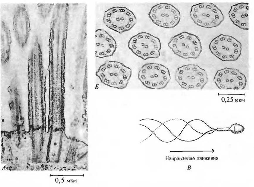
Fig. 2-18. Cilia and flagella share the same internal structure, but cilia are significantly shorter.
The pairs of microtubules are enclosed by the Cell Membrane and an extension of the Cytoplasm. The ATP-driven sliding and bending of microtubules relative to one another produce the wave-like motions of flagella. A. A longitudinal section showing parallel microtubules of a cilium. B. A cross-section of cilia, displaying the 9 + 2 structure formed by nine pairs (doublets) of microtubules surrounding two central single microtubules. C. The MECHANISM OF ACTION of a sperm flagellum propelling the entire cell forward.
The ATP-driven sliding of filaments or microtubules relative to one another is the fundamental process underlying skeletal Muscle contraction, the beating of cilia and flagella, and The formation of characteristic membrane protrusions, invaginations, and folds during cell locomotion, as observed, for example, in amoebae.
Last update: 06/08/2026
Editorial and Educational Adaptation: This material has been compiled based on the primary/original source text. The project team performed an editorial review, corrected technical inaccuracies, structured sections, and adapted the content for an educational format.
What was processed:
- elimination of formatting defects (OCR errors, structural breaks, corrupted characters);
- editorial organization of content;
- standardization of terminology in accordance with academic sources;
- verification of factual statements against the original source text.
All mentions of the author, publication year, and origin of the primary text have been preserved in accordance with the source.