Human Histology - O.D. Lutsyk 2003
Cytology
Cell Membrane and Cytoplasm
Cell membrane (Plasmalemma). The structural basis of the plasmalemma is an elementary biological membrane, The Structure of which is described by the Singer-Nicolson fluid-mosaic model. According to this model, phospholipid molecules contact via their hydrophobic tails while their hydrophilic heads face outward, forming a continuous lipid bilayer in which protein molecules (predominantly Glycoproteins) are partially or fully embedded. Protein molecules that traverse the entire thickness of The Lipid Bilayer or are deeply embedded within it are referred to as integral Proteins, whereas those located exclusively on the lipid surface are known as peripheral or adsorbed proteins (Fig. 1.3). The positioning of protein molecules is not rigidly fixed; depending on the functional state of The Cell, they can undergo lateral diffusion within the plane of the lipid bilayer. This Variability and mosaic-like Topography of the cell surface macromolecular complexes gave name to the fluid-mosaic model of the biological membrane. The lability (fluidity) of the plasmalemma structures depends on its Cholesterol content: the higher the cholesterol concentration, the more readily macromolecular Protein Complexes move within the lipid bilayer. Plasmalemma proteins exhibit a certain degree of functional specialization, including structural, enzymatic, transport, and receptor molecules. A crucial prerequisite for the normal functioning of the biological membrane is the Maintenance of the bilayer continuity (absence of gaps).
The carbohydrate components of plasmalemma glycoproteins and Glycolipids project beyond the outer surface of The cell membrane, forming a specialized supramembrane layer known as the glycocalyx. The oligosaccharide chains of the glycocalyx act as a unique cellular "business card." They mediate cell-Cell Recognition and interactions with the microenvironment. Each cell type is characterized by a specific sequence of monosaccharide residues within its surface oligosaccharide chains of glycopolymers, featuring a unique set and cytotopography of carbohydrate determinants.
On the inner cytoplasmic face, the membrane is in contact with the inner (submembrane) plate, or the cortical layer of the Cytoplasm. This is the most viscous region of the cytoplasm, rich in microfilaments and microtubules that form a highly organized network. These elements participate in the movement of integral plasmalemma proteins, support cytoskeletal and locomotor cellular functions, and facilitate exocytosis. The thickness of the plasmalemma is approximately 10 nm (1 nm=10-9 m).
Thus, the Structural components of the cell coat include the glycocalyx, the inner plate (submembrane or cortical layer of the cytoplasm), and the biological membrane proper, whose properties are best described by the fluid-mosaic model.
Functions of the cell coat. The Main Functions of the plasmalemma include separating the intracellular content from its microenvironment; transporting metabolites, which involves maintaining concentration gradients of sodium and potassium ions across the cell membrane; perimembrane METABOLISM; receiving signals from the external environment; mediating cell recognition and interactions to form Intercellular junctions of varying degrees of complexity; and establishing the characteristic architecture of the cell surface.
Class="center">
Fig. 1.3. Cell membrane (plasmalemma): A - Singer-Nicolson fluid-mosaic model; B - freeze-fracture splitting of the plasmalemma; C - Molecular Organization of the plasmalemma; D - interaction between plasmalemmal components and the submembrane cortical layer of the cytoplasm
Compartmentalization and transport are two opposing yet complementary functions of the plasmalemma. By compartmentalizing itself from the external environment, the cell preserves its individuality, while substance transport enables it to survive and function. Both processes are directed toward maintaining the constancy of the intracellular environment (Homeostasis). Transport from the extracellular medium into the cell (uptake of substances) is called endocytosis, whereas transport in the opposite direction (export of substances) is termed exocytosis. Small molecules can enter the cell either via diffusion (passive transport) or with the assistance of specialized plasmalemma Enzymes known as permeases (Active Transport).
Large molecules and molecular aggregates are taken up by the cell through envelopment by a specific region of the plasmalemma, followed by the invagination (internalization) of the resulting vesicle into the cytoplasm. The uptake of solid particles by this mechanism is called phagocytosis, whereas the ingestion of fluid droplets is termed pinocytosis. The ingested particles are typically degraded, and their chemical building blocks are assimilated by the cell. However, an alternative pathway exists wherein a particle is engulfed at one surface of the cell, traverses the cytoplasm enclosed within a biomembrane, and is expelled unchanged at the opposite cell surface. This phenomenon is known as transcytosis.
The export of cellular Metabolic waste products beyond the cytoplasm (exocytosis) comprises several distinct types: secretion, excretion, incretion/recretion, and clasmatosis. Secretion is the release of synthetic products by the cell that are essential for the normal functioning of body Organs and systems. Excretion is the elimination of toxic or harmful metabolic waste products that must be removed from the Organism. Recretion is the removal of substances from Cells that do not alter their chemical structure during intracellular metabolism (Water, mineral salts). Clasmatosis is the elimination of individual structural components from the cell.
Exocytosis mechanisms are generally the exact reverse of phagocytosis and pinocytosis. For example, the products of cellular synthesis accumulate as biomembrane-enclosed aggregates (within sacs and vesicles of the Golgi complex, or Golgi apparatus), progressively shifting from the central Regions of the cytoplasm to the periphery. Subsequently, the biomembrane of the sac fuses with the plasmalemma, and the contents are released into the intercellular space or the extracellular environment. An alternative pathway involves the export of substances within vesicles (without disrupting their biomembrane integrity), a process known as merocrine-type secretion. When numerous secretory vesicles accumulate at the apical pole of the cell and are released along with the detachment of the apical cytoplasm, this secretory mode is termed apocrine. In holocrine secretion, the cytoplasm gradually fills with synthetic products until the cell degenerates into a secretion droplet enveloped by the plasmalemma.
Perimembrane metabolism is associated with the presence of specialized enzyme systems On the surface of certain cell types capable of hydrolyzing contacting Biopolymers. Such processes are typical, for instance, of the cells lining the Small Intestine, on whose surface membrane Digestion takes place.
The Role of the plasmalemma in cellular reception mechanisms. The reception of chemical signals from the microenvironment by the cell is mediated primarily by specialized receptor Proteins of the plasmalemma. To exert its effect on the cell, a biologically active substance (e.g., a hormone) must bind to a specific plasmalemma protein, with the carbohydrate component (oligosaccharide chains) of the receptor protein playing a decisive role in ensuring the selectivity (Specificity) of this interaction. One potential pathway for subsequent intracellular signal Transduction involves the adenylate cyclase system. For instance, the binding of a receptor to its specific Ligand triggers the activation of adenylate cyclase, an enzyme located on the inner surface of the plasmalemma, which in turn stimulates The production of cyclic adenosine monophosphate (cAMP). cAMP serves as a universal activator of various cytoplasmic enzyme systems through which the cell responds to stimuli. It should be noted that complex reception processes form The basis of cell-cell recognition and are therefore critically essential for the existence of Multicellular Organisms, as they act as a command to inhibit cell proliferation and motility. Notably, the loss of cellular self-control over GROWTH AND REPRODUCTION (the absence of contact inhibition of proliferation) is a hallmark of malignant transformation.
Intercellular junctions. The cell coat, particularly the carbohydrate determinants of its glycocalyx, plays a pivotal role in establishing stable cell-to-cell contacts (Fig. 1.4). The simplest form of intercellular connection is adhesion (attachment, sticking). Recently, specific protein molecules—Lectins, cadherins, and Cell Adhesion molecules (CAMs)—have been shown to play a decisive role in The formation of multicellular aggregates. Specifically, lectin molecules can selectively "recognize" carbohydrate determinants on The surface of neighboring cells, mediating the formation of stable intercellular bridges. The intercellular distance between adjacent plasmalemmas at a simple contact zone is approximately 10-20 nm.
During the evolutionary development of multicellular systems, the forms and types of intercellular junctions became significantly more complex, increasing mechanical strength while enabling a range of specific functions. One way to reinforce intercellular contacts is by expanding the contact surface area between two adjacent cells. In this case, finger-like projections of the plasmalemma and cytoplasm of one cell insert into corresponding invaginations of the neighboring cell's plasmalemma. This type of junction is called an interdigitating, serrated, or interlocking contact. The intercellular gap in these contacts is identical to that of a simple contact, measuring 10-20 nm.
Fig. 1.4. Intercellular junctions: A - diagram of plasmalemma interactions between two adjacent small intestine epithelial cells; B - electron micrograph of the apical region of cells shown in Fig. 1.4,A: TJ, tight junction; AJ, adherens junction; D, desmosome. X 59400; C - graphic reconstruction of the ultrastructure and functional features of various junction types; D - molecular organization of a gap junction area; E - ultrastructure of a synaptic (neuromuscular) junction ensuring unidirectional signal transmission
Further strengthening of intercellular adhesion is achieved by immobilizing the contacting plasmalemmal regions (forming attachment plaques primarily composed of the protein desmoplakin) via Intermediate filaments and the cortical cytoplasmic layer. This type of connection is called a desmosome and occurs where maximum mechanical strength is required, such as in the surface epithelium of the body. The intercellular space within a desmosome is filled with an electron-dense material containing specialized transmembrane fibrillar structures composed of the protein desmoglein. The ends of desmoglein molecules anchor to the attachment plaques, thereby stabilizing this junction type. The intercellular gap at desmosomes is about 25-30 nm wide, and the desmosome diameter is 0.5 µm. At sites where epithelial cells contact the basement membrane, hemidesmosomes are formed. While a desmosome contains two attachment plaques, a hemidesmosome contains only one. The cleft between the epithelial cell and the basement membrane is filled with integrin proteins.
Another junctional form involves the creation of tight sealing plates, known as tight junctions (zonula occludens). In this type of contact, the Plasma Membranes of adjacent cells are brought into extremely close apposition. The extracellular ends of integral Membrane Proteins from neighboring cells dock together, and the remaining space is sealed by Calcium Ions and anastomosing fibrils. The outer hydrophilic layers and the glycocalyx of adjacent plasmalemmas effectively fuse into a continuous layer 2-3 nm thick. Tight junctions are characteristic of the apical surface of cells lining the digestive tract. These sealing belts achieve complete Separation of the intercellular space from the external environment. Tight junctions are found in all types of epithelia (endothelium, mesothelium, ependyma, intestinal epithelium) as well as between fibroblasts, embryonic ectoderm, and mesenchymal cells.
Basal to the tight junction in epithelial cells, adherens junctions frequently form, where the intercellular gap is filled with transmembrane proteins known as E-cadherins. These proteins bind to bundles of Actin filaments that attach to the intracellular membrane surface within the junctional zone.
Gap Junctions, or nexuses, enable direct molecular exchange between neighboring cells. In these junctional areas, which measure 0.5 to 5 µm, hexagonal arrays of particles called connexons (7-8 nm in diameter with a central channel about 1.5 nm wide) are present. Each connexon consists of six subunits of the protein connexin. Connexons span the entire thickness of the membrane. The channels of two apposed connexons align end-to-end, establishing direct chemical communication between the cytoplasms of neighboring cells: cells connected by gap junctions can freely exchange small molecules (inorganic ions, sugars, Amino Acids, NUCLEOTIDES, Vitamins) with a molecular weight not exceeding 1000-1500 daltons. This achieves a form of metabolic cooperation among cells. At gap junction sites, the plasmalemmas of adjacent cells converge to a distance of 2-4 nm. Gap junctions connect, among others, myocardial Muscle cells, smooth myocytes of the uterine wall, and ovarian oocytes and follicular cells.
A synapse is a specialized contact between Nerve Cells or between a nerve cell and a muscle fiber, across which nerve impulses are transmitted. The main structural components of a synapse are the presynaptic membrane (the plasmalemma region of the signaling nerve terminal), the postsynaptic membrane (the plasmalemma region of the target cell receiving the signal), the synaptic cleft measuring 20-30 nm in width (separating the pre- and postsynaptic membranes), and synaptic vesicles filled with neurotransmitter. Synaptic function ensures the unidirectional transmission of information from cell to cell via a chemical mediator.
Based on their structural characteristics, intercellular junctions can be broadly classified into three groups: adhesive, occluding, and communicating. The first group includes simple adhesive contacts, interlocking contacts, and desmosomes. The second group comprises tight junctions, while the third includes gap junctions and synapses.
Cytoplasm. The structural Components of the cytoplasm are the hyaloplasm, Organelles, and inclusions.
The hyaloplasm is the fluid matrix of the cytoplasm that houses organelles and inclusions. It accounts for approximately 50% of the total cytoplasmic volume. The hyaloplasm comprises the Cytosol (water containing dissolved organic and inorganic substances) and the cytomatrix (a trabecular network of protein filaments 2-3 nm in thickness).
Organelles are permanent cytoplasmic structures with a defined architecture and specialized functions. They are generally divided into microscopic organelles, visible under a Light Microscope, and submicroscopic organelles, which can only be resolved with an Electron microscope. Based on the presence of a biological membrane, they are classified as membranous or non-membranous. Membranous organelles include Mitochondria, Lysosomes, Peroxisomes, The Endoplasmic reticulum, and the Golgi apparatus. Non-membranous organelles comprise proteasomes, Ribosomes, microfilaments, microtubules, and the centrosome (cell center). These ten entities are termed general-purpose organelles because they are present in all cell types. General-purpose organelles can aggregate to form characteristic cytoplasmic configurations. Such configurations, marked by the predominant development and unique organization of a specific organelle type, are known as special organelles (e.g., tonofibrils in epithelial cells, myofibrils in muscle cells and fibers, and neurofibrils in nerve cells).
Recent studies have established that cellular protein molecules assemble into multimolecular complexes of characteristic Morphology and specific function—referred to as complexomixes—consisting of anywhere from 5 to over 40 individual molecules. Ribosomes, apoptosomes, and proteasomes serve as classic Examples of complexomixes. A unifying concept is gradually emerging: the same protein "building blocks" can assemble into specific complexomixes to carry out a given function, which then disassemble upon completion of their activity, allowing the individual protein components to be recycled for the Formation of other organelles. Consequently, the intracellular organization increasingly resembles a mosaic in a state of constant dynamic remodeling, where diverse protein elements are recruited to assemble similar Structural motifs across different cell types. Interestingly, despite the inter- and intraspecies diversity of their constituent proteins, individual complexomixes—and particularly their functionally active domains—exhibit remarkable structural conservation, even among evolutionary distant organisms.
Mitochondria are microscopic membranous general-purpose organelles (Fig. 1.5) whose primary function is to generate the energy required for cellular activity and store it within molecules of adenosine triphosphate (ATP). In addition, mitochondria participate in water homeostasis regulation, calcium ion sequestration, and the production of steroid hormone precursors. They were discovered by the German researcher F. Altmann in the late 19th century. Under a light microscope, mitochondria appear as tiny granules and filaments approximately 0.5 µm thick and 1–10 µm long. Electron Microscopy reveals that each mitochondrion has an irregular oval or elongated shape bounded by two membranes: a smooth outer membrane and a folded inner membrane that projects inward to form cristae. The internal cavity is filled with an electron-dense substance known as the matrix. Both the matrix and The inner mitochondrial membrane contain enzyme proteins that facilitate ATP synthesis via Oxidative Phosphorylation of adenosine diphosphate (ADP). Mitochondria are unique among cellular organelles in containing their own deoxyribonucleic acid (DNA) molecules, alongside various RNA species and ribosomes housed within the matrix.
Lysosomes are submicroscopic membranous general-purpose organelles (Fig. 1.6) discovered in 1955 by Christian de Duve. Their primary function is The breakdown of biopolymers of diverse chemical composition (a process known as cellular digestion). To this end, lysosomes contain a battery of hydrolytic enzymes (over 60 are currently known), with acid phosphatase serving as the marker enzyme. These enzymatic complexes are sequestered within a closed membranous vesicle approximately 0.2–0.4 µm in diameter, which prevents lysosomal enzymes from leaking into the hyaloplasm and protects the cell from autodigestion. Depending on their ultrastructural and functional characteristics, lysosomes are categorized into primary lysosomes (where enzymes remain inactive), secondary lysosomes or phagosomes (where activated enzymes directly interact with target biopolymers), and residual bodies (membrane-bound undigested remnants). Notably, lysosomes are involved in both the degradation of the cell's own macromolecular complexes (autophagocytosis) and the digestion of internalized exogenous particles (heterophagocytosis). A deficiency in a specific lysosomal enzyme leads to the intracellular accumulation of abnormal biopolymers, precipitating so-called lysosomal storage diseases (thesaurismoses). To date, over 30 distinct lysosomal storage disorders have been described (Table 2).
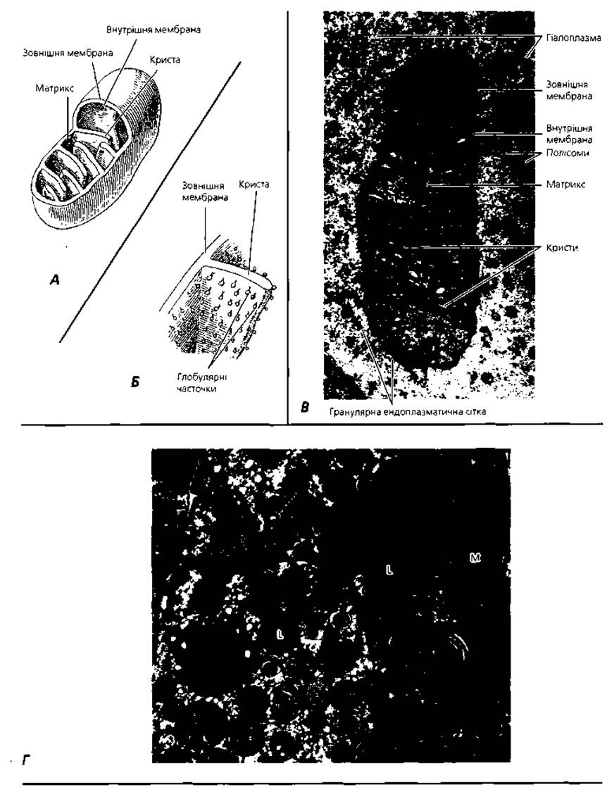
Fig. 1.5. Mitochondria: A – 3D reconstruction of the ultrastructure; B – detail of Fig. 1.5, A: arrangement scheme of globular particles involved in energy conversion and storage; C – electron micrograph of an epithelial cell mitochondrion, ×75,000; D – lipid droplets (L) and associated mitochondria (M) involved in lipid synthesis within an Adrenal gland cell, ×19,000
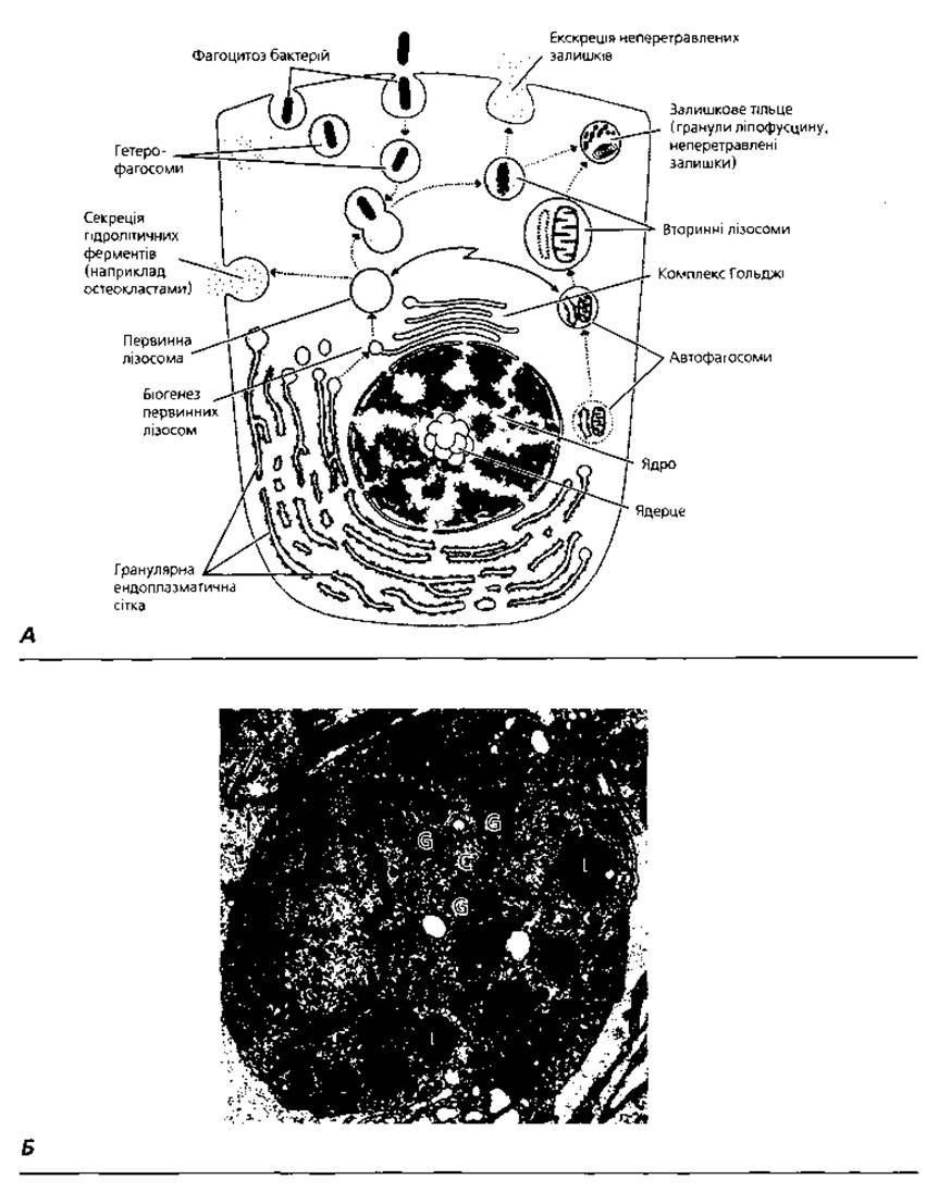
Fig. 1.6. Lysosomes: A – diagram illustrating the biogenesis of primary lysosomes from the rough Endoplasmic reticulum and the Golgi apparatus, their maturation into secondary lysosomes and residual bodies, and their role in secretion and excretion; B – electron micrograph of secondary lysosomes (L) in the cytoplasm of a peritoneal macrophage. Arrows indicate numerous cytoplasmic projections: C – centriole; G – Golgi apparatus, ×15,000
Table 2. Examples of storage DISEASES ASSOCIATED WITH lysosomal enzyme defects
|
Disease |
Deficient lysosomal enzyme |
Affected cell type |
Primary organs affected |
|
Hurler syndrome |
a-L-iduronidase |
Fibroblasts and osteoclasts accumulate dermatan sulfate |
Bones and Nervous system |
|
Sanfilippo syndrome A |
Heparan N-sulfatase |
Fibroblasts accumulate heparan sulfates |
Bones and nervous system |
|
Hexosaminidase A |
Neurons accumulate glycolipids |
Nervous system |
|
|
Gaucher disease |
a-D-glucosidase |
Macrophages accumulate glycolipids |
|
|
I-cell disease |
Phosphotransferase |
Fibroblasts and osteoclasts accumulate dermatan sulfate |
Bones and nervous system |
Proteasomes. Evidence emerged in the 1970s indicating that protein degradation is not restricted to lysosomes. In the late 1980s, two independent research groups demonstrated that proteins are degraded by large polyprotease complexes termed proteasomes. It is now established that every human cell contains approximately 30,000 proteasomes, with a molecular mass of about two million daltons. Each proteasome consists of a barrel-shaped core particle and one or two regulatory particles situated at one or both ends of the organelle (Fig. 1.1). The core particle comprises four stacked rings, each composed of seven subunits surrounding a central channel. The regulatory particles recognize and bind proteins targeted for destruction, unfold the protein molecules, and thread them into the central channel of the core, where proteases cleave them into fragments of varying lengths. These fragments are subsequently broken down by Other Enzymes into amino acids for the synthesis of new proteins.
The recognition of proteins destined for proteasomal degradation is primarily governed by ubiquitination—the covalent attachment of ubiquitin molecules to the protein. Ubiquitination proceeds in three sequential steps involving three classes of enzymes: E1, E2, and E3. In the first step, ubiquitin is activated by E1. Next, the activated ubiquitin is transferred from E1 to an E2 conjugating enzyme. The third step involves The transfer of activated ubiquitin from E2 to the target protein, a reaction catalyzed by an E3 ligase. Hundreds of distinct E3 enzymes exist, each recognizing specific Amino acid sequences on target proteins to direct their ubiquitination.
Numerous studies have demonstrated that protein ubiquitination and subsequent proteasomal degradation are essential for numerous physiological processes: regulating intracellular metabolism, immune surveillance, clearing aberrant protein molecules, Cell Division, Intercellular Communication, organismal GROWTH AND DEVELOPMENT, and circadian rhythms. Conversely, disruptions in ubiquitination pathways (such as Mutations in E3 ligases) and the resulting impairment or blockade of proteasomal degradation underlie several hereditary disorders (e.g., cystic fibrosis), neurodegenerative conditions (Parkinson's and Alzheimer's diseases), viral pathologies, and carcinogenesis.
Peroxisomes are submicroscopic membranous general-purpose organelles (Fig. 1.7) discovered in the early 1960s through the collaborative efforts of biochemists and morphologists. Peroxisomes play a pivotal role in cellular detoxification by neutralizing toxic metabolic byproducts. These roughly spherical, membrane-bound vesicles, measuring about 0.2–0.5 µm in diameter, are filled with an enzymatic matrix wherein catalase serves as the marker enzyme. Electron microscopy often reveals a dense core, or crystalloid, composed of fibrous and tubular macromolecular structures located at the center of the peroxisomal matrix.
The enzymatic systems of peroxisomes are geared toward scavenging reactive atomic oxygen (primarily by regulating hydrogen peroxide metabolism and breakdown), as well as degrading ethanol and uric acid, and regulating Lipid Metabolism.
The endoplasmic reticulum is a submicroscopic membranous general-purpose organelle forming an interconnected intracytoplasmic circulatory network, first described by K. Porter in 1945. It constitutes a continuous system of tubules, vesicles, and cisternae enclosed by a single uninterrupted biomembrane (Figs. 1.1 and 1.8). The endoplasmic reticulum membrane is in direct continuity with The Plasma Membrane and the nuclear envelope. A distinction is made between agranular (smooth) and granular (rough) endoplasmic reticulum. The smooth endoplasmic reticulum, featuring tubules 50–100 nm in diameter, consists exclusively of lipid membranes. The rough endoplasmic reticulum is characterized by ribosomes attached to its cytosolic surface (Fig. 1.8), with tubular diameters ranging from 20 to 1,000 nm. The smooth endoplasmic reticulum is implicated in lipid and Carbohydrate Metabolism, the detoxification of xenobiotics, and calcium ion sequestration. The function of the rough endoplasmic reticulum is dictated by the presence of ribosomes and centers on the Biosynthesis of Proteins destined for both intracellular utilization and export. Beyond its metabolic and circulatory roles, the endoplasmic reticulum serves as the primary site for de novo synthesis of Cellular Membrane Structures. Components synthesized in the rough endoplasmic reticulum can be incorporated into lysosomes, peroxisomes, the Golgi apparatus, the plasma membrane, and the nuclear envelope, or utilized for the self-renewal of the endoplasmic reticulum itself.
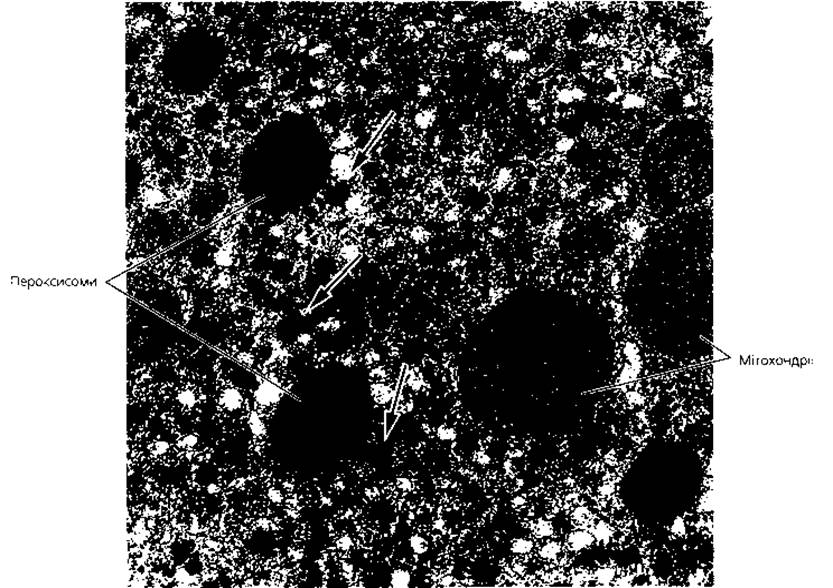
Fig. 1.7. Peroxisomes. Electron micrograph showing peroxisomes and mitochondria in a hepatocyte cytoplasm. Arrows indicate Glycogen inclusions, ×30,000.
The Golgi apparatus, or Golgi complex, is a microscopic membranous general-purpose organelle (Fig. 1.9) that completes the Processing of cellular secretory products, notably executing their terminal glycosylation. The Golgi complex concentrates secretory substances and directs their export from the cell. It is named after the Italian histologist Camillo Golgi, who first described this complex in nerve cells in 1898. Morphologically, the Golgi complex consists of an array of interconnected cisternae, approximately 25 nm thick, flattened centrally and dilated at their peripheral margins. The intercellular space between individual cisternae is 20–25 nm. Small vesicles bud off from the dilated rims of the cisternae (Fig. 1.6). An individual stack of such cisternae and associated vesicles is called a dictyosome, often metaphorically compared to a stack of plates with their convex surfaces facing The Nucleus. A single cell may contain multiple dictyosomes separated by layers of hyaloplasm. The synthesis and maturation of secretory products are finalized as they transit from cisternae located near the nucleus (the cis face) toward the plasma membrane (the trans face). Vesicles pinching off from the cisternal margins contain fully formed secretory products ready for release. These concentrated macromolecular complexes are expelled either via fusion of the vesicle membrane with the plasmalemma (exocytosis) or by the release of mature vesicles from the cell.
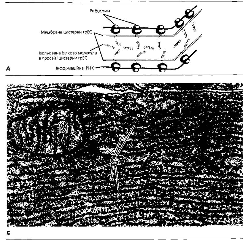
Fig. 1.8. Rough endoplasmic reticulum (rER): A – diagram of Protein Biosynthesis and accumulation within the rER lumen; B – electron micrograph of rER in a gastric chief cell: C – rER cisternae; M – mitochondrion
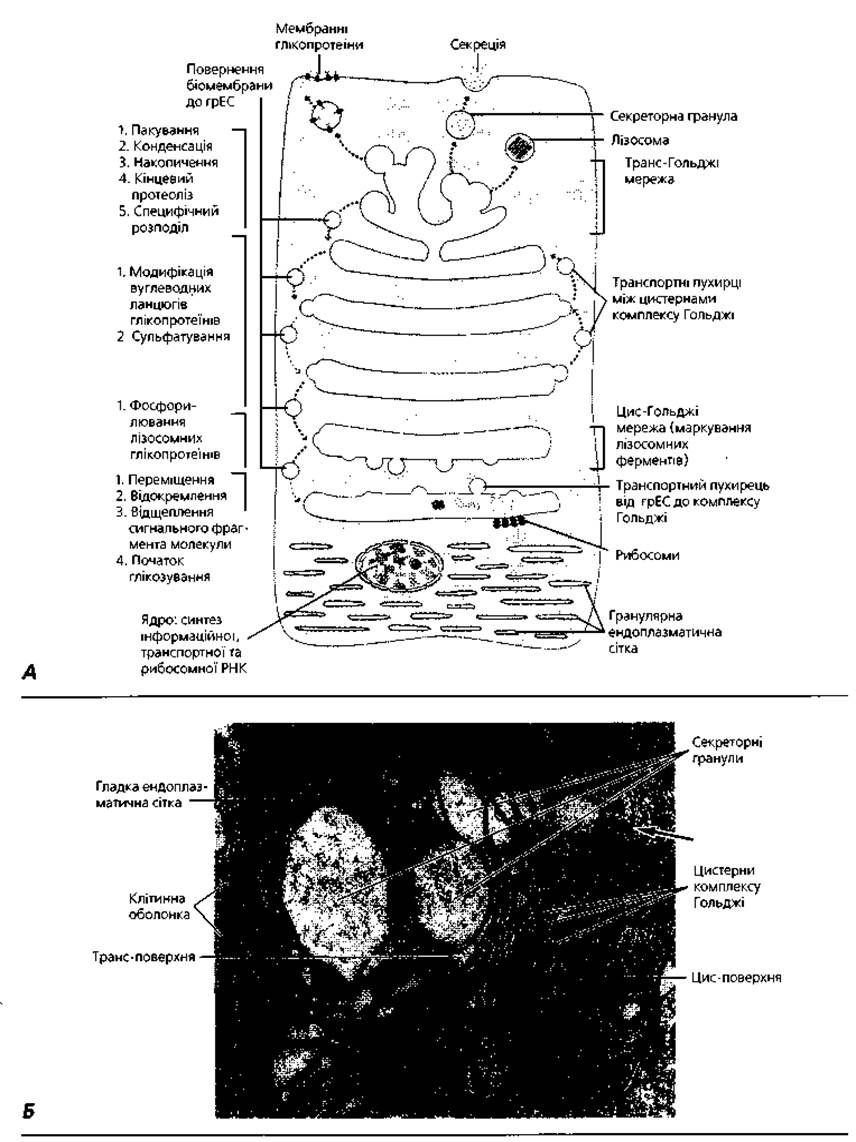
Fig. 1.9. Golgi apparatus: A – diagram illustrating the interrelationships among the Golgi apparatus, rough endoplasmic reticulum, lysosomes, secretory granules, and vesicles; B – electron micrograph of the Golgi complex in a mucus-secreting cell. The arrow indicates a rough endoplasmic reticulum cisterna, ×30,000.
Ribosomes are submicroscopic non-membranous general-purpose organelles (Fig. 1.10) where Amino acids are linked into peptide chains, effectively synthesizing protein molecules. Ribosomes within the rough endoplasmic reticulum were first described by G. Palade. Morphologically, ribosomes consist of two subunits whose assembly forms a mushroom-like structure approximately 20 nm in diameter. Chemically, a ribosome is a ribonucleoprotein complex comprising ribosomal RNA and proteins in a 1:1 ratio. Exposure to damaging agents or disturbances in cellular electrolyte homeostasis (such as magnesium ion depletion) causes ribosomes to dissociate into their constituent subunits (disaggregation), resulting in a loss of biological activity. Multiple ribosomes threaded along a common strand of Messenger RNA are termed polyribosomes (Polysomes). Polysomes suspended freely in the hyaloplasm predominantly synthesize proteins for intracellular consumption, whereas membrane-bound polysomes on the endoplasmic reticulum mainly synthesize proteins destined for export.
Microfilaments are submicroscopic non-membranous general-purpose organelles (Fig. 1.11) that function as both Structural elements of the Cytoskeleton and the cell's contractile apparatus. They are delicate fibers composed of actin (thin filaments, ~5 nm in diameter), Myosin (thick filaments, ~25 nm in diameter), Tropomyosin, or alpha-actinin, and are located predominantly in the cortical (submembranous) zone of the cell and within cytoplasmic projections. Their primary function is motor-contractile. Intermediate microfilaments have a diameter of 10–15 nm. The specific protein composition of intermediate filaments serves as a reliable histochemical marker for particular cell types; for instance, keratin typifies epithelial cells, vimentin marks Connective Tissue, desmin characterizes muscle, and neurofilaments and glial fibrillary acidic protein identify neural tissue. Intermediate filaments are largely responsible for maintaining cell shape. Recent studies have also confirmed their involvement in The regulation of genome activity and cellular differentiation processes (Table 3). In specialized cells, microfilaments can organize into more complex bundles, such as tonofibrils in epithelial cells, myofibrils in muscle cells, and neurofibrils in neurons. Due to this specialized organization, tonofibrils, myofibrils, and neurofibrils are classified as specialized organelles of their respective cell types.
Microtubules are submicroscopic, non-membranous general-purpose organelles (Fig. 1.12) whose primary function is to facilitate the motility of cellular organelles and form the cytoskeleton (Fig. 1.1 ). Microtubules are composed of Globular proteins called tubulins, the molecules of which are capable of polymerization. By "stringing" together in a specific manner, individual molecules form bead-like structures. Thirteen parallel strands of these "beads" form a hollow cylinder with a diameter of about 25 nm and an inner lumen of 15 nm. The thickness of the cylinder wall corresponds to the diameter of a single tubulin molecule, which is 5 nm. The polymerization of tubulin molecules is a dynamic process that halts under unfavorable environmental conditions (such as a drop in Temperature or colchicine Treatment). Partial depolymerization leads to microtubule shortening, while complete depolymerization results in their disintegration (dissociation) into individual tubulin molecules. Microtubules serve as the structural basis of the centrosome, as well as specialized structures such as Cilia and flagella.
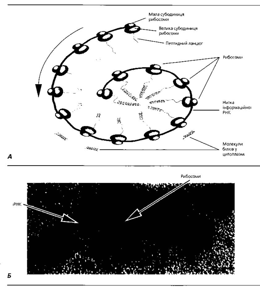
Fig. 1.10. Ribosomes: A — schematic representation of free ribosomes (polysomes) involved in The biosynthesis of cytoplasmic proteins; B — electron micrograph of a polysome comprising five ribosomes attached to a messenger RNA strand synthesizing the Hemoglobin globin chain, x 400,000
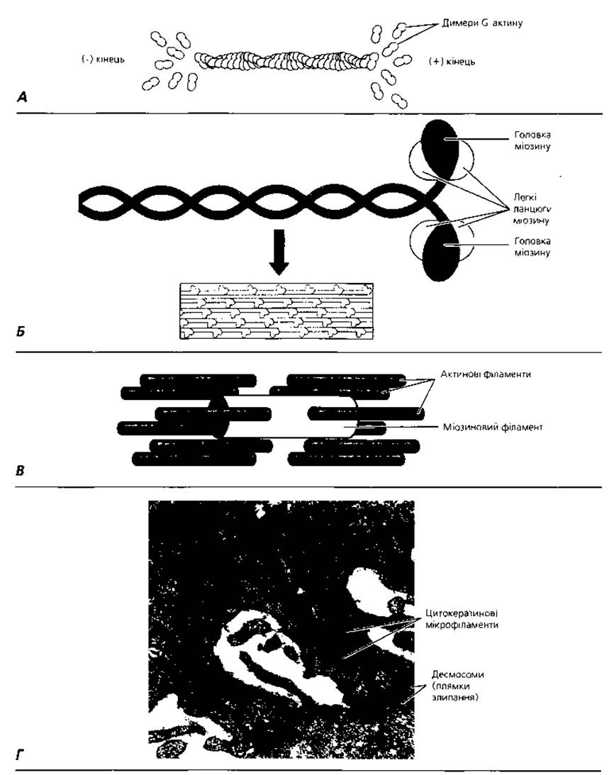
Fig. 1.11. Microfilaments: A — actin (thin) filament: The addition of G-actin dimers at the (+) end and dissociation at the (-) end allows the filament length to adjust to cellular demands; B — self-assembly of a myosin (thick) filament from myosin molecules; C — ratio of actin to myosin filaments in a contracted muscle fiber; D — electron micrograph of cytokeratin (intermediate) filaments in the region of desmosomal cell-to-cell contacts
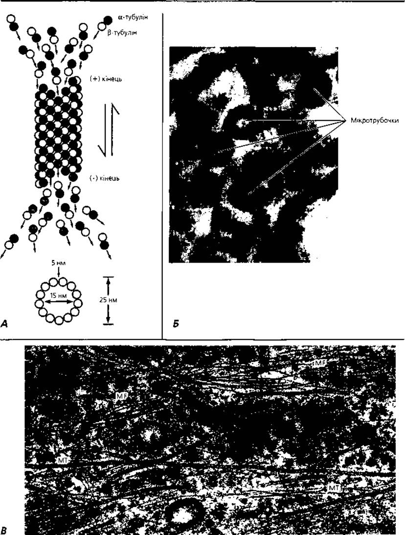
Fig. 1.12. Microtubules: A — diagram of self-assembly at the (+) end and dissociation at the (-) end of a microtubule through the addition or detachment of α- and β-tubulin dimers, ensuring microtubule elongation, shortening, or self-disintegration according to cellular needs; B — electron micrograph of cross-sectioned microtubules: 13 tubulin dimers arranged in a spiral; C — electron micrograph of longitudinally oriented microfilaments (MF) and microtubules (MT) in a fibroblast cytoplasm, x60,000
Table 3. List of the most common Antigens used for immunocytochemical Diagnostics and subsequent treatment
|
Antigens |
Type of Pathology |
|
Cytokeratins |
Undifferentiated epithelial tumors, carcinomas, adenocarcinomas |
|
Glial fibrillary acidic proteins |
Glial tumors |
|
Vimentin |
Connective tissue tumors |
|
Desmin |
Muscle tissue tumors |
|
Tumors producing protein or peptide hormones |
|
|
Carcinoembryonic antigen |
Glandular tumors, predominantly of the digestive tract and Airways |
|
Prostate-specific antigen |
Prostate tumors |
|
Steroid Hormone Receptors |
Airway tumors |
|
Viral antigens |
Viral infections |
The centrosome (cell center) is a microscopic, non-membranous general-purpose organelle (Fig. 1.13) described by W. Flemming in 1875, which ensures chromosome segregation during cell division. In a cell not preparing for division, the centrosome is located near the nucleus and consists of two fully formed centrioles surrounded by the centrosphere. Two adjacent centrioles are referred to as a diplosome. The base of each centriole contains nine triplets of parallel-oriented microtubules, which spatially form a cylinder with a diameter of 200 nm and a length of about 500 nm (Fig. 1.1). In addition to microtubules, the centriole contains specific macromolecular structures known as arms, through which the triplets are interconnected. These "arms" contain the protein dynein, which exhibits ATPase activity and plays a decisive role in the motor functions of centrioles. The long axes of both centrioles are located in mutually perpendicular planes. The centrosphere represents an organelle-free hyaloplasm surrounding the centrioles, radially penetrated by microfilaments and microtubules. During CELL PREPARATION FOR division, centriole duplication occurs, followed by the migration of each newly formed pair to opposite poles of the cell. The functional activity of the centrosome is driven by a mechanism that stimulates tubulin polymerization, promoting the growth of existing microtubules and the formation of new ones.
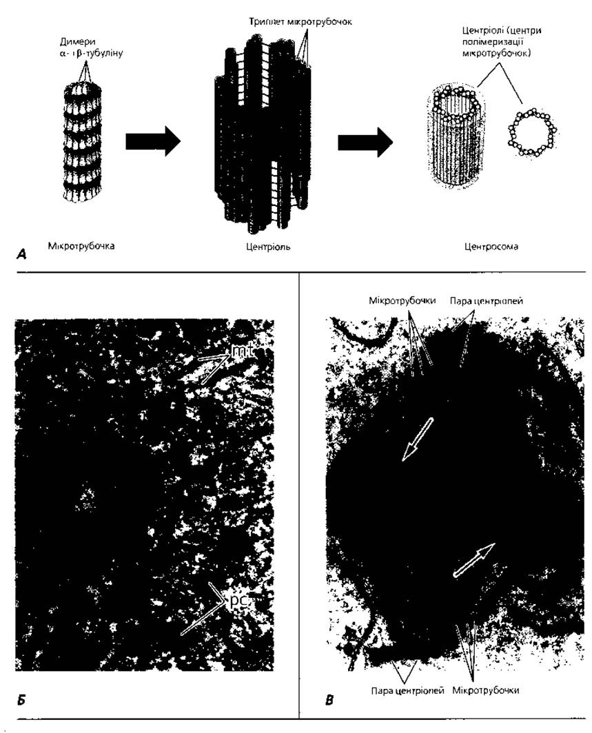
Fig. 1.13. Centrosome: A — diagram of microtubule self-organization (9x3) into a centriole and the cell center (centrosome) (not to scale); B — electron micrograph of a cross-sectioned fibroblast centriole: mt, microtubules; pc, pericentriolar material; C — electron micrograph of a spermatocyte in mitotic metaphase: microtubules extend from the pair of centrioles at each cell pole to the equatorial plate of the Chromosomes. Arrows indicate attachment sites of microtubules to the centromeric regions of chromosomes, x19,000
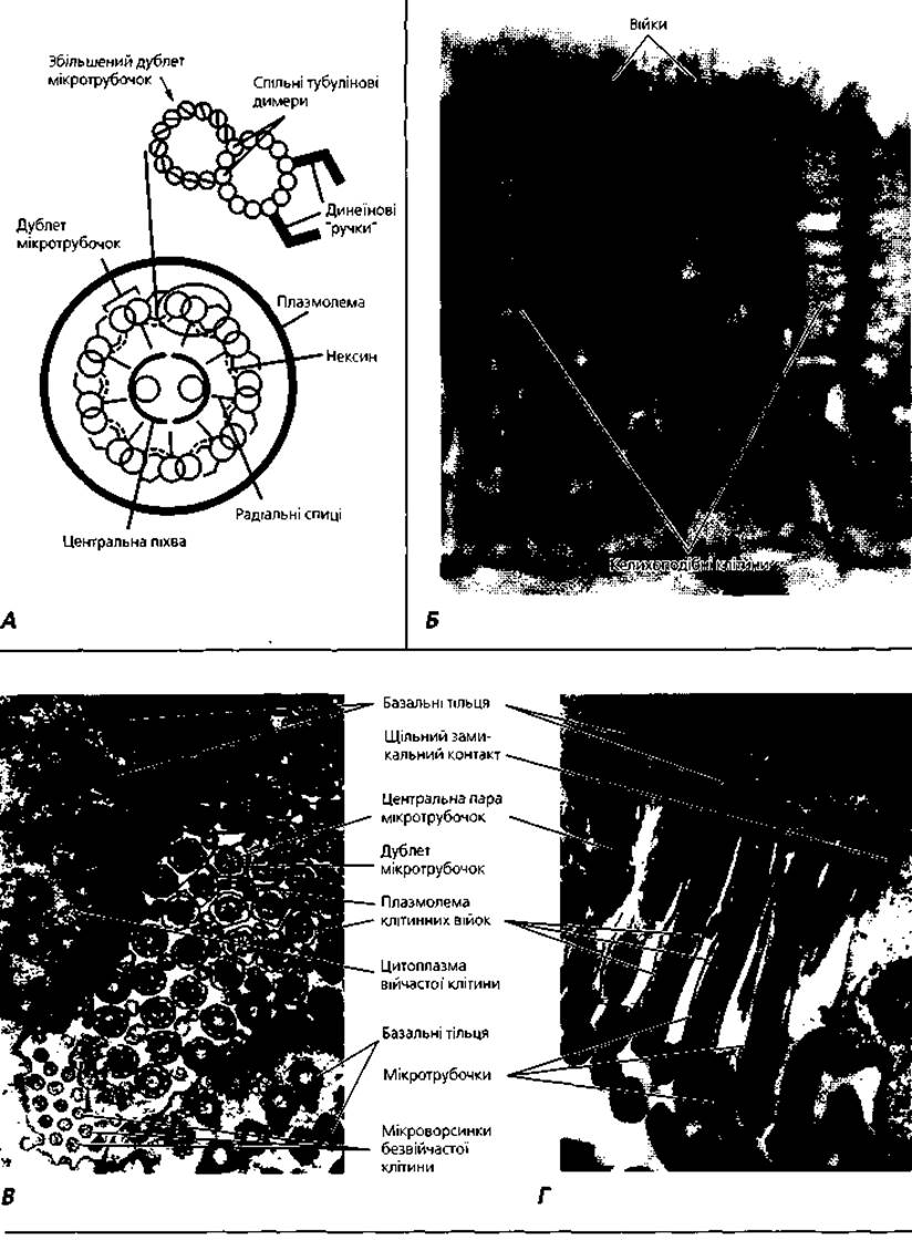
Fig. 1.14. Cilia: A — diagram of the axonemal microtubule organization (9x2+2) of a cellular cilium; B — light microscopy of the respiratory ciliated epithelium of the Trachea, x1200. Cytology/cytology/93.html">ELECTRON MICROGRAPHS OF cross-sectioned (C) and longitudinally sectioned (D) cilia of female oviduct epithelial cells, x25,000
Cilia (Fig. 1.14) and flagella are slender cytoplasmic extensions based on a highly organized microtubular system. Cilia are 5-10 µm long, whereas flagella can reach up to 150 µm in length. The diameter of both structures is roughly identical, measuring about 200 nm. Externally, a cilium is covered by the plasmalemma, enclosing a central axial filament known as the axoneme. The axoneme contains nine pairs (doublets) of microtubules forming a hollow cylinder with a diameter of about 150 nm. A central pair of microtubules is localized at the center of this cylinder. The axonemal microtubule system is described by the formula (9×2)+2. At the Base of the cilium, where it transitions into the cytoplasm, lies the basal body, which structurally resembles a centriole and consists of nine microtubule triplets. The formula for the basal body microtubule system is (9×3)+0, identical to that of a centriole. Often, two basal bodies are located at the base of a cilium, with their long axes positioned at right angles to each other. The basal body and axoneme are interconnected: two microtubules from each triplet of the basal body extend into the microtubule doublet of the axoneme. Minor shifts in the microtubule doublets can cause the entire cilium or flagellum to bend. In various cells, the movement of cilia and flagella may resemble a pendulum stroke, a conical (whirlpool) motion, a wave-like motion, etc. Free-living ciliated and flagellated cells can migrate through space (e.g., spermatozoa). Fixed cells with cilia on their apical surface utilize ciliary beating to transport fluid, mucus, or suspended particles and cells (e.g., ciliated Cells of the respiratory tract, oviducts, etc.).
Table 4. Selected Human and Animal diseases caused by damage to cytoplasmic components
|
Damaged Cellular Component |
Disease |
Molecular Defect |
Morphological Changes |
Characteristic Symptoms |
|
Mitochondria |
Mitochondrial cytopathy |
Impairment of oxidative phosphorylation |
Increased size and number of mitochondria in muscle tissue cells and their derivatives |
High basal metabolic rate without hyperthyroidism |
|
Microtubules |
Kartagener syndrome |
Absence of dynein in flagella and cilia |
Absence of arms in microtubule doublets |
Immobility of flagella and cilia, leading to male sterility and chronic respiratory infections |
|
Murine diabetes |
Decreased tubulin content in pancreatic islet ß-cells |
Reduction of microtubules in pancreatic islet ß-cells |
Diabetes |
|
|
Lysosomes |
Metachromatic leukodystrophy |
Absence of lysosomal phosphatase |
Decreased motor activity and intellect |
|
|
Hurler syndrome |
Absence of lysosomal α-L-iduronidase |
Accumulation of dermatan sulfates in fibroblasts and osteoblasts |
Growth retardation and mental impairment |
|
|
Golgi complex |
I-cell disease |
Phosphotransferase deficiency |
Accumulation of dermatan sulfates in fibroblasts and osteoblasts |
Bone defects, psychomotor retardation |
|
Secretory granules |
Proinsulin diabetes |
Defect in the proinsulin-cleaving enzyme |
None |
Diabetes |
Damage to specific cellular organelles leads to various disorders (Table 4).
Inclusions, unlike organelles, are not permanent structural components of the cytoplasm and lack a strictly defined structure. Inclusions can be exogenous or endogenous. Depending on their functional purpose, endogenous inclusions are subdivided into excretory, trophic, pigmentary, etc. Essentially, inclusions can be regarded as macromolecular aggregates accumulated by the cell in the cytoplasm under various physiological conditions.
Terms to Remember
1. Plasmalemma. 2. Glycocalyx. 3. Cortical (submembrane) layer. 4. Endocytosis. 5. Phagocytosis. 6. Pinocytosis. 7. Transcytosis. 8. Secretion. 9. Merocrine secretion type. 10. Apocrine secretion type. 11. Holocrine secretion type. 12. Excretion. 13. Recretion. 14. Clasmacytosis (clasmatosis). 15. Lectin. 16. Cadherin. 17. Adhesion. 18. Desmosome. 19. Nexus (gap junction). 20. Connexon. 21. Connectin. 22. Synapse. 23. Hyaloplasm. 24. Cytosol. 25. Cytomatrix. 26. Organelle. 27. Mitochondrion. 28. Lysosome. 29. Phagosome. 30. Residual body. 31. Autophagy (autophagocytosis). 32. Heterophagy (heterophagocytosis). 33. Proteasome. 34. Peroxisome. 35. Endoplasmic reticulum. 36. Golgi apparatus (lamellar complex). 37. Dictyosome. 38. Ribosome. 39. Polysome. 40. Microfilament. 41. Tonofibril. 42. Myofibril. 43. Neurofibril. 44. Keratin. 45. Vimentin. 46. Desmin. 47. Microtubule. 48. Tubulin. 49. Centrosome (cell center). 50. Centriole. 51. Diplosome. 52. Centrosphere. 53. Cilium. 54. Axoneme. 55. Basal body. 56. Flagellum. 57. Inclusion.
Last update: 09/08/2026
Editorial and Educational Adaptation: This material has been compiled based on the primary/original source text. The project team performed an editorial review, corrected technical inaccuracies, structured sections, and adapted the content for an educational format.
What was processed:
- elimination of formatting defects (OCR errors, structural breaks, corrupted characters);
- editorial organization of content;
- standardization of terminology in accordance with academic sources;
- verification of factual statements against the original source text.
All mentions of the author, publication year, and origin of the primary text have been preserved in accordance with the source.