Biochemical Foundations of Human Vital Activity - Volkov, N.I., Nesen, E.N. 2000
Biochemical Foundations of Human Vital Activity
Biochemistry of Proteins
Protein Biosynthesis and Its Regulation
Amino Acids formed during The breakdown of dietary Proteins and delivered to Tissues are used primarily for The Biosynthesis of Organism-specific proteins. Every day, The Human Body produces about 1.3 g of protein per 1 kg of body weight, which determines the daily dietary requirement. Cellular proteins are constantly synthesized because they have a limited lifespan. For instance, the half-life of Liver proteins is approximately 9 days, Muscle Proteins last about 120 days, and the body's total protein content is completely renewed roughly every 130–150 days. METABOLISM/35.html">Protein Biosynthesis plays a vital role in GROWTH AND DEVELOPMENT, as well as in tissue repair and adaptation during athletic training.
Protein biosynthesis is a complex, multistep process. Significant contributions to uncovering its mechanisms were made by scientists A.M. Belozersky, A.S. Spirin, A.A. Bayev, F. Crick, S. Ochoa, M. Nirenberg, and others. However, many aspects of this intricate process remain to be fully elucidated.
DNA plays the primary role in determining The Structure of synthesized proteins. The sequence and arrangement of amino acids in a protein molecule are dictated by The nucleotide sequence in the DNA molecule. Each amino acid is encoded by three adjacent NUCLEOTIDES (triplets), known as codons. The four nucleotides (A, G, T, C) that make up DNA can form 64 different triplets, capable of encoding all 20 standard amino acids (see Chapter 11).
DNA molecules do not participate directly in Protein Synthesis. They reside within the Cell Nucleus and contain the Genetic information governing Protein Structure. Protein synthesis itself takes place in Ribosomes—cellular structures located in the Cytoplasm.
Stages of Protein Synthesis. The complex process of protein biosynthesis in tissues can be divided into several major stages, including Transcription, Amino Acid Activation, and translation (Fig. 96).
Transcription is The process of synthesizing a Messenger RNA molecule using a specific segment of a DNA molecule (a Gene) as a template, which encodes the protein's structural information. First, specialized Enzymes (DNA polymerases) break the Hydrogen Bonds between the nitrogenous bases of the two complementary DNA strands. The DNA double helix then unwinds locally, and an mRNA molecule is synthesized along one of the strands by the enzyme RNA polymerase according to THE PRINCIPLE OF complementary base pairing (see Fig. 96, a). In this way, the genetic information regarding protein structure is transcribed. Complexed with Nuclear Proteins, the mRNA leaves The Nucleus and enters the cytoplasm, while the DNA restores its original structure. This stage occurs in the nucleus and serves as the initial step in triggering the synthesis of a specific protein, which is ultimately carried out on the ribosomes.
Amino acid activation is the process of interaction with tRNA molecules. Since There are 20 standard amino acids, There are also more than 20 distinct types of tRNA. The activation process proceeds with the participation of aminoacyl-tRNA synthetase and an ATP molecule.
The overall reaction of amino acid activation and their attachment to tRNA can be represented as follows:
Class="center">
Each amino acid has its own specific enzymes involved in its activation. They exhibit high activity in the presence of Mg2+ ions. A disruption in the Specificity of these enzymes can introduce errors into the Introduction/19.html">Primary Structure of the protein during polypeptide chain formation, subsequently leading to mutational Changes in the organism.
tRNA molecules feature two specific triplets. One is the codon recognition site that binds to The amino acid, and the other is the anticodon, which corresponds to the specific amino acid's codon on the mRNA. Due to this mechanism, Amino acids are arranged during Protein synthesis in the exact sequence dictated by the codon sequence in the mRNA. The activated amino acids are then delivered to the ribosomes.
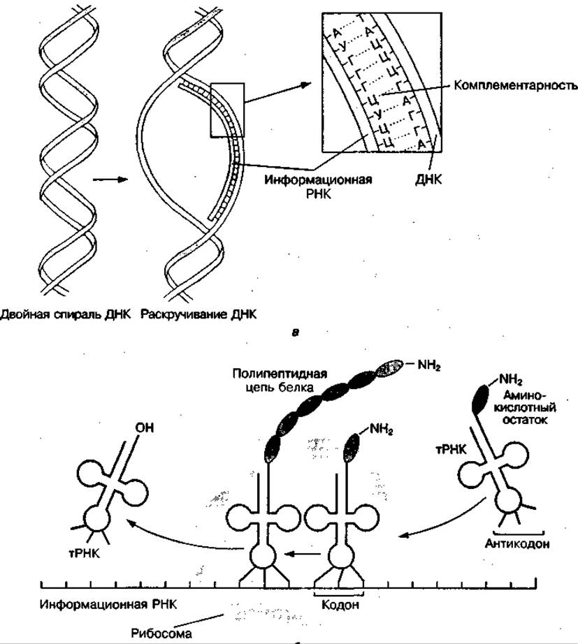
Fig. 96 Diagram of the stages of protein biosynthesis: transcription (a), translation (b)
Translation is the process of synthesizing a protein polypeptide chain on ribosomes, during which genetic information is transferred from the mRNA molecule into the specific Amino Acid Sequence of the synthesized protein. The mRNA molecule moves between the two ribosomal subunits—the small (30S) and large (50S) subunits. The mRNA attaches to the small subunit, while the protein-synthesizing enzyme (peptidyl transferase) is located on the large subunit. As the mRNA moves between the ribosomal subunits, its codons interact with tRNA anticodons via complementary base pairing. Meanwhile, specialized enzymes catalyze the attachment of the amino acid residue to the growing polypeptide chain. This process is activated by rRNA.
The termination of protein biosynthesis is directed by termination codons (stop signals) in the mRNA—UAA, UAG, and UGA—which cannot be bound by any tRNA. Consequently, the completion phase of Protein synthesis is called termination. A release factor is then recruited, and the polypeptide chain is detached from the ribosomes. The newly synthesized protein folds into a specific Spatial Structure characteristic of that particular protein. The complex Tertiary Structure of the protein molecule forms spontaneously in the cytoplasm and is determined by its Primary structure as well as its local environmental conditions.
Protein synthesis requires a vast expenditure of ATP; for instance, at least five ATP molecules are consumed just to add a single amino acid to the growing polypeptide chain. Consequently, the Rate of protein synthesis directly depends on the rate at which cellular ATP levels are replenished.
Regulation of Protein Biosynthesis
All Cells in the organism share an identical genome and synthesize between 10,000 and 20,000 different proteins, yet they differ from one another by the presence of cell-specific proteins. Red Blood Cells are characterized by a high Hemoglobin content, Skin cells by Collagen, the Pancreas by digestive enzymes, and skeletal Muscles by the contractile proteins Actin and myelinated structures like Myosin. The concentration of various proteins—and sometimes their entire spectrum—changes with age, as well as under The Influence of internal and external environmental factors and pathological metabolic shifts. Even relatively minor alterations in THE SPECTRUM OF proteins synthesized within a cell can significantly impact its function and structure. All of this indicates that living organisms possess mechanisms for controlling protein synthesis. Regulatory mechanisms play a crucial role in the organism's adaptation to muscular activity by ensuring the upregulation or emergence of new adaptive proteins in muscles and other tissues.
The Regulation of Protein Synthesis can occur at all of its stages: at the level of transcription of a given gene, the Selection and transport of mRNA from the nucleus to the cytoplasm, and the incorporation of mRNA into the translation process on ribosomes. The most thoroughly studied mechanism of protein biosynthesis regulation is at the transcriptional level, i.e., during mRNA formation.
According to the prevailing concept of genetic-level regulation of protein biosynthesis (transcription), the DNA molecule contains various types of genes: a system of structural genes carrying genetic information for protein structures, operator and promoter genes that together constitute an Operon, and regulatory genes. The genetic apparatus is controlled through specific substances known as Inducers and repressors.
The activity of an operon as an mRNA supplier is controlled by an operator gene, whose function is governed by a spatially isolated regulator gene responsible for synthesizing the repressor protein. It is this free repressor protein that binds to the operator gene, either blocking operon function or switching it into an inactive (repressed) state.
The repressor protein, in turn, is subject to the influence of allosteric inducers, which bind to it and alter its conformation, thereby stimulating or inhibiting the interaction between the repressor and the operator gene. Substrates (such as inducers of enzyme synthesis) or reaction products frequently act as allosteric inducers. Their regulatory role is illustrated in Fig. 97.
The Mechanism of induction of protein synthesis is as follows: inducer molecules bind to the repressor protein, which promotes the release of the operator gene and initiates the synthesis of a specific mRNA (transcription). Cyclic AMP—whose levels fluctuate under hormonal influence—along with certain Hormones (such as Steroids) and specific metabolites, play a vital role in regulating the activity of RNA synthetase enzymes.

Fig. 97 Diagram of protein biosynthesis regulation
The provided scheme of protein biosynthesis regulation does not fully reveal the complexity and diversity of control systems, since regulation also occurs at the level of amino acid activation and transport to ribosomes, at the level of DNA biosynthesis, various types of tRNA and ribosomes, as well as at the level of Organization of subcellular structures (formation of Polysomes, protein-lipid membranes, etc.), cells (nuclear-cytoplasmic interactions, etc.), Organs, and organisms (hormonal regulation, dependence of the accuracy of protein synthesis code reading on Temperature).
In medical and sports practice, pharmacological agents that act as protein synthesis inducers are widely used. They are referred to as anabolics. Anabolics are subdivided into exogenous (extracellular) and endogenous (intracellular). Exogenous anabolics are typically hormonal anabolic steroids that stimulate protein synthesis at the transcription level. Endogenous anabolics are generally metabolic products or structural Materials for the synthesis of nucleotides and Nucleic Acids; that is, they exert an anabolic effect at the level of nucleic acid synthesis. Potassium orotate, inosine, and adenylic acid are used as such protein synthesis inducers.
Last update: 06/08/2026
Editorial and Educational Adaptation: This material has been compiled based on the primary/original source text. The project team performed an editorial review, corrected technical inaccuracies, structured sections, and adapted the content for an educational format.
What was processed:
- elimination of formatting defects (OCR errors, structural breaks, corrupted characters);
- editorial organization of content;
- standardization of terminology in accordance with academic sources;
- verification of factual statements against the original source text.
All mentions of the author, publication year, and origin of the primary text have been preserved in accordance with the source.