BIOTECHNOLOGY - V. H. Gerasymenko - 2006
Part I. General Biotechnology
Chapter 3. BASICS OF MOLECULAR BIOLOGY
3.2. PROTEIN BIOSYNTHESIS AND ITS REGULATION
3.2.2. Stages of Protein Biosynthesis
Protein Synthesis involves about three hundred different macromolecules, represented in Eukaryotic Cells by more than 70 ribosomal Proteins, 20 Amino Acid Activation Enzymes, over ten auxiliary enzymes, nearly 100 enzymes participating in protein maturation (Processing), as well as transfer and Ribosomal RNAs exceeding seven dozen. Current understanding of The Mechanism of protein synthesis was preceded by discoveries made in the early 1950s by a team led by P. Zamecnik: protein molecules are formed from Amino Acids within the fraction of ribonucleoprotein particles located in the Cytoplasm (Ribosomes). In 1957, P. Zamecnik and M. Hoagland discovered that amino acid activation and their attachment to the tRNA molecule are an enzymatic process involving specific Aminoacyl-tRNA synthetases. Furthermore, F. Crick substantiated The Role of tRNA in this process: with one end, it binds to the carboxyl group of the activated amino acid, and via another region (the anticodon) to the triplet nucleotide sequence of mRNA encoding the corresponding amino acid. The complex process of METABOLISM/35.html">Protein Biosynthesis can be examined in stages (Table 3.5).
Class="center">Table 3.5.
Components involved in various Stages of Protein biosynthesis in E. coli (according to A. Lehninger, 1985)
Stages of Translation |
Required Components |
Amino acid activation |
20 amino acids, 20 aminoacyl-tRNA synthetases, 20 or more tRNAs, ATP, Mg2+ |
mRNA, N-formylmethionine tRNA, initiating codon of mRNA (AUG), 30S ribosomal subunit, 50S ribosomal subunit, GTP, Mg2+, initiation factors (IF-1, IF-2, IF-3) |
|
Elongation |
70S ribosome (initiation complex), set of aminoacyl-tRNAs corresponding to mRNA codons, Mg2+, elongation factors (Tu, Ts, G), GTP, peptidyl transferase |
Termination |
ATP, terminating codon of mRNA, polypeptide release factors (R1, R2, S) |
Folding and processing |
Specific enzymes and Cofactors |
Amino acid activation. At The First stage of translation, which takes place in the Cytosol across all Cell types, an ATP-dependent conversion of amino acids into aminoacyl-tRNA occurs. The reaction proceeds in two stages: 1. an aminoacyl adenylate is formed from an Amino Acid and ATP—an activated compound representing a mixed anhydride in which the carboxyl group of The amino acid is linked to the phosphate group of adenylic acid (AMP); 2. the aminoacyl group (amino acid residue) of the aminoacyl adenylate (aminoacyl-AMP) is transferred to the corresponding tRNA molecule to form aminoacyl-tRNA, an activated intermediate compound involved in protein synthesis. The process of amino acid activation and their subsequent attachment to tRNA are catalyzed by specific aminoacyl-tRNA synthetases, also referred to as activating enzymes. Each enzyme is strictly specific both with respect to the tRNA and its corresponding amino acid. The two-stage process of amino acid activation takes place within the catalytic center of the enzyme:
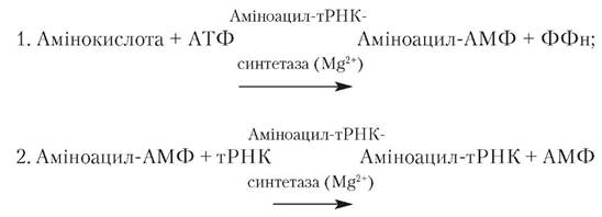
In all cases, during the Second Stage, the activated amino acid attaches to the adenylic acid residue or adenine nucleotide (A) within the CCA triplet at the 3'-end of the tRNA molecule.
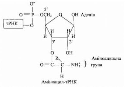
The transfer of the aminoacyl group to the 2'- or 3'-OH group of the ribose residue of the adenine nucleotide in the CCA sequence at the 3'-end of the tRNA depends on the amino acid and the aminoacyl-tRNA synthetase catalyzing aminoacyl-tRNA formation. The activated amino acid can rapidly shift between the 2'- and 3'-positions and back. The stage of activation and transfer of a specific amino acid is catalyzed by the same aminoacyl-tRNA synthetase. The activation of each amino acid consumes the energy of two high-energy phosphate bonds, rendering the overall amino acid activation reaction virtually irreversible. During The conversion of amino acids into aminoacyl-tRNA complexes, the reactivity of the monomeric components used in the polymerization reaction increases; upon interaction of the amino acid with a specific tRNA, Selection of the appropriate amino acids required for Polypeptide chain synthesis is achieved. Lacking The ability to recognize codons in mRNA, amino acids acquire this function as part of aminoacyl-tRNA; simultaneously, the activated carboxyl groups of amino acids become reactive, forming peptide bonds 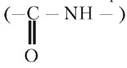 with the amino groups of neighboring amino acids.
tRNA molecules play the role of terminal adaptors that translate the information encoded in The nucleotide sequence of mRNA into the language of protein. No less important in the decoding process is the second set of adaptors—aminoacyl-tRNA synthetase molecules.
Thus, The Genetic Code is deciphered with the aid of two interdependent sets of adaptors performing a highly specific function, As a result of which each amino acid can occupy the position determined by its triplet nucleotide sequence in the mRNA molecule, i.e., its codon.
Ribosomes are required to carry out the reactions of protein synthesis during the stages of polypeptide chain initiation, elongation, and termination.
The ribosome. E. coli ribosomes have been studied the most. Their number in a Introduction/4.html">Prokaryotic Cell exceeds 1.5·104, their diameter is 18–20 nm, their mass ranges between 2500–2800 kDa, and their sedimentation coefficient is 70S. Prokaryotic ribosomes contain 65% rRNA and 35% protein. Ribosomes account for nearly a quarter of The Cell's dry mass. Eukaryotic ribosomes are significantly larger than bacterial ones (diameter is approximately 21 nm, mass is nearly 4000 kDa, sedimentation coefficient is 80S, the protein-to-rRNA ratio is approximately 1:1, and their number in a Eukaryotic Cell is ~105). The synthesis of proteins comprising the ribosomal Structure occurs in the cytoplasm, while the self-assembly of ribosomal subunits takes place in the nucleolus through the interaction of protein molecules and rRNA involving magnesium ions. Prokaryotic and eukaryotic ribosomes consist of two unequal subunits. In prokaryotic ribosomes, the sedimentation coefficient of the larger subunit is 50S and the smaller one is 30S, with masses of 1800 and 1000 kDa, respectively; in eukaryotic ribosomes, the sedimentation coefficient of the larger subunit is around 60S, and the smaller one is 40S.
The large subunit (50S) of a prokaryotic ribosome contains one molecule of 23S rRNA (~3200 NUCLEOTIDES), one molecule of 5S rRNA (~120 nucleotides), and 34 proteins; the small subunit (30S) contains 21 proteins and one molecule of 16S rRNA.
Much like bacterial ribosomes, eukaryotic ribosomes dissociate into large (60S) and small (40S) subunits, which, in turn, can dissociate into their constituent proteins and RNAs. The large subunit comprises three RNA molecules (28S, 7S, and 5S) and over 40 different ribosomal proteins; the small subunit contains one molecule of 18S rRNA and approximately 33 different ribosomal proteins.
The Proteins of the large and small subunits of prokaryotic ribosomes are numbered. In the 50S subunit, they range from L1 to L34 (from Large); in the 30S subunit, from S1 to S21 (from Small). All proteins constituting E. coli ribosomes have been individualized; for most of them, the Amino Acid Sequence, Primary Structure, and molecular mass (ranging within 6.5–75 kDa) have been established, and The nucleotide sequences of E. coli rRNAs have also been determined.
All rRNAs and most eukaryotic ribosomal proteins have also been isolated in pure form and studied. Ribosomal 30S and 50S subunits can, under appropriate conditions, be reconstructed
into functionally active structures from a set of individual components (proteins and rRNA) obtained by subunit dissociation via self-assembly of these macromolecules; for the spontaneous formation of 50S subunits, the presence of the pre-assembled 30S subunit in the system is required.
It has been suggested (A. Lehninger, 1985) that rRNAs act as frameworks for the orderly arrangement of ribosomal Polypeptides, whose enzymatic and other specific Functions have not yet been established for all proteins. According to X-Ray Structural Analysis and Electron Microscopy data, the subunits within the ribosome are arranged asymmetrically, possess an irregular geometric shape, and are connected to each other in such a way that a groove remains between them through which the mRNA molecule passes during polypeptide chain synthesis, along with a second groove that holds the growing polypeptide chain. The first groove accommodates 35 RNA nucleotides, and the second accommodates approximately 30 amino acids. Mitochondria and chloroplast ribosomes differ from Eukaryotic cytoplasmic ribosomes; they bear a greater resemblance to the 70S particles of prokaryotic organisms.
Protein synthesis in mitochondria, Chloroplasts, and Bacteria follows a general scheme (A. Lehninger, 1985; L. Stryer, 1985; B. Alberts et al., 1986).
Polypeptide chain initiation. It has been established that in E. coli and other prokaryotes, the N-terminal amino acid during polypeptide chain assembly is always an N-formylmethionine residue. This and other facts formed the basis for the hypothesis regarding The Significance of formylated Methionine as an initiator in polypeptide chain synthesis. Formylated methionine is produced through two sequential reactions. In this context, the existence of two tRNAs—tRNAMet and tRNAfMet—which accept methionine, should be noted, along with the fact that the enzyme formyltransferase (transformylase) is unable to formylate free methionine. However, formylation of the methionine residue is not always possible even in a complex with tRNA. This reaction can occur only when the tRNA is the specific tRNAfMet:
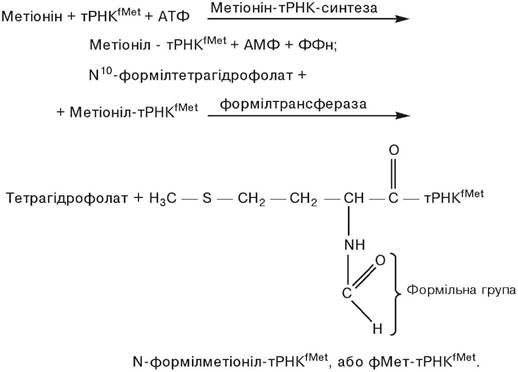
The complex of methionine with another tRNA—methionyl-tRNA (tRNAMet)—is not formylated and is used to incorporate methionine into internal Regions of the synthesized polypeptide chain. Although the Primary structure of tRNAfMet and tRNAMet differs, they share the same UAC anticodon, enabling both tRNAs to interact with the AUG codon. However, the anticodon triplet of the fMet-tRNAfMet complex interacts with the AUG codon only if the latter is located at the beginning of the mRNA coding sequence. The presence of the AUG codon within the internal nucleotide sequence of mRNA involves interaction with unformylated Met-tRNAMet. The presence of the N-formyl group in the methionine residue within the fMet-tRNAfMet initiator complex determines its interaction with a specific initiation site on the ribosome, which neither Met-tRNAMet nor any other aminoacyl-tRNA can react with; blocking the amino group of methionine with the formyl residue prevents the incorporation of such an amino acid into internal regions of the polypeptide chain.
In polypeptide chains synthesized in eukaryotic cell ribosomes, the N-terminal amino acid is always methionine, attached via a specialized initiating methionyl-tRNA; in eukaryotic Mitochondria and chloroplasts, just as in bacteria, protein synthesis begins with N-formylmethionine, which confirms the prevailing view regarding the bacterial origin of these subcellular structures found within eukaryotic cells.
At the stage of polypeptide chain initiation, a crucial step is the three-stage process of initiation complex formation (Fig. 3.8). First, the interaction between the 30S ribosomal subunit and the IF-3 initiation factor yields a structure in which the IF-3 protein prevents its association with the 50S subunit. Attachment of mRNA to the 30S subunit is achieved via a specific initiation signal, which is a purine-rich (A, G) sequence centered approximately 10 nucleotides away from the 5'-end of the mRNA initiation codon (5')AUG(3'), meaning translation cannot begin directly at the 5'-end of the mRNA. The first translated codon is typically located about 25 nucleotides away from the 5'-end. The initiation signal, represented by a short region (6–10 nucleotides) of the mRNA, interacts with a complementary nucleotide sequence located at the 3'-end of the 16S rRNA of the 30S subunit, helping to anchor the mRNA in the proper position for initiation. This interaction ensures the correct positioning of the AUG initiation codon on the 30S subunit. Next, protein initiation factor IF-2 and GTP, previously bound to formylmethionyl-tRNAMet, join the complex consisting of the 30S subunit, IF-3 factor, and mRNA (stage 2). Subsequent attachment of the 50S ribosomal subunit to the previously formed macromolecular complex structure—consisting of the 30S subunit, IF-3 protein initiation factor, mRNA, GTP, IF-2 protein initiation factor, and N-formylmethionyl-tRNAMet—results in a functionally active 70S ribosome. During its formation at the 50S subunit attachment stage, the GTP molecule bound to IF-2 is hydrolyzed into GDP and Pi, which, together with IF-3 and IF-2, form a complex. Thus, the exact site of protein synthesis initiation (the AUG codon, genetic initiation signal) is determined by base-pairing between the leader sequence of nitrogenous bases upstream of the mRNA AUG codon and the nucleotide sequence located at the 3'-end of the 16S rRNA of the 30S subunit, as well as the Complementary interaction between the mRNA AUG codon and the anticodon (3')UAC(5') of N-formylmethionyl-tRNAfMet.
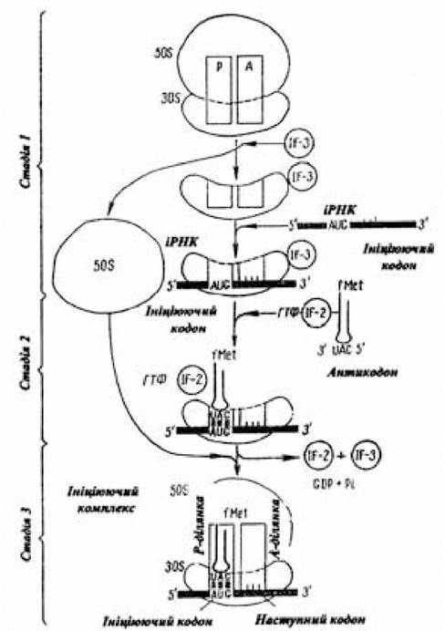
Fig. 3.8. Scheme of the three-stage process of initiation complex formation
(after A. Lehninger, 1985)
To properly position N-formylmethionyl-tRNAfMet within the functionally active 70S initiation complex, it binds to the peptidyl site (P-site) of the 70S complex; the second site for aminoacyl-tRNA attachment is called the aminoacyl site (A-site). These sites are formed by the joining of specific regions of the 30S and 50S subunits. In this state (the P-site is occupied by the initiating fMet-tRNAfMet, and the A-site is free), the initiation complex is ready to continue the translation process.
Elongation. Polypeptide chain elongation is a cyclical process comprising three stages. The cycle begins with the interaction of GTP with one of the three elongation factors—Tu, which are soluble cytoplasmic proteins. An aminoacyl-tRNA whose anticodon is complementary to the codon following the initiation codon in the 5'-3' direction of mRNA interacts with GTP-Tu to form a ternary aminoacyl-tRNA–Tu–GTP complex, which is delivered into the free A-site of the functionally active 70S ribosome by the activated elongation factor (Tu–GTP) and binds to this site. The correct positioning of the aminoacyl-tRNA is checked twice (Fig. 3.9). On the one hand, this involves complementary codon-anticodon interaction; on the other hand, it involves specific bonding between regions of the tRNA and rRNA molecules. Only when this condition is met can the elongation process proceed.
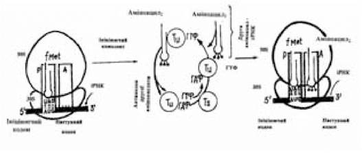
Fig. 3.9. Scheme of the first stage of elongation
(after A. Lehninger, 1985)
The next reaction is GTP Hydrolysis and the elimination of the Tu–GDP complex from the 70S ribosome. The GDP residue in the latter is displaced by another elongation factor, Ts. The new Tu–Ts complex is disassembled under The Influence of GTP, and the newly formed GTP-Tu once again delivers a complementary aminoacyl-tRNA to the vacant A-site of the 70S ribosome. GTP-Tu does not react with fMet-tRNAfMet, meaning the latter does not enter the A-site and internal AUG codons are not read by the initiator tRNA. By THE START OF the second stage of the elongation cycle, the P-site is occupied by fMet-tRNAfMet, and the corresponding aminoacyl-tRNA is located in the A-site. Everything is prepared for the amino acid residues to react and form a peptide bond (Fig. 3.10).
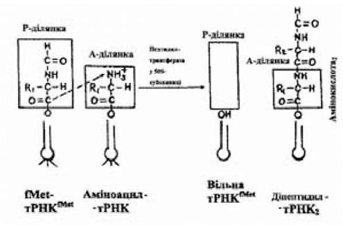
Fig. 3.10. Scheme of peptide bond formation
(after A. Lehninger, 1985)
This reaction is catalyzed by peptidyl transferase, which is one of the proteins of the 50S subunit and whose activity is manifested in the presence of K+. The activated formylmethionine residue, located within fMet-tRNAfMet at the P-site, is transferred to the amino group of the aminoacyl-tRNA occupying the A-site. The new compound resulting from this reaction (dipeptidyl-tRNA) contains two amino acid residues connected by a peptide bond and is located in the A-site of the 70S ribosome; the initiating tRNAfMet, freed from the activated amino acid residue, remains in the P-site.
The Third Stage of elongation—translocation—involves three movements (Fig. 3.11). As the 70S ribosome moves a distance of one codon along the mRNA toward its 3'-end, the dipeptidyl-tRNA located in the A-site shifts to the P-site, causing the free tRNA to detach from the P-site and enter the cytoplasm. Thus, dipeptidyl-tRNA ends up in the peptidyl site, and the aminoacyl site is once again ready to bind the next aminoacyl-tRNA, whose anticodon is complementary to the subsequent mRNA codon located in the aminoacyl site zone of the 70S ribosome.
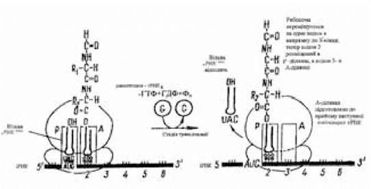
Fig. 3.11. Scheme of translocation
(after A. Lehninger, 1985)
A new three-stage elongation cycle begins, upon completion of which tripeptidyl-tRNA will be formed. The movement of the ribosome along the mRNA by one codon is called
translocation. This movement is carried out with the participation of a third elongation factor, G, or translocase, and the energy generated by the hydrolysis of another GTP molecule. The formation of a single peptide bond (The addition of one amino acid) consumes the energy of hydrolysis of two GTP molecules.
Termination. A point is reached in polypeptide chain synthesis where the A-site of the ribosome is occupied by one of the stop codons: UAA, UGA, or UAG. In this case, codon-anticodon interaction does not occur because normal cells do not contain tRNAs with anticodons complementary to termination signals. Termination triplets do not code for Amino Acids and are therefore referred to as nonsense codons.
With high Specificity, mRNA terminating triplet sequences interact with protein release factors R1, R2, and S (releasing factors). The first recognizes the UAA or UAG codons, while the second recognizes UAA or UGA. The interaction of one of the release factors with the termination codon at the aminoacyl site of the 70S ribosome activates peptidyl transferase and alters its specificity. This results in the hydrolytic Cleavage of the polypeptide from the peptidyl-tRNA, the acceptance of H2O by the activated peptidyl residue, the release of the newly synthesized protein molecule, the dissociation of the tRNA from the vacated P-site, and the splitting of the 70S ribosome into its 30S and 50S subunits, preparing them for the synthesis of a new protein molecule (Lehninger A., 1985; Stryer L., 1985).
Several functionally active ribosomes can simultaneously reside on a single mRNA molecule, with each ribosome occupying a space equivalent to 80 nucleotides. This capability significantly enhances the efficiency of mRNA utilization. Multiple ribosomes simultaneously translating the same mRNA molecule form a polyribosome structure, or polysome, in which each ribosome functions autonomously to synthesize its own polypeptide chain.
Folding and Processing of the polypeptide chain.
The primary structure (amino acid sequence) is the decisive factor in forming the three-dimensional structure that renders a protein functionally active. However, a protein molecule frequently acquires its biologically active conformation only as a result of processing or covalent (post-translational) modification. This process varies among different proteins and involves reactions of cleavage or addition of atomic groups. For instance, in proteins of bacterial origin, the formyl group part of the N-terminal N-formylmethionine is deformylated during processing. Special aminopeptidases catalyze the hydrolytic removal of one or more N-terminal amino acid residues, which is why they are often undetectable in fully formed mature proteins. During post-translational maturation, N-terminal polypeptide leader sequences are cleaved; these act as specific signals guiding the protein to its cellular destination. Very frequently, the N-terminus of the polypeptide chain undergoes modification while the Synthesis of the remainder of the chain is still ongoing.
Other widespread reactions involved in the Modification of the polypeptide structure—and which thereby influence the formation of its final conformation—include the Acetylation of the amino group of the N-terminal amino acid, the phosphorylation of the OH groups of hydroxyamino acid residues (Serine, Threonine, Tyrosine), which increases the negative charge of the respective proteins, the carboxylation of aspartic and glutamic acid residues, the methylation of Lysine residues and the carboxyl groups of certain glutamic acid residues, the attachment of side carbohydrate chains, the addition of prosthetic groups, and the formation of Disulfide Bonds.
Last update: 11/08/2026
Editorial and Educational Adaptation: This material has been compiled based on the primary/original source text. The project team performed an editorial review, corrected technical inaccuracies, structured sections, and adapted the content for an educational format.
What was processed:
- elimination of formatting defects (OCR errors, structural breaks, corrupted characters);
- editorial organization of content;
- standardization of terminology in accordance with academic sources;
- verification of factual statements against the original source text.
All mentions of the author, publication year, and origin of the primary text have been preserved in accordance with the source.