Principles of Biochemistry, Volume 3 - A. Lehninger 1985
Molecular Mechanisms of Genetic Information Transfer
DNA: Structure of Chromosomes and Genes
The nucleotide sequences of several DNAs have already been deciphered
In 1977, the complete nucleotide sequence of the DNA of bacteriophage фX174 was determined (p. 850). This remarkable achievement marked the beginning of a new era in the biochemistry of genes and Chromosomes. Since then, the nucleotide sequences of a great many genes have been deciphered, and it is now possible—in principle, at least—to determine The base sequence of virtually any DNA.
Prior to 1977, the nucleotide sequences of many tRNAs and several small mRNAs had been deciphered. Robert Holley and his colleagues were the first to determine The nucleotide sequence of a nucleic acid: Yeast Alanine tRNA. This study, completed in 1965, required several years of work. Although tRNA molecules consist of fewer than 100 nucleotide residues, they contain many unusual modified bases that had to be identified. DNA Sequencing (i.e., the determination of nucleotide sequence) presents additional challenges. We have already mentioned that an average E. coli Gene consists of about 1,200 nucleotide pairs, and the entire DNA molecule of bacteriophage фX174 contains more than 5,000 pairs. Previously, there were no Methods available to selectively cleave DNA at a specific nucleotide—say, at all A residues. Even if such a method existed, Cleavage would yield a very large set of smaller fragments that would be extremely difficult to separate. Furthermore, even if these fragments could be separated and sequenced, reconstructing the complete sequence from them would be practically impossible.
Decisive success was achieved thanks to three major breakthroughs. The first of these was the discovery of Restriction Endonucleases, which cleave DNA molecules only at a relatively small number of specific sites. The Use of two or more restriction endonucleases (Table 27-7) made it possible to cleave DNA molecules into individual fragments in different ways, generating overlapping sequences—much like the application of two different Proteolytic Enzymes (such as Trypsin and Chymotrypsin) once made it possible to cleave polypeptide chains into different sets of fragments and establish Amino acid sequences in overlapping regions (Section 6.7, e). For example, the DNA of simian virus 40 (SV40) (Fig. 27-30), which is capable of transforming certain Cells into malignant ones, was cleaved by restriction endonucleases at a series of specific sites to yield fragments convenient for mapping the locations of individual genes. Figure 27-31 schematically illustrates the cleavage sites of SV40 DNA by three restriction endonucleases.
Class="center">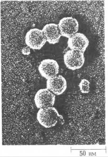
Fig. 27-30. Simian virus 40 (SV40) causes Cancer in hamsters and other small animals. It is one of the smallest carcinogenic Viruses. The protein coat of SV40 has the shape of an icosahedron, i.e., a twenty-sided polyhedron.
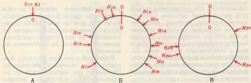
Fig. 27-31. Restriction sites in the circular DNA molecule of SV40 for three different restriction endonucleases—EcoRI, Hin, and HpaI—each of which recognizes and catalyzes the cleavage of The Double Helix at specific sites. A. EcoRI cleaves both strands of circular SV40 DNA at only a single site, converting it into a linear form. This site is taken as the reference point (zero coordinate), from which the positions of SV40 DNA Restriction sites for other restriction endonucleases are determined. B. Hin (a mixture of HindII and HindIII) cleaves the DNA at eleven sites, yielding twelve fragments. C. HpaI cleaves the DNA at only four locations, yielding five fragments. These enzymes were the first restriction endonucleases used by Daniel Nathans and his colleagues to map genes in SV40 DNA. Restriction sites in the SV40 genome are now known for numerous other endonucleases.
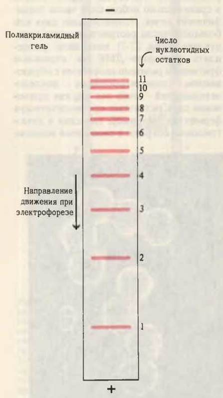
Fig. 27-32. Electrophoretic Separation of oligonucleotides by chain length on a polyacrylamide gel slab. The shorter the oligonucleotides, the faster they migrate toward the positive electrode. By adjusting the porosity of the polyacrylamide gel, this procedure can be used to separate fairly long oligonucleotides containing up to two hundred or more residues, even if these oligonucleotides differ by as little as a single residue.
The second major breakthrough was the refinement of electrophoretic methods for separating DNA fragments According to the number of nucleotide residues they contain. These methods have such high resolution that they can separate DNA fragments up to 200 NUCLEOTIDES in length, even if they differ in length by as little as a single nucleotide (Fig. 27-32).
The third breakthrough was The Development of DNA Cloning METHODS (Chapter 30), which made it possible to obtain sufficiently large quantities of pure genes—the Starting Material for sequencing. Two principal approaches to DNA sequencing were proposed, each with A number of variations. Frederick Sanger, who was the first to determine the Amino Acid Sequence of a protein, namely Insulin (Section 6.8), also became the first scientist to establish the nucleotide sequence in a DNA molecule (that of bacteriophage фX174); this occurred in 1977. Sanger and his colleagues developed a very elegant procedure known as the chain-termination method, or the "plus-minus" system. Independently, Alan Maxam and Walter Gilbert in the USA proposed a somewhat different approach, termed the chemical method. Both approaches utilize fragments obtained by cleaving the starting DNA with restriction endonucleases. Box 27-1 describes the principles of the nucleotide sequencing method developed by Maxam and Gilbert.
Box 27-1. Sequencing a Short DNA Fragment Using the Maxam-Gilbert Chemical Method
In the description below, some details are omitted in order to focus attention on the fundamental Principle of the method. Suppose we have a restriction endonuclease-generated DNA fragment of 10 nucleotide residues with the sequence
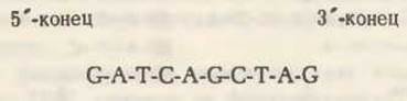
The first step consists of introducing a radioactive label into the 5'-terminal residue (G), which is shown against a red Background:

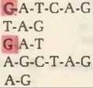
The 5'-end-labeled oligonucleotide is then divided into four portions. The first portion is subjected to a chemical Treatment that causes the oligonucleotide to break down into smaller pieces via the statistical removal of C residues. Such Cleavage of the oligonucleotide at C residues may yield the following mixture:
In this set of fragments, the labeled 5'-terminal residue is shown against a red background. Note that two fragments are labeled, meaning they contain the 5'-end of the original oligonucleotide, whereas the fragments T—A—G, A—G—C—T—A—G, and A—G are not labeled, i.e., they lack the original 5'-end. We will be interested only in the labeled fragments.
The second portion of the original labeled oligonucleotide is subjected to a different chemical treatment that specifically removes only G residues, resulting in the generation of another set of labeled fragments (Fig. 1). The third portion of the original labeled oligonucleotide undergoes a similar treatment, fragmenting As a result of the removal of only A residues. Likewise, the fourth portion is cleaved by the removal of only T residues. Ultimately, four distinct mixtures of labeled fragments are obtained using four different chemical Procedures (Fig. 1).
Each of the four fragment mixtures is subjected to gel Electrophoresis under conditions that ensure the separation of fragments according to the number of nucleotide residues they contain, regardless of the identity of those residues. In such a separation, smaller fragments migrate faster. The exact position of each labeled fragment in the gel is determined by autoradiography. The positions of the labeled fragments for each of the cleavage variants are shown in Fig. 1. The positions of unlabeled fragments are not detected, but these fragments are not needed to decipher the sequence.
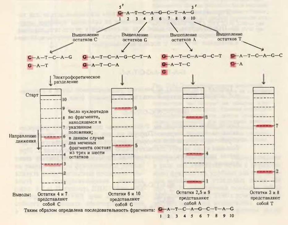
In Fig. 1, the results of the first fragmentation method, in which C residues were removed, are compared with the electrophoretic pattern showing the positions of the resulting fragments ranging from 1 to 10 residues in length. Two labeled fragments were detected here. Obviously, these are precisely the fragments containing the 5'-end of the original oligonucleotide. The labeled fragments are found at positions corresponding to oligonucleotides 3 and 6 residues long. It is clear that in the original nucleotide sequence, a C residue must have immediately followed these labeled fragments in the direction of the 3'-end, because the chemical procedure that led to the cleavage of the original oligonucleotide specifically removed only this residue. Thus, as a result of the first chemical treatment, we learned that C residues must be located at positions 4 and 7 (counting from the 5'-end) of the original oligonucleotide.
The same procedure is now applied to the other three sets of fragments obtained by the specific removal of G, A, and T residues, respectively, from the original oligonucleotide (Fig. 1). From the diagram, we can see that the labeled fragments generated by the removal of G residues migrate at rates corresponding to oligonucleotides 5 and 9 nucleotide units long; thus, residues 6 and 10 in the original oligonucleotide must be G. The third set of fragments, obtained by the removal of A, indicates that residues at positions 2, 5, and 9 were A; the fourth set of fragments, obtained by the cleavage of T residues, points to the presence of T residues at positions 3 and 8. The bottom part of Fig. 1 shows the nucleotide sequence established using this simple procedure and logical deduction. By this method, the nucleotide sequence of oligonucleotides containing 200 or even more residues can often be determined in less than 2 days.
To decipher the nucleotide sequence of an entire DNA molecule, it is first fragmented using a restriction endonuclease. Then each of the resulting fragments is sequenced individually according to the scheme shown in Fig. 1. In a second aliquot, the original DNA is cleaved at different sites using a different restriction endonuclease, yielding a second set of fragments. Once the sequencing of all fragments In the second set is complete, comparing the two sets makes it possible to find the overlapping regions required to assemble the fragments of the first set in the correct order. As a result, the nucleotide sequence of the natural DNA of interest can be established. Sometimes, in order to resolve ambiguities in certain Regions of the sequence remaining after the first two cleavages, it is necessary to analyze a third or fourth set of fragments.
Last update: 06/08/2026
Editorial and Educational Adaptation: This material has been compiled based on the primary/original source text. The project team performed an editorial review, corrected technical inaccuracies, structured sections, and adapted the content for an educational format.
What was processed:
- elimination of formatting defects (OCR errors, structural breaks, corrupted characters);
- editorial organization of content;
- standardization of terminology in accordance with academic sources;
- verification of factual statements against the original source text.
All mentions of the author, publication year, and origin of the primary text have been preserved in accordance with the source.