LEHNINGER PRINCIPLES OF BIOCHEMISTRY - VOLUME 1. THE FOUNDATIONS OF BIOCHEMISTRY: STRUCTURE AND CATALYSIS - 2011
PART I. STRUCTURE AND CATALYSIS
5. PROTEIN FUNCTION
5.2. Reversible Binding of a Protein to a Ligand: Oxygen-Binding Proteins
We have already described how protein conformation depends on the binding of small ligands (O2 and CO2) to heme, and how it, in turn, affects that binding. However, many Protein-Ligand interactions occur without the participation of prosthetic groups. The ligand-binding site most often resembles the BPG-binding site on a Hemoglobin molecule—simply a cavity within the protein molecule containing specific amino acid residues that ensure binding Specificity. A common property of these binding sites is the effective recognition of even very structurally similar ligands.
All vertebrates possess an immune system capable of distinguishing "self" molecules from "non-self" (foreign) molecules and destroying those perceived as foreign. In this way, The Immune System combats Viruses, Bacteria, and other pathogens, as well as molecules capable of harming the Organism. At the physiological level, the Immune Response to a foreign particle or organism represents a complex and coordinated process of interactions among various classes of protein and other molecules and diverse Cell types. At the level of individual Proteins, the immune response serves as a prime example of how reversible protein-ligand binding builds an extraordinarily sensitive and specific biochemical system.
The immune response is mediated by specialized Cells and proteins
The immune response involves various types of leukocytes (white Blood Cells), including macrophages and lymphocytes, which derive from undifferentiated stem cells in the Bone Marrow. Leukocytes can exit the bloodstream and patrol Tissues, with each cell capable of synthesizing one or more proteins that recognize and bind foreign molecules.
Immunity relies on two complementary systems—humoral and cellular. Humoral immunity (from the Latin humor, meaning fluid) targets bacterial infections and extracellular viruses (those present in Body Fluids), but can also react to the Introduction of individual proteins into the organism. Cellular immunity destroys host cells infected by viruses, as well as certain parasites and foreign tissues.
The principal agents of humoral immunity are soluble proteins called Antibodies or IMMUNOGLOBULINS (Ig). Immunoglobulins bind to bacteria, viruses, and large foreign molecules destined for destruction. Comprising up to 20% of Blood Plasma protein, immunoglobulins are produced by B cells (B lymphocytes), so named because they complete their maturation in the bone marrow.
The key players in cellular immunity are specific T cells (T lymphocytes, named after the Thymus, where they complete their maturation), specifically cytotoxic T cells (CTLs), or killer cells. Proteins located On the surface of cytotoxic cells—called T-cell receptors—participate in recognizing infected cells or parasites. Receptors are proteins, typically situated on the outer surface of The Cell and extending beyond The Plasma Membrane, that recognize and bind extracellular ligands, thereby triggering specific cellular processes.
In addition to cytotoxic T cells, there are T helper cells (TH cells). Their function is to produce soluble signaling proteins called cytokines, which include interleukins. TH cells interact with macrophages; they participate only indirectly in the destruction of infected cells and pathogens by selectively stimulating those cytotoxic T cells and B cells that bind a specific antigen. This process, known as clonal Selection, increases the population of immune cells capable of reacting to the appearance of that antigen. The critical role of TH cells is most vividly demonstrated in the case of the HUMAN IMMUNODEFICIENCY VIRUS (HIV), which causes Acquired Immunodeficiency Syndrome (AIDS). TH cells are the primary target of HIV; the destruction of these cells progressively dismantles the entire immune system. The Functions of various leukocyte types are summarized in Table 5–2.
Class="center">Table 5–2.
Cell type |
Functions |
Macrophages |
Engulf large particles and cells via phagocytosis |
B lymphocytes (B cells) |
Synthesize and secrete antibodies |
T lymphocytes |
Interact with infected host cells via T-cell receptors |
T helper cells (TH) |
Interact with macrophages and secrete cytokines (interleukins) that stimulate the proliferation of CTLs, TH cells, and B cells |
Each protein of the immune system, whether an antibody produced by a B cell or a T-cell receptor, specifically binds molecules with a particular chemical Structure, distinguishing them from all other molecules. The Human Body can produce more than 108 different antibodies, each characterized by a distinct specificity. This extraordinary diversity makes possible the recognition and binding of virtually any chemical structure on The surface of viruses or foreign cells. The vast repertoire of antibodies results from the assembly of immunoglobulin genes from individual segments via genetic recombination (Chapter 25, Figure 25–26).
Certain Features of the interaction between antibodies or T-cell receptors and their target molecules are unique to the immune system and require specialized terminology. Any molecule or pathogen capable of eliciting an immune response is called an antigen. An antigen can be a virus, a Bacterial Cell wall, an individual protein, or another macromolecule. A complex antigen may bind to several different antibodies. Each antibody and each T-cell receptor binds to only one specific molecular structure on the antigen, known as an antigenic determinant, or epitope.
The immune system generally does not react to small molecules that typically serve as intermediates or End products of cellular METABOLISM. Molecules with a molecular mass of less than 5,000 usually do not elicit an immune response. However, under laboratory conditions, small molecules can be covalently attached to large carrier proteins, and the resulting complex as a whole will provoke an immune response. Such small molecules are called haptens. Antibodies generated in response to the administration of a protein-hapten complex are also capable of binding free haptens. Such antibodies are sometimes used in The Development of analytical assays, discussed later, or as catalytic antibodies (Box 6–3). We now return to a more detailed examination of Antibodies and Their antigen-binding capabilities.
Antibodies Have Two Identical Antigen-Binding Sites
The predominant class of antibodies is immunoglobulin G (IgG); these account for a substantial fraction of the total protein in blood serum. IgG molecules are constructed from four polypeptide chains—two large ones, called heavy chains, and two light chains—which are linked together by noncovalent interactions and Disulfide Bonds into a large complex with a molecular mass of 150,000. The heavy chains of IgG interact with each other at one end of the molecule and then diverge, each associating with a light chain, giving the molecule a characteristic Y-shaped structure (Figure 5–21).
Figure 5–21. Structure of immunoglobulin G (IgG). (a) In an IgG molecule, pairs of light and heavy chains associate to form a Y-shaped structure. Two antigen-binding sites are formed by the combination of the variable domains of the light (VL) and heavy (VH) chains. Treatment with Papain cleaves the molecule into Fc and Fab fragments. The Fc fragment contains a bound carbohydrate moiety (see b). (b) Ribbon model of the first complete IgG molecule to be crystallized (PDB ID 1IGT). Although the molecule contains two identical heavy chains (shown in two shades of blue) and two identical light chains (two shades of red), the crystal structure exhibits an asymmetric conformation. Conformational flexibility likely plays a crucial role in immunoglobulin function.

Proteases can cleave the immunoglobulin molecule at hinge regions that separate the "stem" of the IgG from its "arms." Cleavage by papain yields the stem, designated the Fc fragment because it typically crystallizes readily, and two arms, designated Fab fragments (for antigen-binding fragment). Each Fab fragment possesses a single antigen-binding site.
The structure of immunoglobulins was first elucidated by Gerald Edelman and Rodney Porter. Each immunoglobulin chain contains clearly identifiable domains, some of which share a conserved structure across all types of IgG, while others vary. The constant domains exhibit characteristic Structural motifs known as the immunoglobulin fold, which are found in all proteins with a β structure (Chapter 4). Each heavy chain contains three constant domains, whereas the light chain contains one. Both heavy and light chains possess a single variable domain, which exhibits the highest degree of Amino Acid Sequence variation. Together, the variable domains of the heavy and light chains form the antigen-binding site (Figures 5–21, 5–22).
Figure 5–22. Binding of an antigen to IgG. To achieve optimal antigen binding, the binding site on the IgG molecule often undergoes subtle conformational changes. Such induced fit is a general phenomenon in protein-ligand interactions.

Many vertebrates have five classes of immunoglobulins, of which IgG is just one. Each class features its own characteristic heavy chain: α, δ, ε, γ, and μ in IgA, IgD, IgE, IgG, and IgM, respectively. All immunoglobulins contain one of two classes of light chains: κ or λ. The overall structure of IgD and IgE resembles that of IgG. IgM exists either as a monomeric, membrane-bound form or as a secreted pentamer (Fig. 5-23). IgA is found predominantly in secretions such as saliva, tears, and milk, and may exist as monomers, dimers, or trimers. IgM is the first class of antibodies produced by B lymphocytes during maturation and serves as the principal antibody appearing in the Cytology/cytology/16.html">Early stages of the primary immune response. Some B cells soon switch to producing IgD (with the same antigen-binding site as the IgM previously synthesized by that cell), although the precise physiological functions of IgD remain incompletely understood.
Figure 5-23. Pentameric structure of IgM. The pentameric assembly is held together by disulfide bonds (shown in yellow). The J chain is a polypeptide with Mr = 20,000; this chain is found in both IgA and IgM.

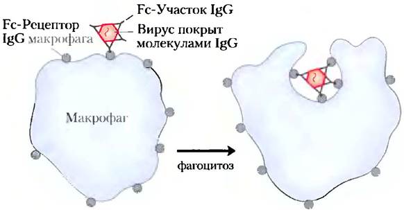
Immunoglobulin G (IgG) constitutes the principal antibody synthesized during a secondary immune response initiated by memory B cells—that is, during the immune response to Antigens previously encountered by the immune system. Upon binding to a bacterium or virus that has invaded the organism, IgG stimulates certain leukocytes, such as macrophages, which are capable of engulfing and destroying pathogens, and also activates Other components of the immune response. Furthermore, a distinct class of receptors on the macrophage surface recognizes and binds the Fc region of IgG. Once these Fc receptors bind to the antibody-pathogen complex, the macrophage engulfs the complex via phagocytosis (Fig. 5-24).
Figure 5-24. Phagocytosis of an antibody-coated virus. The Fc regions of antibodies bind to Fc receptors on the macrophage surface, leading to the engulfment and destruction of the virus.
IgE plays a pivotal role in mediating allergic reactions: it interacts with basophils (phagocytic leukocytes) in the blood and with mast cells, which are abundant in tissues and secrete histamine. Through its Fc region, immunoglobulin E binds to Fc receptors on basophils or mast cells. In this state, IgE functions effectively as an antigen receptor. Upon binding an antigen, these cells secrete histamine and other biologically active amines that induce vasodilation and increased vascular permeability. This vascular response likely facilitates the recruitment of immune cells and proteins to sites of inflammation, but it also triggers the classic symptoms of allergy. Pollen and other harmless environmental allergens are perceived by the organism as foreign invaders, provoking an immune response that is normally directed against pathogens. ■
Antibodies bind antigens with high affinity and specificity
The specificity of antigen-C binding by antibodies is determined by The amino acid residues in the variable regions of both heavy and light chains. While many residues in these regions vary from one protein to another, the degree of variation differs. Certain residues, particularly those forming the antigen-binding site, are hypervariable, meaning they exhibit an exceptionally high degree of Variability. Binding specificity is governed by the chemical complementarity between the antigen and its binding site on the antibody molecule—specifically, molecular shape and the spatial arrangement of charged, nonpolar, and hydrogen-bonding groups. For instance, a binding site bearing a negatively charged group can bind an antigen with a positive charge at the corresponding position. In many cases, this complementarity is achieved through a mutual adaptation between the antigen and the binding site as they approach one another. The conformational adjustments that occur within the antibody and/or antigen molecules allow the complementary groups to interact in the most energetically favorable manner. This phenomenon is well illustrated by The formation of a complex between an HIV peptide (model antigen) and a Fab fragment (Fig. 5-25), which clearly demonstrates the structural changes accompanying antigen binding.
Figure 5-25. Induced fit in the binding of an antigen to IgG. The surface of an IgG Fab fragment is shown interacting with a small peptide from the human immunodeficiency virus (HIV). To facilitate tracking structural Changes in the molecule, two heavy-chain residues are colored blue, and one light-chain residue is colored red. (a) Top view of the antigen-binding site of the IgG Fab fragment (PDB ID 1GGC). (b) View in the same orientation, but with the Fab fragment in the conformation that binds the antigen (PDB ID 1GGI). The antigen molecule itself is omitted here to provide a clear view of the changes occurring within the binding site. Note that the ligand-binding cavity has widened and certain groups have shifted spatially. (c) The same view in the presence of the bound antigen (depicted as a red stick model).

Antibody-antigen interactions are typically characterized by very high affinity—the dissociation constant Kd is roughly 10-10 M (recall that the lower the Kd, the tighter the binding). This magnitude is determined by the cumulative energy of various ionic, hydrophobic, Van der Waals interactions, and Hydrogen Bonds that stabilize the complex. The binding energy required to achieve a Kd of 10-10 M is approximately 65 kJ/mol.
Antibody-antigen interactions form The basis of numerous analytical techniques
The extraordinarily high affinity and specificity of antibody-antigen binding have enabled their widespread application in analytical biochemistry and clinical Diagnostics. Two MAIN TYPES OF antibodies are utilized: polyclonal and monoclonal. Polyclonal antibodies are produced by multiple different B lymphocyte clones in response to a specific antigen, such as a foreign protein injected into an animal. These B lymphocytes generate antibodies that bind to specific, yet distinct, epitopes on the antigen. Consequently, polyclonal preparations represent a heterogeneous mixture of antibodies recognizing various Regions of the protein. In contrast, Monoclonal Antibodies are synthesized by a population of identical B cells (a clone) cultured in vitro. These antibodies are homogeneous in that they all bind to the exact same epitope. The pioneering Methods FOR PRODUCING monoclonal antibodies were developed by Georges Köhler and César Milstein.
Georges Köhler, 1946–1995

César Milstein, 1927–2002

The remarkable specificity of antibodies finds extensive practical utility. Specific antibodies can be covalently attached to a solid support matrix and used for affinity Column Chromatography (Fig. 3-17c). When a complex mixture of proteins is passed through the column, the antibodies specifically bind only their target proteins and retain them on the matrix, while all other proteins pass through unretarded. The protein of interest can subsequently be eluted from the column using a salt solution or another dissociating reagent. This represents a powerful and widely used method for Protein Purification.
In another versatile analytical approach, antibodies are covalently conjugated with a radioactive label, a fluorophore, or an enzyme marker that allows for their straightforward detection. When such labeled antibodies bind to a target protein, the marker reveals the protein's presence in solution, its localization within a gel, or even its distribution inside a living cell. Several methods relying on this general strategy are illustrated in Figure 5-26.
Figure 5-26. Analytical techniques employing antibodies. The specific antibody-antigen interaction forms the basis of numerous methodologies utilized for the identification and quantification of proteins in complex mixtures. (a) Schematic representation of the general approach. (b) Detection of antibodies against Herpes simplex virus (HSV) in blood using an enzyme-linked immunosorbent assay (ELISA). The microplate wells are coated with HSV antigen, which captures anti-HSV antibodies from the sample. Horseradish peroxidase-conjugated antibodies directed against human IgG are used as secondary antibodies following the steps outlined in (a); the greater the concentration of anti-HSV antibodies in the blood sample, the more intense the yellow color developed in the well. (c) Immunoblot (Western blot). Lanes 1–3 contain protein kinase samples obtained at successive stages of purification. Following SDS-Polyacrylamide gel Electrophoresis, the gel was stained with Coomassie brilliant blue. Lanes 4–6 contain identical samples that, after electrophoretic Separation, were electroblotted onto a nitrocellulose membrane. The membrane was subsequently probed with anti-protein kinase antibodies under specific conditions. The numbers between the gel and membrane photographs indicate the positions of marker proteins of known molecular masses (kDa).

The enzyme-linked immunosorbent assay, commonly known as ELISA, enables rapid Quantitative determination of an antigen within a sample (Fig. 5-26b). The protein sample is immobilized on an inert solid surface, most commonly a 96-well polystyrene microtiter plate. The wells are then blocked by incubation with a solution of an inexpensive, nonspecific protein (such as casein derived from nonfat dry milk) to prevent any non-specific adsorption of proteins added in subsequent steps. Following this blocking step, a solution of primary antibodies—directed against the target protein—is added to the wells. Unbound antibodies are washed away, and a solution of enzyme-conjugated secondary antibodies (which recognize the primary antibodies and catalyze the formation of a colored product) is introduced. Remaining secondary antibodies are washed away, and an appropriate substrate for the enzyme label is added. The amount of product formed, measured spectrophotometrically by its color intensity, is directly proportional to the concentration of the target protein in the sample.
In immunoblotting (also known as Western blotting), proteins previously separated by polyacrylamide gel electrophoresis are transferred electrophoretically from the gel onto a nitrocellulose or PVDF membrane (Fig. 5-26c). The membrane is blocked with a nonspecific protein, much like in an ELISA, and then sequentially probed with primary antibodies, enzyme-conjugated secondary antibodies, and a suitable substrate. A colored or chemiluminescent product is generated exclusively at the precise position (band) containing the target protein. Immunoblotting allows for the detection of minor protein components within complex mixtures and the approximate Determination of the molecular mass of the protein under investigation. Immunoblotting.
In the following chapters, we will encounter further aspects of antibody function and diversity. They play an indispensable role in modern medicine and provide profound insights into the structure and regulation of genes and proteins.
Summary of Section 5.2 Complementary Interactions Between Proteins and Ligands: The Immune System and Immunoglobulins
■ The immune response is driven by specialized white blood cells and their associated proteins. T lymphocytes synthesize T-cell receptors. B lymphocytes synthesize immunoglobulins. Through clonal selection, helper T cells induce the proliferation of B cells or cytotoxic T cells, which produce immunoglobulins or T-cell receptors, respectively, capable of binding a specific antigen.
■ Humans possess five classes of immunoglobulins, each serving a distinct biological function. Quantitatively, IgG is the most abundant. IgG molecules are Y-shaped and consist of two heavy chains and two light chains. At the two tips of the molecule—each formed by one heavy and one light chain—lie hypervariable regions that constitute two antigen-binding sites.
■ Each immunoglobulin typically binds to only a single site on a large antigen (such as a protein), known as an epitope. This binding process is frequently accompanied by conformational changes, leading to an induced fit between the antibody and the antigen.
Last update: 06/08/2026
Editorial and Educational Adaptation: This material has been compiled based on the primary/original source text. The project team performed an editorial review, corrected technical inaccuracies, structured sections, and adapted the content for an educational format.
What was processed:
- elimination of formatting defects (OCR errors, structural breaks, corrupted characters);
- editorial organization of content;
- standardization of terminology in accordance with academic sources;
- verification of factual statements against the original source text.
All mentions of the author, publication year, and origin of the primary text have been preserved in accordance with the source.