BIOLOGY Volume 2 - A Guide to General Biology - 2004
14. TRANSPORT IN ANIMALS
14.9. The Immune System
14.9.2. T Cells and Cell-Mediated Immune Response
The Thymus gland is located in the chest cavity, slightly above The Heart and directly behind the manubrium of the Sternum. It begins to function during embryonic development, reaching peak activity shortly before and immediately after birth. Once a child is weaned, the gland decreases in size, and its function soon ceases.
The crucial role of the thymus in The Development of The Immune System is demonstrated by the following experimental findings:
1. Removal of the gland in newborn mice leads to their death due to a deficiency of lymphocytes in tissue fluid and Blood.
2. Thymus tissue from an adult mouse, transplanted into a newborn mouse to replace the removed thymus, is incapable of reacting to Antigens.
3. Removal of the thymus in older mice does not cause any adverse effects.
Bone Marrow stem Cells that give rise to T lymphocytes must migrate to the thymus while it is still approaching peak activity. There, they develop into so-called thymocytes. At this stage, any cells that recognize "self" antigens are destroyed, ensuring that the Organism does not subsequently attack its own Tissues. As they mature, a portion of the thymocytes differentiate into T cells. They leave the thymus and enter the bloodstream. Some remain in Circulation, while others migrate into tissue fluid, Lymph Nodes, and various other Organs, such as the Spleen.
The outer membrane of T cells bears specific receptors structurally similar to Antibodies. Unlike antibody receptors, however, these receptors cannot recognize whole antigen molecules. They bind only to fragments of antigens or other foreign molecules presented to them by other cells, usually macrophages. Based on surface membrane features (the "self" antigens present on them), T cells are subdivided into T4 and T8 cells, which differ in function. T4 cells are known as helper cells. The HUMAN IMMUNODEFICIENCY VIRUS (HIV), which causes AIDS, primarily infects these cells. There are two types of T8 cells, namely suppressor cells and killer cells (cytotoxic cells). Each type of T Cell produces its own specific Class of lymphokines. Lymphokines are small peptide molecules with diverse Functions, which are briefly described below. They are also known as cytokines or interleukins.
T4 cells work in conjunction with macrophages. First, a macrophage engulfs the antigen-bearing organism. It then cleaves off an antigen fragment (a peptide) and displays it on its surface, effectively presenting it to immune cells. A T4 lymphocyte with a matching receptor Structure recognizes this peptide as foreign and responds by producing large amounts of lymphokines that perform various functions, specifically: 1) stimulating T-cell proliferation; 2) initiating the inflammatory process; 3) stimulating antibody production by B cells.
Killer cells (a type of T8 lymphocyte) produce fewer lymphokines, but they are capable of destroying virus-infected and malignant cells (Fig. 14.39). This occurs via chemical attack (cytotoxicity) or by perforating the target cell's outer membrane. Killers recognize not fragments of external antigens, but foreign Peptides generated within the affected organism itself—such as a viral fragment produced inside an infected cell and displayed on its surface, or a mutant protein produced by a malignant cell. In addition, they attack and gradually destroy transplanted organs (Section 25.7.13).
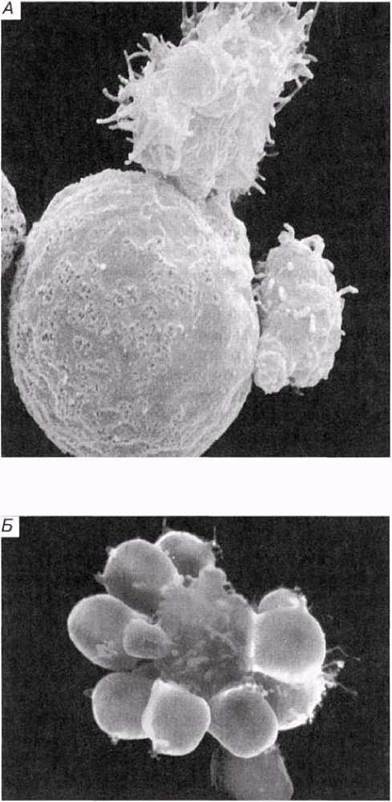
Fig. 14.39. A. Scanning electron micrograph of T killer cells (relatively small cells at the top and right) attacking a larger cell (typically either a Cancer cell, a virus-infected cell, or a cell from a transplanted organ or tissue). B. Scanning electron micrograph showing a T killer cell (relatively large cell in the center) attached to a group of foreign red Blood Cells and destroying them.
Lymphokines secreted by suppressor cells inhibit The activity of all leukocyte types, including phagocytes, whereas lymphokines secreted by helper cells stimulate it. Thus, the numerical ratio between these Two Types of lymphocytes regulates the overall strength of the Immune Response (Fig. 14.40).
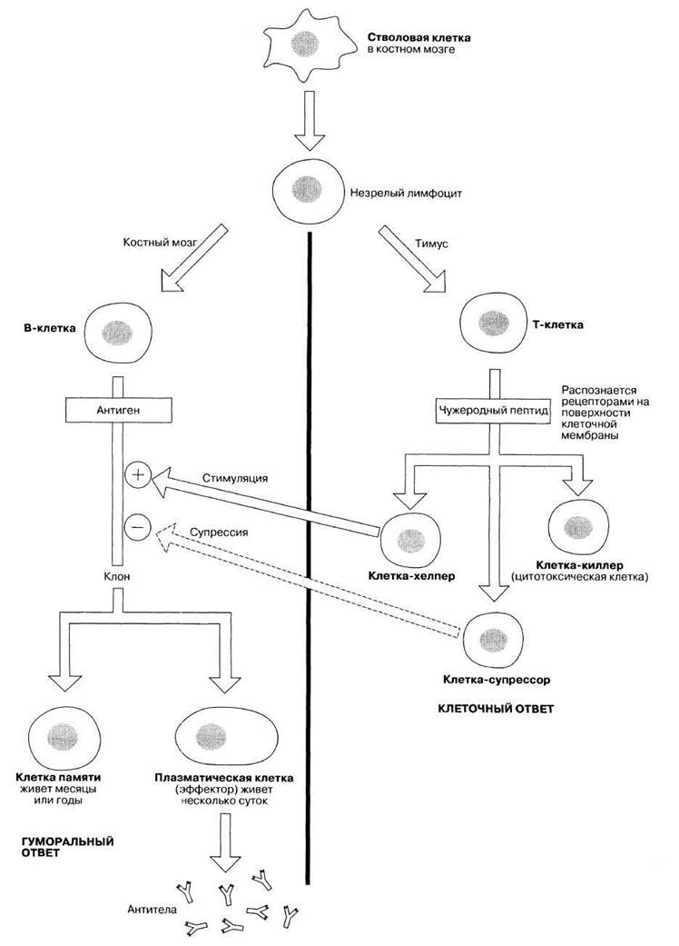
Fig. 14.40. An Overview of the Main stages in the development of cell-mediated and humoral immune responses in humans.
Last update: 06/08/2026
Editorial and Educational Adaptation: This material has been compiled based on the primary/original source text. The project team performed an editorial review, corrected technical inaccuracies, structured sections, and adapted the content for an educational format.
What was processed:
- elimination of formatting defects (OCR errors, structural breaks, corrupted characters);
- editorial organization of content;
- standardization of terminology in accordance with academic sources;
- verification of factual statements against the original source text.
All mentions of the author, publication year, and origin of the primary text have been preserved in accordance with the source.