IMMUNOLOGY - Roitt I. - Mir 2000
Chapter 6. Antibodies and Their Cellular Receptors
■ Circulating Antibodies recognize Antigens in the Blood and tissue fluid.
■ In most mammalian species, there are five antibody classes: IgG, IgA, IgM, IgD, and IgE.
■ The basic structural unit of immunoglobulins consists of two light and two heavy chains. The classes are distinguished by their heavy chains. IgA and IgM are oligomers of the basic four-chain unit.
■ Immunoglobulin chains are folded into several globular structures called domains; light chains form two domains, and heavy chains form four or five depending on the Ig Class.
■ Proteolytic Enzymes can be used to generate immunoglobulin fragments for research or medical purposes. Papain cleaves the IgG molecule into three fragments: two antigen-binding (Fab) and one Fc; Pepsin cleaves a large F(ab')2 fragment containing both antigen-binding sites.
■ Antigen-binding sites are formed by hypervariable (V) regions of Ig chains. The V domains of any light or heavy chain contain three such regions. The folding of chains into domains brings the hypervariable regions together at the protruding tips of the molecule, forming two antigen-binding sites in each four-chain unit.
■ All antibodies serve two Functions. In addition to antigen binding, they carry out one or more effector functions. The structural Regions of the immunoglobulin molecule responsible for effector activity (such as Complement activation or Cell binding) are spatially separated from the antigen-binding sites and are located primarily in the Fc region.
■ Receptors for immunoglobulins are present On the surface of mononuclear leukocytes, neutrophils, natural killer Cells, eosinophils, basophils, and mast cells. By interacting with the Fc region of immunoglobulins of various isotypes, these receptors stimulate processes such as phagocytosis, antitumor cytotoxic activity, and mast cell degranulation. Most Fcγ receptors belong to the immunoglobulin superfamily and possess two or three extracellular immunoglobulin-like domains.
The primary function of the specific Immune Response is the specific recognition of foreign antigens. Two Different types of molecules participate in recognition: immunoglobulins and T-cell antigen receptors (TCRs) (Fig. 6.1). The structural diversity of these molecules (see Chapter 8), which enables them to recognize a vast array of different antigens, arises from numerous Gene rearrangements. T-cell receptors are discussed in detail in Chapter 7.

Fig. 6.1. T- and B-cell antigen-recognition receptors likely share a common phylogenetic ancestor and belong to the immunoglobulin superfamily. The core of the B-cell receptor is formed by two identical heavy (H) and two identical light (L) chains. Directly associated with the main part of the receptor are additional components (Igα and Igβ), which presumably couple it to Intracellular Signaling pathways. Circulating antibodies are structurally similar to the core of these B-cell receptors but lack their transmembrane and intracytoplasmic segments. The antigen-binding site of the T-cell receptor consists of one a-chain and one β-chain (or one γ- and one δ-chain) associated with four structurally distinct transmembrane Peptides (γ, δ, ε, and ζ).
Immunoglobulins are a group of Glycoproteins found in the Blood Plasma and tissue fluid of all mammals. Some immunoglobulin molecules are anchored to The Plasma Membrane of B cells and function as antigen-specific receptors. Others (antibodies) exist as free molecules in plasma or Lymph. Antibody synthesis is carried out by B cells, a process that requires contact with an antigen and its-induced maturation of B cells into antibody-forming cells (AFCs). AFCs include, in particular, plasma cells that secrete large amounts of antibodies (this is how histologists originally named AFCs detected in blood and Tissues). The membrane-bound immunoglobulins of immature B cells (precursors) share the exact same antigen-binding Specificity as the antibodies produced by mature AFCs.
IMMUNOGLOBULINS: A DISTINCT FAMILY OF PROTEINS
Most higher mammals possess five classes of immunoglobulins—IgG, IgA, IgM, IgD, and IgE—which differ in molecular size, charge, Amino Acid Composition, and carbohydrate content.
In addition to interclass differences, there is also considerable heterogeneity within each class. For instance, the electrophoretic properties of immunoglobulins are so diverse that they are found in all normal serum fractions, ranging from α to γ (Fig. 6.2).

Fig. 6.2. Human serum Electrophoresis gel showing the distribution of the four main immunoglobulin classes. In an electric field, Serum Proteins are separated according to their molecular charge into α1, α2, β, and γ fractions based on their mobility. (IgE is similar in mobility to IgD, but due to its low serum concentration, it is not quantitatively represented here.) The IgG class exhibits the greatest molecular charge heterogeneity; other immunoglobulin classes occupy a narrower mobility range, primarily in the β and "fast" γ regions of the electrophoretogram.
Immunoglobulins are bifunctional molecules
Each immunoglobulin performs two functions. One region of the molecule is dedicated to antigen binding, while the other mediates so-called effector functions. These include the binding of the immunoglobulin to body tissues, various cells of The Immune System, specific phagocytic cells, and the first component of complement (C1q) upon activation of the classical pathway.
An immunoglobulin's class and subclass are determined by The Structure of its heavy chain
The basic structural unit of an immunoglobulin of any class consists of two identical light and two identical heavy polypeptide chains held together by Disulfide Bonds (Fig. 6.3). The type of heavy chain dictates the immunoglobulin class and subclass. For example, the four human IgG subclasses (IgG1, IgG2, IgG3, and IgG4) possess heavy chains designated γ1, γ2, γ3, and γ4, respectively; all of these are immunochemically identified as γ chains yet exhibit minor structural differences from one another.

Fig. 6.3. The basic structural unit of immunoglobulins consists of two identical light and two identical heavy polypeptide chains joined together by disulfide bonds (red lines). Note the positions of the amino-terminal (N) and carboxy-terminal (C) ends of the peptide chains.
IgG subclasses 1, 2, 3, and 4 account for approximately 66%, 23%, 7%, and 4% of the total molecules in this class, respectively. Two IgA subclasses (IgA1 and IgA2) are also known, but human IgM, IgD, and IgE subclasses have not yet been discovered. The Diversity of immunoglobulin classes and subclasses is driven by the isotypic variation of their molecules.
Evolutionarily, immunoglobulin subclasses apparently emerged later than classes. Consequently, human IgG subclasses differ markedly from the four IgG subclasses identified in mice.
Each immunoglobulin class has a distinct set of functions
All immunoglobulins are glycoproteins, with a carbohydrate content ranging from 2–3% in IgG to 12–14% in IgM, IgD, and IgE. The PHYSICOCHEMICAL PROPERTIES OF immunoglobulins are summarized in Fig. 6.4.
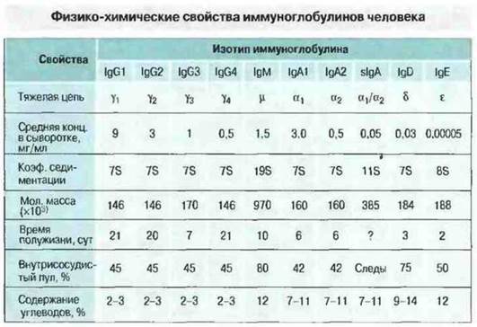
Fig. 6.4. Immunoglobulins of each class—IgG, IgM, IgA, IgD, and IgE—possess a characteristic heavy-chain type: γ, μ, α, δ, and ε, respectively. Within certain classes, different variants of heavy chains exist, which determine the division of the class into subclasses. For example, the human IgG pool comprises four subclasses, the differences between which lie in the STRUCTURE OF THE γ-chain. Immunoglobulin classes (isotypes) differ in their properties. Notably, in body secretions, IgA is represented by a secretory form (sIgA)—a dimer joined to an additional peptide chain (referred to as the secretory component). The concentration of sIgA in blood serum is very low, whereas in intestinal juice it can be quite significant.
IgG. This is the major Ig isotype in normal human serum, accounting for 70–75% of total serum immunoglobulins. The IgG molecule is a four-chain monomer with a sedimentation coefficient of 7S and a Molecular Weight of 146 kDa. Notably, IgG3 proteins are somewhat larger than those of other subclasses due to a slightly longer γ-chain. Class G immunoglobulins are evenly distributed between the intra- and extravascular pools and constitute the majority of secondary immune response antibodies, as well as the bulk of antitoxins. Furthermore, maternal IgG provides infant resistance to infections During the first few months of life. In humans, antibodies of all IgG subclasses cross the Placenta into the fetal body, establishing robust passive Immunity throughout the neonatal period. In mammalian species characterized by The transfer of maternal immunoglobulin to offspring exclusively after birth, such as pigs, IgG acquired from milk selectively crosses from the gastrointestinal tract into the newborn's bloodstream.
IgM. This class accounts for approximately 10% of the total serum immunoglobulin pool. The IgM molecule is a pentamer of the basic four-chain unit. An individual heavy chain has a molecular weight of ~65 kDa, and the entire molecule is 970 kDa. Antibodies of this class are predominantly located in the intravascular immunoglobulin pool and dominate as "early" antibodies, most frequently appearing in immune responses against structurally complex pathogenic microorganisms.
IgA. Proteins of this class constitute 15–20% of total immunoglobulins in human serum, where more than 80% exist as a monomer—a four-chain unit. However, in the serum of most other mammals, IgA is predominantly present in a polymeric form, most commonly as a dimer of the four-chain unit. IgA is the principal immunoglobulin class in seromucous secretions, such as saliva, colostrum, and milk, as well as in the mucosal discharges of the respiratory and urogenital tracts.
Secretory IgAs (sIgA) belong to the IgA1 or IgA2 subclass and are predominantly represented by a dimeric form with a sedimentation coefficient of 11S and a molecular weight of 385 kDa. They are present in high concentrations in seromucous secretions, where they are associated with another protein known as the secretory component.
IgD. This class makes up less than 1% of all plasma immunoglobulins, but it is abundantly expressed on the membrane of many B cells. The Biological Role of this immunoglobulin class is not yet fully understood; it is hypothesized to participate in the antigen-dependent differentiation of lymphocytes.
IgE. The concentration of this immunoglobulin class in serum is extremely low, but it is detectable on the surface membranes of basophils and mast cells in every individual. Additionally, IgE sensitizes mucosal cells, particularly those of the Nasal cavity, Bronchi, and conjunctiva. IgE likely plays a crucial role in anti-helminthic immunity, although in developed countries it is most commonly associated with the Pathogenesis of allergic diseases, such as Bronchial Asthma and hay fever.
Last update: 13/08/2026
Editorial and Educational Adaptation: This material has been compiled based on the primary/original source text. The project team performed an editorial review, corrected technical inaccuracies, structured sections, and adapted the content for an educational format.
What was processed:
- elimination of formatting defects (OCR errors, structural breaks, corrupted characters);
- editorial organization of content;
- standardization of terminology in accordance with academic sources;
- verification of factual statements against the original source text.
All mentions of the author, publication year, and origin of the primary text have been preserved in accordance with the source.