Introduction to Molecular Biology: From Cells to Atoms - Anthony Rees, Michael Sternberg 2002
Cells and Molecules at Work
Antibodies and Their Functions
Class="center">Introduction/introduction.files/image108.jpg" width="604"/>
Fig. 40.1. Immunoglobulin G molecule.
Immunity refers to the body's ability to recognize and destroy foreign elements that have entered it, such as microorganisms or Viruses. The Immune System is most complexly organized in mammals—the penetration of foreign substances serves as a signal for triggering a series of processes occurring at the CELLULAR AND MOLECULAR levels, collectively referred to as the Immune Response.
IMMUNOGLOBULINS, or Antibodies, are molecules circulating in the body responsible for recognizing foreign, or "non-self," elements. This recognition is followed by a series of reactions constituting the humoral, or antibody-mediated, response.
Antigens are foreign molecules that can bind to antibodies. The formation of an antigen–antibody complex is typically accompanied by the initiation of a specific process that ultimately leads to a significant increase in the concentration of the corresponding antibodies. An antibody specifically binds to a particular region of the antigen. This region is called an epitope. A single antigen may contain numerous epitopes. Typically, antigens are either individual macromolecules, such as Proteins, Nucleic Acids, Lipids, or CARBOHYDRATES, or combinations of macromolecules, exemplified by the outer surfaces of viruses and Bacteria.
Immunoglobulin G (IgG), also referred to as y-globulin, is the most abundant immunoglobulin. Its molecular mass is 150,000. An IgG molecule consists of four polypeptide chains—two identical heavy (H) chains and two identical light (L) chains. The H chain (molecular mass 50,000) contains approximately 450 amino acid residues, while the L chain (molecular mass 25,000) contains 220. The four chains are covalently linked together by disulfide bridges (Fig. 40.1).
Variable and constant regions of H and L chains. A comparison of the primary structures of several IgGs belonging to the same species reveals that the sequences of the N-terminal halves of the L chains vary widely. Therefore, it is customary to say that the first (i.e., N-terminal) 110 amino acid residues form the variable, or V, region. The C-terminal halves frequently have identical sequences. These 110 residues at the C-terminus constitute the constant, or C, region. The sequences of the L chain C region can be divided into two classes. Accordingly, L chains are subdivided into two types: kappa (κ) and lambda (λ). In each of the H chains, the variable region, approximately 110 amino acid residues long, is also located in the N-terminal portion, while the remaining 340 Amino Acids form the constant region.
Hypervariable Regions of the V regions of L and H chains. A comparison of the Amino acid sequences of various L chain V regions has shown that within the V region there are segments characterized by an extremely high degree of Variability, and thus termed hypervariable. The L chain contains three such spatially separated segments (L1, L2, and L3 in Fig. 40.1)—each consisting of approximately 6 residues. Similarly, 3 hypervariable regions (H1, H2, and H3) are also found in the V region of the H chain. The hypervariable regions of the L and H chains make up the antigen-binding site, i.e., the part of the Ig molecule that recognizes the antigen. Due to the high degree of Variability of the hypervariable regions, a vast number of different antigen-binding sites can exist.
An immunoglobulin domain is a structural unit consisting of approximately 110 residues. The L chain is built from two domains corresponding to the variable (V) and constant (C) regions. These domains are designated VL and CL. In the H chain, four domains can be distinguished: the variable domain (VH) and three domains forming the constant region (CH1, CH2, and CH3). The amino acid sequences of the domains exhibit a certain degree of similarity to one another. In particular, in each domain, the polypeptide chain forms a loop (in all domains these loops are of approximately the same length) closed by an intrachain disulfide bond.
Fab and Fc fragments are obtained from the intact IgG molecule by Limited proteolysis of the polypeptide chain using Papain. Each fragment has a molecular mass of about 50,000. Two of the resulting fragments possess antigen-binding properties and are therefore designated Fab (fragment antigen binding). The third fragment crystallizes readily and is denoted as Fc (fragment crystallised). Fab consists of a single L chain (VL and CL domains) and the first half of a single H chain (VH and CH1), whereas Fc consists of the second halves of two H chains, i.e., two CH2 and two CH3 domains.
The three-dimensional Structure of IgG was determined by X-Ray Diffraction Analysis. The IgG molecule has a Y shape—each branch of the Y corresponds to the Fab region, and the Base of the Y to the Fc region. The tertiary structures of all domains show a high degree of similarity. Each 110-residue domain is a tightly packed globule consisting of a pair of superimposed β-sheet layers (see Fig. 10.1). The individual domains are in close contact. For example, the 4 domains of the Fab region interact pairwise with each other—VH with VL, and CH1 with CL. In the Fc region, the CH3 domains are situated opposite one another, while two branched carbohydrate chains are localized between the CH2 domains. The portions of the polypeptide chain connecting any two domains are generally less tightly packed than the domains themselves. The region between Fab and Fc is called the hinge region, because its inherent flexibility allows the Fab regions to move relative to Fc.
The antigen-binding sites are located at the tips of both Branches of the Y-shaped molecule. In each variable domain, the hypervariable regions are brought into spatial proximity. The close contact between the VH and VL domains, in turn, results in all hypervariable regions being positioned adjacently, thereby forming the antigen-binding site. It has been demonstrated that the binding of an antigen to an IgG molecule can be accomplished through Hydrogen Bonds, salt bridges, Van der Waals interactions, and hydrophobic interactions. Fig. 40.1 schematically illustrates the localization of vitamin K1 within the antigen (vitamin K1)–antibody (IgG) complex.
The Fc region plays a crucial role in triggering those immune response processes that ultimately lead to the destruction of foreign elements. One of these processes involves the sequential activation of the Components of the so-called Complement system. The first step consists in the binding of a C1q molecule to the CH2 domain of IgG. C1q binds exclusively to IgG that has formed a complex with an antigen.
C1q is an integral part of the first component (C1) of The Complement System and resembles a bouquet of tulips in shape. The molecule contains 18 polypeptide chains. Each "tulip" consists of 3 chains. The tulip HEAD represents a globule, whereas the elongated stem apparently has a triple-helical Collagen structure (Ch. 11). In any event, The sequence of each chain in the stem is constructed from repeating triplets —Gly—X—Y—. To activate subsequent components of the complement system, several IgG molecules must attach to the antigen and thereby approach each other closely enough to enable the binding of these IgGs to the heads of the same C1q molecule (Fig. 40.2).
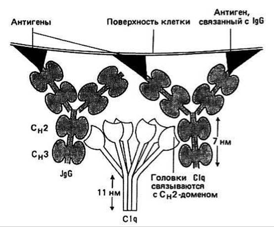
Fig. 40.2.
Antibody diversity Throughout its lifetime, the Organism produces a vast number of different antibodies (>108) against all kinds of antigens. This capacity of the organism is currently explained by somatic Gene rearrangement. This means that at birth There is a limited number of immunoglobulin genes in the organism (significantly less than 108), and the required diversity is achieved through the rearrangement of these genes.
The decoding of immunoglobulin gene nucleotide sequences has provided insight into The Mechanism of somatic rearrangement. Each V and C domain is encoded by separate genes. Furthermore, the variable region gene is divided into different segments. The light chain V region contains two such segments, designated V and J (from joining). The calculation of the number of possible VL regions is based on the currently established facts that there are 100 different V segments and 5 different J segments, and
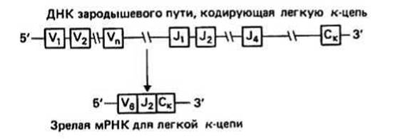
Fig. 40.3.
V/J splicing can occur in 10 ways. Consequently, the formation of 100 × 5 × 10 = 5 × 10³ VL regions is possible. The heavy chain V region contains three segments, designated V, J, and D (from diversity). There are approximately 100 V segments, 5 J segments, and 50 D segments, and each of the V/J and J/D junctions can be formed in 10 ways. Thus, the formation of 100 ∙ 5 ∙ 50 ∙ 10 ∙ 10 = 2.5 ∙ 106 VH regions is possible. Together with 5 ∙ 103 VL regions, the total number of possible VH–VL pairs, and consequently of different antigen-binding sites, amounts to 5 × 103 × 2.5 × 106 = 1010.
Immunoglobulin classes. All known immunoglobulins are subdivided into 5 classes—IgG, IgA, IgM, IgD, and IgE—depending on The Nature of the Amino Acid Sequence of the HEAVY CHAIN CONSTANT region. There are correspondingly 5 classes of these sequences—γ, α, μ, δ, and ε. Immunoglobulins of different classes also vary in the number of H and L chain pairs and, in some cases, in the length of the H chain (IgM and IgG possess an additional CH4 domain located downstream of the CH3 domain).
|
Class |
Chains |
Molecular mass |
|
IgG |
y2κ2 or y2λ2 |
150,000 |
|
IgA |
(a2κ2)1-3 or (a2λ2)1-3 |
160,000, 320,000, or 480,000 |
|
IgM |
(μ2κ2)5 or (μ2λ2)5 |
900,000 |
|
IgD |
δ2κ2 or δ2λ2 |
185,000 |
|
IgE |
ε2κ2 or ε2λ2 |
200,000 |
Last update: 13/08/2026
Editorial and Educational Adaptation: This material has been compiled based on the primary/original source text. The project team performed an editorial review, corrected technical inaccuracies, structured sections, and adapted the content for an educational format.
What was processed:
- elimination of formatting defects (OCR errors, structural breaks, corrupted characters);
- editorial organization of content;
- standardization of terminology in accordance with academic sources;
- verification of factual statements against the original source text.
All mentions of the author, publication year, and origin of the primary text have been preserved in accordance with the source.