Introduction to Molecular Biology: From Cells to Atoms - Anthony Rees, Michael Sternberg 2002
Cells and Molecules at Work
Movement in Non-Muscle Cells
Class="center">Introduction/introduction.files/image106.jpg" width="609"/>
Fig. 39.1.
Cell MOTILITY is a phenomenon characteristic of the vast majority of unicellular eukaryotic organisms as well as many cell types in Multicellular Organisms. Although protists, plant Cells, and animal cells differ significantly in phenotype, the mechanisms governing their motility exhibit a remarkable degree of similarity. The same cellular machinery that controls whole-cell movement is also capable of directing the movement of Organelles within The Cell. All types of cellular motility can be classified depending on whether they are driven by microtubules or microfilaments.
MICROTUBULES are long, tubular structures. Each microtubule has a constant diameter and consists of molecules of the globular protein tubulin, which is a dimer with a Molecular Weight of 110,000–120,000. There are two types of tubulin monomers, α and β, and the dimer is typically a heterodimer, αβ. A microtubule is a hollow cylinder with an outer diameter of approximately 24 nm and an inner diameter of 15 nm. Microtubule lengths vary from several micrometers in the Cytoplasm of most cells and several tens of micrometers in flagella and cilia, to several millimeters in nerve axons (Ch. 36). The cylinder wall, which is about 5 nm thick, is composed of 13 circularly arranged protofilaments in lateral contact with one another, each being a polymer of the tubulin heterodimer. A side view of such a microtubule is shown in Fig. 39.2. In Eukaryotic cells, microtubules can form part of a characteristic Structure known as the flagellar and ciliary axoneme, which differs from structures found in Prokaryotic Cells (Ch. 2). Because flagella and cilia are present in both unicellular organisms (e.g., protists) and multicellular organisms (e.g., humans) and play an essential role in cell motility, The structure of the axoneme will be examined in detail (see below). Located within the cytoplasm of eukaryotic cells, microtubules play a key role in processes such as mitotic spindle formation and endocytosis (Ch. 3), whereas in flagella and cilia they participate in generating cell movement.
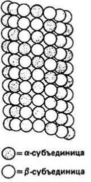
Fig. 39.2.
Microtubule-associated Proteins are integral Components of the microtubule system; they play a crucial role in regulating processes such as microtubule assembly and disassembly, as well as The formation of cross-bridges that link microtubules to one another and to other cellular components.
The axoneme is a predominantly microtubule-based structure that serves as the primary motility engine of flagella and cilia. It is surrounded by a membrane that is a direct continuation of the cell's Plasma Membrane. Cilia are short (1–5 µm) motile cellular protrusions present on the cell surface in large numbers. In contrast, when there are only a few protrusions and they are long (up to 1–2 mm), they are referred to as flagella. A cross-section of the axoneme reveals a central pair of tubules surrounded by a ring of nine doublet tubules. Structurally, axonemal tubules are either classic microtubules with a wall composed of 13 protofilaments (A-tubules) or incomplete tubules made of 10 protofilaments, forming an open, crescent-shaped cylinder in cross-section (B-tubules). Closely associated with the two central tubules, which belong to type A, is a surrounding protein sheath. A short distance away lie the nine doublet tubules, each consisting of an A-tubule and an attached B-tubule. The axoneme contains Two Types of permanent bridges: interdoublet bridges, which are composed of the protein nexin and connect the A-tubules of doublets to the B-tubules of adjacent doublets, and radial spokes, which consist of a yet-unnamed protein and link each doublet to the central sheath. Dynein molecules are periodically distributed along the A-tubules of the doublets; under certain conditions, these form interdoublet bridges that generate the force driving flagellar and Ciliary movement. Although the movement patterns of Cilia and flagella differ slightly—the former exhibiting a whip-like beat and the latter a wave-like motion—their molecular basis is identical in both cases.
The sliding filament model proposed to explain ciliary beating has much in common with the similarly named model describing Muscle contraction (Ch. 37). In the presence of ATP, the dynein protein, which forms the "arms" on the A-tubule of each doublet, binds to the B-tubule of the adjacent doublet. This protein is a Ca2+- and Mg2+-dependent ATPase of high molecular weight (300,000–400,000) and can exist in at least two conformational states. It adopts one state upon binding to the neighboring B-tubule and transitions to another during subsequent ATP Hydrolysis. This transition, accompanied by the movement of the dynein arms along the plane of the long axis of the tubules, results in the sliding of some tubules relative to others. Through the formation and dissociation of numerous cross-bridges, the tubules slide—or more accurately, "crawl"—along adjacent tubules. If sliding occurs predominantly on one side of the axoneme, the cilium bends. The upper part of Fig. 39.1 illustrates a sequence of successive positions of a cilium bending during a beat cycle. By beating, the cell pushes against the liquid medium and thereby propels itself through it.
MICROFILAMENTS are thread-like organelles approximately 5 nm in diameter found in most eukaryotic cells; they are polymers of the globular protein Actin (molecular weight 43,000). Microfilaments are either distributed randomly throughout the cytoplasm or form fibrillar bundles, such as in the leading edges of moving cells. In many mammalian cells, microfilament bundles, sometimes referred to as "stress fibers," form a network spanning the entire intracellular space during interphase (Ch. 29). During mitosis, numerous microfilaments are found in the region where the cell divides into daughter cells, forming the so-called "contractile ring." Non-muscle Cells also contain the protein Myosin, and their movement is likely driven by the same type of actin-myosin interaction underlying muscle contraction. One hypothesis regarding amoeboid movement—specifically the solation-contraction coupling model proposed by Taylor—is discussed below.
The solation-contraction coupling model, which describes The Mechanism of amoeboid movement, postulates that the amoeba cytoplasm can exist in two states: gel and sol. The gel-like cytoplasm is localized exclusively in regions directly adjacent to The Plasma Membrane and contains actin filaments cross-linked by one or more types of actin-binding proteins (e.g., α-actinin or filamin). Because of these cross-links, the gel actin filaments, despite interacting with myosin, cannot participate in contraction. In the rear of the amoeba, There is a region of high Ca2+ concentration that triggers the dissociation of actin-binding proteins from actin filaments, marking the transition from gel to sol. Following the detachment of cross-linking proteins, other proteins (such as villin and gelsolin) destabilize the long actin filaments, resulting in short filaments capable of interacting with myosin. Interaction with myosin leads to contraction occurring via the classical sliding filament mechanism (Ch. 37). The contraction at the rear of the amoeba, where the gel transitions into the sol, generates an increase in hydraulic pressure that forces the cytoplasm to flow toward the leading edge into the advancing pseudopodium. As soluble actin and actin-binding proteins reach the front of the cell, they re-form a gel, thereby maintaining a constant balance between the gel and sol states in the amoeba. Similar models involving cytoplasmic streaming—driven by contractile pushing forces—have been proposed to explain movement in many cells of higher organisms.
Last update: 13/08/2026
Editorial and Educational Adaptation: This material has been compiled based on the primary/original source text. The project team performed an editorial review, corrected technical inaccuracies, structured sections, and adapted the content for an educational format.
What was processed:
- elimination of formatting defects (OCR errors, structural breaks, corrupted characters);
- editorial organization of content;
- standardization of terminology in accordance with academic sources;
- verification of factual statements against the original source text.
All mentions of the author, publication year, and origin of the primary text have been preserved in accordance with the source.