Molecular Biology of the Cell - Volume 2 - Alberts B., Bray D., Lewis J., Raff M., Roberts K., Watson J. 1993
Cytoskeleton
Ciliary movement
Following Muscle contraction, the most thoroughly studied form of cellular motility is ciliary beating. Cilia are miniature, Hair-like structures about 0.25 µm thick, built from microtubules (microtubules being the second of the three main fibrous elements of the Cytoskeleton). Cilia are found in many Cell types and occur in most animals and some lower plants. Their primary function is to generate fluid flow near The Cell surface or propel the cell forward through the Water. Protozoa, for instance, use cilia both for locomotion and for gathering food particles. In humans, a vast number of cilia (106 or more per 1 cm2) belonging to the epithelial Cells of the Lower Respiratory Tract continuously move mucus, laden with dust particles and dead cell debris, upward toward the Oral Cavity, where the mucus is swallowed and eliminated. Cilia also ensure The transport of the oocyte through the oviduct, while a structurally similar appendage—the flagellum—drives vertebrate spermatozoa.
Just as The Study of muscle contraction contributes greatly to our understanding of Actin- and Myosin-based motility processes in nonmuscle cells, so our knowledge of the ciliary beating mechanism helps us understand how microtubule systems generate Other types of movement, such as Intracellular Transport or mitosis-related processes.
11.3.1. Cilia and flagella are characterized by oscillatory movements—bending waves
Cilia move in a coordinated fashion, forming unidirectional traveling waves across the cell surface (Fig. 11-49). Each cilium operates like a tiny whip: an effective forward stroke, during which the cilium straightens completely and overcomes the resistance of the surrounding fluid, is followed by a recovery phase, during which it returns to its initial position by bending in a way that minimizes environmental resistance (Fig. 11-50, A). The beating cycles of adjacent cilia are subtly out of phase, resulting in the wavelike pattern visible under a Microscope.
Class="center">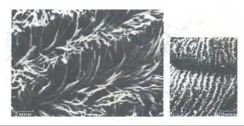
Fig. 11-49. Scanning Cytology/cytology/93.html">ELECTRON MICROGRAPHS OF the ciliated epithelium from the gut of a marine worm. Although cilia are distributed relatively evenly across the surface, their beating is coordinated to form rows of waves traveling in the same direction. These waves are more clearly visible at lower magnification on the right.
Flagella of sperm and many protozoa are very similar in their internal Structure to cilia, but they are usually much longer and exhibit a different movement pattern: instead of whip-like strokes, they display propagating quasi-sinusoidal waves (Fig. 11-50, B). Nevertheless, the Molecular Basis of motility is identical in both cases. It should be noted, however, that bacterial flagella are fundamentally different from the cilia and flagella of Eukaryotic cells (see Section 12.5.4).
11.3.2. A cilium contains a bundle of parallel microtubules forming a 9+2 structure
Ciliary beating is driven by the bending of its axial structure, known as the axoneme. This is a complex assembly of microtubules and associated Proteins. Microtubules typically appear as hollow protein cylinders with an outer diameter of 25 nm (see below). In the axoneme, they are modified and arranged in a highly characteristic pattern. The discovery of this system was one of the most striking results of early electron microscopic studies: nine doublet tubules are arranged in a circle around a central pair of single microtubules (Fig. 11-51). This 9 + 2 structure is characteristic of the cilia and flagella of almost all eukaryotic organisms, ranging from protozoa to humans. Microtubules extend along the entire length of the axoneme, which is typically about 10 µm and can reach up to 200 µm in some cells.
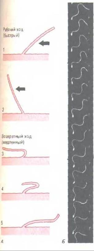
Fig. 11-50. Different beating patterns of cilia and flagella. A. The stroke of a cilium (e.g., respiratory epithelium cilium) resembles the rowing motion of a swimmer. The effective stroke (steps 1 and 2), which propels fluid across the cell surface, is followed by a markedly different recovery stroke (steps 3, 4, 5). Each cycle typically lasts 0.1–0.2 s and generates a force perpendicular to the axonemal axis. B. Shown for comparison is the wave-like flagellar movement of a tunicate sperm. The cell was photographed under stroboscopic illumination at a flash frequency of 400 s-1. Note that the waves, which maintain a constant amplitude, continuously propagate from the Base of the flagellum to its tip. As a result, the cell moves straight ahead, as if driven by its axoneme—a mechanism entirely different from that of a cilium. (B—courtesy of S. J. Brokaw.)
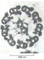
Fig. 11-51. Electron micrograph of the flagellum of the green alga Chlamydomonas (cross section) (courtesy of Lewis Tilney). The characteristic 9 + 2 structure typical of almost all eukaryotic cilia and flagella is visible. A diagram showing the Main Components of this structure is presented in Fig. 11-53.
While the two central microtubules are identical and each forms a separate complete cylinder, the outer doublets consist of two fused microtubules—one complete and one incomplete (subfibers A and B, respectively). Along the line of contact, these subfibers share a portion of a common wall. Cross sections show that the complete microtubule of the doublet is formed by a ring of 13 subunits, whereas the incomplete one (subfiber B) consists of only 11.
11.3.3. Microtubules are hollow cylinders formed by tubulin molecules [31]
Microtubules are composed of tubulin molecules, each of which is a heterodimer formed by two tightly bound globular subunits. These subunits are related proteins (about 450 Amino Acids each) designated as a- and ß-tubulins. Although tubulin is present in virtually all eukaryotic cells, the primary source for biochemical studies is vertebrate Brain: 10–20% of the soluble protein extracted from the brain by most Methods is tubulin, which is unsurprising given the high Abundance of microtubules in the long axons and dendrites of Nerve Cells.
During microtubule assembly, tubulin molecules form linear protofilaments in which the a-tubulin of one dimer contacts the ß-tubulin of the next. An intact microtubule contains 13 such protofilaments arranged parallel side-by-side around a central region that appears empty in electron micrographs (Fig. 11-52). Because all protofilaments are aligned in parallel with the same orientation, microtubules—much like actin filaments—are polar structures possessing fast-growing plus ends and slow-growing minus ends (see Scheme 11-2). The plus ends of microtubules are located at the tip of the cilium.
Like actin and many other cytoskeletal proteins, tubulin in most organisms is encoded by a family of closely related genes. The unicellular green alga Chlamydomonas has two a-tubulin and two ß-tubulin genes, whereas the fruit fly Drosophila has four of each. The Amino acid sequences of tubulins from different organisms are also quite similar, though they are not as evolutionarily conserved as actin: Yeast ß-tubulin, for instance, is 70% identical to chicken ß-tubulin, whereas the actins of these species share over 90% identical amino acid residues.
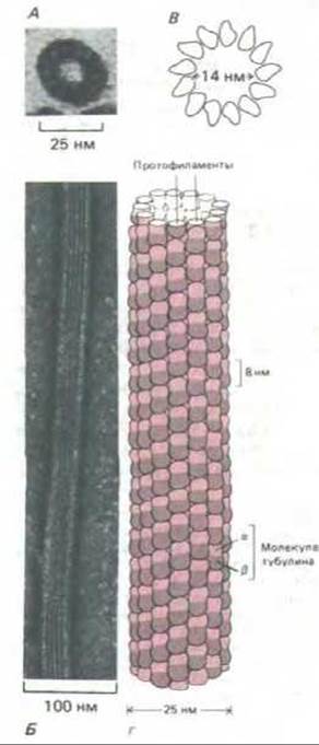
Fig. 11-52. A. Electron micrograph of a microtubule cross section, showing a ring of 13 subunits, each corresponding to an individual tubulin molecule. B. Electron micrograph of a microtubule (negative staining). C and D. Diagrams of microtubule structure showing how tubulin molecules form the cylinder wall. C—13 molecules in a microtubule cross section; D—side view of a short microtubule segment with tubulin molecules arranged in longitudinal rows (protofilaments). Each of the 13 protofilaments consists of a chain of tubulin molecules representing aß-heterodimers. A microtubule is a polar structure; different ends of the tubulin molecule (a and ß) face opposite ends of the microtubule. [Courtesy of Richard Linck (A) and Rolley Williams (B); D—based on data from Linda Amos.]
The remarkable evolutionary conservation of actin and tubulin may be due, at least in part, to structural constraints imposed by their interactions with numerous (and diverse) accessory proteins. Tubulin molecules, much like actin, interact not only with each other but also with many auxiliary proteins. As we will see, these proteins modify microtubule properties and link them to other cellular structures. It appears that most random mutational changes disrupt at least one of the Functions of microtubules or Actin filaments and are therefore detrimental to the Organism.
All currently known tubulins, when mixed in vitro, form identical microtubules. Nevertheless, it seems likely that certain variations in tubulin structure are functionally significant for the cell. In particular, in higher vertebrates, regions of both tubulin types (a and ß) containing an unusually high number of acidic amino acid residues exhibit clear tissue-specific differences. This region in tubulin molecules is thought to be involved in binding accessory proteins, and alterations in its Amino Acid Sequence could modulate microtubule function by affecting these Protein Interactions.
11.3.4. A long, thin filament runs along the wall of the microtubule doublet [32]
Although most microtubules appear to consist solely of tubulin subunits, specialized types of microtubules (such as the microtubule doublets in a cilium) utilize additional proteins for their construction. If cilia or flagella microtubules are induced to dissociate in a dilute salt solution, particularly stable fragments of subfiber A ribbons, consisting of two to four protofilaments, can be isolated from the mixture. In addition to tubulin, these fragments contain the protein tektin, which forms long filaments 2–3 nm thick, apparently related to Intermediate filaments. Tektin filaments run alongside the microtubule doublet wall and likely contribute to The formation of the shared wall of subfibers A and B. It is believed that these filaments, or other as yet unidentified fibrous molecules, determine the periodic arrangement of specialized structures on microtubules, as described below.
11.3.5. The axoneme of cilia and flagella contains protein links, “arms”, and “spokes” [33]
The axoneme is associated with many other protein structures that interact to supply the cilium with energy and enable it to perform wave-like movements. Perhaps the most important of these structures are the short lateral projections ("arms") extending from each outer-ring microtubule doublet toward the adjacent doublet (Fig. 11-53). Pairs of these projections are arranged along the entire length of subfiber A at intervals of 24 nm. They consist of a protein called dynein and, as we will see, play a crucial role in the movement of cilia and flagella. Another protein, nexin, forms cross-links between adjacent doublets located at slightly greater intervals than the dynein arms; these appear to be quite elastic and hold the axoneme circumferentially, restricting the sliding of neighboring microtubules.

Fig. 11-53. Diagram of a cross-section of a cilium (corresponding to the photomicrograph in Fig. 11-51). Various structures extending from the microtubules are located along the entire cilium with a specific periodicity (see Table 11-2).
A radial "spoke" extends inward from each outer doublet, reaching the central sheath that surrounds the central pair of single microtubules (Fig. 11-53). When viewing the axoneme from the side, all these structures—dynein arms, nexin links, radial spokes, and central sheath projections—appear as lateral protrusions repeating with a periodicity characteristic of each structure (Table 11-2).
Table 11-2. Main Protein Structures of the Axoneme
|
Axonemal components (periodicity along the axoneme) |
Function |
|
Tubulin (8 nm) Dynein arms (24 nm) |
Major component of microtubules Project from microtubule doublets, interact with neighboring doublets, causing bending |
|
Nexin links (86 nm) |
Hold adjacent microtubule doublets together |
|
Radial spokes (29 nm) |
Extend from each of the 9 outer doublets toward the central pair |
|
Central sheath projections (14 nm) |
Emerge as a row of lateral arms from the central pair of microtubules; together with radial spokes, they regulate the beat pattern of the cilium |
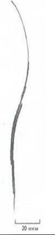
Fig. 11-54. Transmission electron micrograph of an isolated axoneme (from a Tetrahymena cilium) briefly treated with Trypsin to partially disrupt the protein bonds maintaining its normal structure. Following ATP Treatment, individual microtubule doublets slide relative to each other, leading to a dramatic increase in axoneme length—up to 9-fold. (F.D. Warner, D.R. Mitchell, J. Cell Biol. 89: 35-44, 1981. Reproduced by permission of Rockefeller University Press.)
11-18
11.3.6. Microtubule Sliding Drives Axonemal Movement [34]
If a flagellum is severed from the cell using a laser beam, it retains The ability to produce wave-like movements. This means that the motor apparatus resides within the flagellar axoneme itself rather than at its base (as in bacterial flagella, see Section 12.5.4). Indeed, even an isolated axoneme can move in a salt solution containing ATP.
The bending of cilia and flagella is driven by the mutual sliding of microtubules. This was demonstrated in experiments where isolated axonemes were treated with Proteolytic Enzymes that destroy nexin links and radial spokes while leaving the dynein arms and microtubules intact. If ATP at a concentration of just 10 µM is added to such a partially "digested" axoneme, it begins to elongate (reaching up to 9 times its original length); this occurs because the constituent fibers (doublets) of the axoneme telescope out of the weakened structure (Fig. 11-54). Apparently, adjacent outer doublets actively slide relative to each other once freed from stabilizing cross-links (such as nexin). In an intact structure, however, this sliding movement is converted into a bend, as shown schematically in Fig. 11-55.
11.3.7. Dynein Is Responsible for Sliding [35]
If microtubule doublets are capable of actively sliding past one another, a force must exist to drive this movement. This force cannot be generated by nexin cross-links, since sliding ability is retained after their destruction by proteolysis. Meanwhile, dynein arms are not destroyed by proteolysis. Normally, the arms extending from each outer doublet of the axoneme do not reach the adjacent doublet, but they come into contact with it when the cilium exhausts its ATP supply (Fig. 11-56).
Dynein is a large protein complex containing two or three (depending on the source) globular heads connected to a common base by thin, flexible stalks (Fig. 11-57). Each globular HEAD possesses ATPase activity, which is stimulated approximately sixfold upon association with a microtubule. An entire dynein arm consists of a single dynein molecule. Kinetic studies indicate that the heads likely drive microtubule sliding in the cilium through a mechanism fundamentally similar to the action of myosin heads in muscle (Section 1.1.10): unidirectional movement of dynein heads along a microtubule—from the plus end to the minus end—is powered by repeated cycles of Conformational Changes in each head, driven by ATP binding and Hydrolysis. This movement generates the force that tends to push the neighboring microtubule doublet toward the tip of the axoneme (see Fig. 11-55).
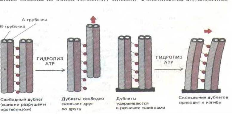
Fig. 11-55. Mutual sliding of two outer microtubule doublets (left) results in bending if the doublets are anchored at one end (right). The bases of the dynein molecules are attached exclusively to the A-tubule, leaving the heads free to contact the adjacent B-tubule. Presumably, the distinct STRUCTURE OF THE B-tubule prevents the dynein molecule base from binding to it. Such Asymmetry in the Organization of dynein molecules is essential to prevent a futile "tug-of-war" between adjacent microtubules; this is likely why each of the nine outer microtubules is an A-B doublet.
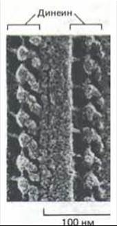
Fig. 11-56. Electron micrograph of a cilium (deep-etched). Dynein arms extending from the microtubule doublet at regular intervals are visible. (Courtesy of John Heuser.)
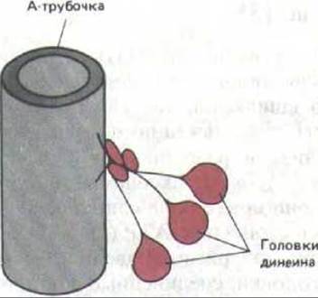
Fig. 11-57. Dynein is a large protein complex (Molecular Weight of about 2 million) composed of 9 to 12 polypeptide chains, the largest of which has a molecular weight of approximately 450,000. The base of the molecule is firmly bound to the A-tubule (this binding is ATP-independent), whereas the large globular heads contain sites for ATP-dependent attachment to the neighboring microtubule. When the heads hydrolyze their bound ATP, they move toward the minus end of this second microtubule, thereby causing a relative longitudinal Displacement of the two adjacent doublets in the cilium or flagellum (see Fig. 11-55).
11-17
11-19
11.3.8. Microtubule Sliding Must Be Regulated to Produce Ciliary Bending [36]
If all dynein arms were activated simultaneously, like myosin heads in a contracting muscle, the axoneme would simply coil into a tight spiral. Therefore, for a local ciliary bend to form and propagate as a wave from the base to the very tip, specialized regulatory mechanisms must exist to coordinate dynein activity. This regulation cannot depend on Ca2+ or other ion fluxes, as the axoneme retains its ability to bend even after The Plasma Membrane has been removed. Evidently, the activation of individual dynein arms depends on the mechanical movements of other axonemal components, and the signal is transmitted to dynein via Protein-Protein Interactions. The elasticity of the flagellum also plays an important role, as it tends to restore the equilibrium configuration of the entire structure in the absence of active contraction.
11-20
11.3.9. Axonemes Can Be Studied Using Genetic Methods [37]
When isolated axonemes in purified form are subjected to protein analysis by two-dimensional Polyacrylamide gel Electrophoresis (Section 4.4.5), about 200 different Polypeptides can be detected in them. Investigating the functions of these proteins and their localization within the axoneme is greatly facilitated by using mutant organisms for this purpose. A favorite subject for such research is the unicellular green alga Chlamydomonas reinhardtii, which possesses two flagella that it uses to swim (Fig. 11-58). Many motility mutants of this alga have been isolated. Some of them have defects in the flagellar assembly mechanism, and therefore flagella either fail to form or turn out to be rudimentary; others have flagella, but they are immotile or move very slowly. Various structural anomalies can be seen in electron micrographs of such mutant flagella. In one class of immotile mutants, the only noticeable change was the loss of dynein arms. Another class lacks only radial spokes, whereas a third class simultaneously lacks the central pair of microtubules and the central sheath. Isolated, membrane-free axonemes from mutants of all three classes are incapable of movement in the presence of ATP.
The most valuable mutants for understanding flagellar operation proved to be those that can move despite the loss of certain axonemal components. For example, slowly swimming mutants have been isolated that lack either inner or outer dynein arms; consequently, the presence of either single type of dynein is sufficient to generate the driving force, although normally each of them contributes to flagellar motility. More unexpected were secondary Mutations that restore motility to immotile flagella lacking the central doublet or radial spokes without restoring the missing components. The existence of such mutants suggests that the absence of the central doublet or the radial spoke complex somehow turns off The activity of dynein arms, "freezing" the flagellar motor, while a mutation in a second Gene "turns off the switch" and restores axonemal beating. Interestingly, motile double mutants with a defective core can only move in a flagellar manner and are incapable of the whip-like strokes typical of Chlamydomonas that are characteristic of cilia. Apparently, the ciliary type of movement requires a more complex structure than the undulating movement of a typical flagellum (see Fig. 11-50).
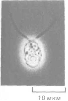
Fig. 11-58. The unicellular green alga Chlamydomonas reinhardtii. This organism swims using two flagella that work together to produce whip-like movements resembling a swimmer's stroke (unlike a sperm flagellum). (Courtesy of John Hopkins.)
Defects in cilia and flagella also occur in humans, for example, in Various Forms of hereditary Male Infertility caused by sperm immobility. Depending on the type of genetic anomaly, the sperm flagellum may lack dynein arms, radial spoke heads, or the central sheath with one or both central microtubules. Exactly the same defects are found in the ciliated epithelial cells of these individuals, and they frequently suffer from chronic respiratory diseases—recurrent Bronchitis and chronic sinusitis—because impaired ciliary epithelial function prevents proper mucus clearance from the Bronchi and Paranasal Sinuses. Notably, about half of the people suffering from this immotile cilia syndrome also exhibit a very rare feature: situs inversus viscerum, i.e., complete reversal of body Symmetry, where The Heart is located on the right, The Liver and Appendix on the left, etc. (the entire complex of anomalies is known as Kartagener syndrome). In this regard, it has been suggested that the unidirectional beating of cilia in the Early stages of organism development may play a key role in establishing normal body asymmetry.
Having examined ciliary and flagellar movement, we now turn to the question of how they are formed.
11.3.10. Centrioles Perform Two Different Functions in the Cell [38]
It is possible to detach a pair of flagella from Chlamydomonas while leaving the centrioles intact; in this case, the flagella rapidly regenerate. Almost all the protein components necessary for this are present in soluble form within the Cell Cytoplasm, and new flagella are built from them. Certain assembly stages can also occur in cell-free extracts: tubulin molecules polymerize into microtubules (a process we will examine in more detail in Section 11.4), and dynein arms can reattach to the axoneme from which they were previously washed off with a high-ionic-strength solution. However, axonemal proteins by themselves are incapable of restoring the characteristic 9 + 2 structure. This requires a "seed" or primer that acts as a template upon which further growth takes place. In the cell, this role is played by the centriole.
A centriole is a small cylindrical organelle about 0.2 µm thick and 0.4 µm long. The centriolar wall is formed by nine groups of three fused microtubules (9 triplets), with each triplet tilted toward the central axis at a 45° angle to the circumference—like turbine blades (Fig. 11-59). Adjacent triplets... [The centriole, also called the basal body, forms the lower part of the ciliary axoneme. On the right is a schematic representation of the centriole. The centriole consists of nine microtubule triplets, with each triplet (c b a) containing one complete microtubule and two incomplete microtubules attached to it. Special proteins form cross-links that support the cylindrical structure (highlighted in color). (Courtesy of D. T. Woodrum, R. W. Linck.)] are interconnected at regular intervals, and electron micrographs often reveal pale protein "spokes" radiating toward each triplet from the central region, resembling a wagon wheel (Fig. 11-59, A). Centrioles are frequently paired, positioned at right angles to each other (Fig. 11-60).
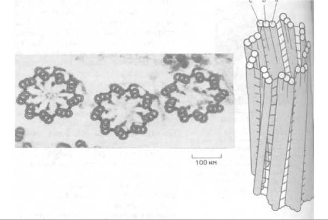
Fig. 11-59. Electron micrograph of a cross section through three centrioles in the cortex of a protozoan.
The centriole is an invariant component of the ciliary axoneme, in which context it is traditionally referred to as a basal body. Specialized extensions known as striated rootlets connect this centriole to Other components of the cytoskeleton. During formation or regeneration, each doublet of axonemal microtubules originates from two of the three microtubules of the centriole triplet, preserving the ninefold symmetry of the centriole in The structure of the axoneme. Autoradiographic data indicate that tubulin and other axonemal proteins are added to the distal end of the entire structure, i.e., to the plus ends of the microtubules. How the central pair of microtubules arises in the axoneme remains unknown; this pair is absent in the centriole.
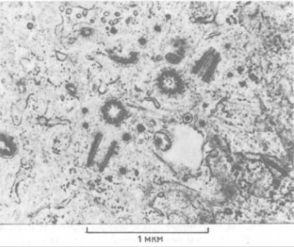
Fig. 11-60. This electron micrograph shows two new centrioles formed as a result of the Replication of two mother centrioles. In this section, one centriole in each pair is seen in cross section and the other in longitudinal section, meaning that the centrioles in each pair are positioned perpendicular to one another. (M. McGill, D. P. Highfield, T. M. Monahan, R. B. Brinkley. J. Ultrastruct. Res. 57: 43-53, 1976.)
Those centrioles that form the basal bodies of cilia perform a highly specialized function within the cell, given that cilia themselves are specialized structures. Alongside this, almost all animal cells contain a pair of centrioles that serves as the central element of the centrosome, or cell center. The centrosome (Section 13.5.2) organizes Cytoplasmic microtubules in interphase cells, and in dividing cells it duplicates to give rise to the two poles of the mitotic spindle (as we will discuss in the next section). Sometimes centrioles can alternately perform one function and then the other: in Chlamydomonas, for example, both flagella disappear prior to each mitosis, and the basal bodies leave their Location to become the spindle poles.
11.3.11. New centrioles typically arise via the duplication of pre-existing ones [39]
Against the backdrop of the continuous increase in animal cell mass during the Cell Cycle, two discrete duplication events stand out: the doubling of chromosome number (METABOLISM/36.html">DNA replication) and the duplication of centrioles. In cultured fibroblasts, the latter process roughly coincides in time with the onset of DNA Synthesis. First, the two "halves" of the centriolar pair separate, after which a daughter centriole is assembled on each such "half"—once again at a right angle to the original (Fig. 11-60). An immature centriole contains 9 single microtubules; each microtubule apparently subsequently acts as a template for the assembly of the triplets characteristic of a mature centriole.
In vertebrates, ciliated cells may bear hundreds of cilia, and the centrioles of precursor cells ensure the Formation of the required number of basal bodies. For instance, during the Differentiation of the ciliated epithelial cells of the oviducts and Trachea, the pair of centrioles shifts from its usual location near The Nucleus to the apical region of the cell where the cilia will form. There, rather than producing a single daughter centriole as usual, each mother centriole gives rise to numerous electron-dense "satellites." Basal bodies subsequently form from these satellites and migrate to the plasma membrane to initiate cilia growth there.
However, instances are known where centrioles apparently arise de novo. Thus, although unfertilized eggs of many animals lack functioning centrioles and utilize sperm centrioles for the first mitotic division following Fertilization (Section 15.4.8), under specific experimental conditions (such as a drastic disruption of ion balance or electrical stimulation) they can generate varying numbers of centrioles. Each such centriole initiates the formation of a small aster, and one of these asters can subsequently be utilized by the cell for division, leading to The Development of a haploid organism from the unfertilized egg (a course of events known as parthenogenesis—see Section 13.4.8). Presumably, the cytoplasm of unfertilized eggs contains certain centriole precursors that, under exceptional circumstances, can transform into new, genuine centrioles.
An unusual mode of centriole duplication and their continuity across a long Lineage of cell generations led researchers at one time to suggest that centrioles are fully autonomous, self-replicating Organelles. Although we now know this is not the case and that they can form de novo in the cytoplasm under certain conditions, it remains possible that some of the information required for centriole formation is contained within the centrioles themselves (much like how the reproduction of Mitochondria and Chloroplasts depends on their own extrachromosomal genes—see Section 7.5). In Chlamydomonas, the group of genes encoding the proteins involved in building the structure of basal bodies and axonemes resides in a discrete genetic element, and this element is passed on to daughter cells independently of the main Chromosomes; its nature and localization, however, remain to be elucidated.
Eukaryotic cilia and flagella contain a cylindrical bundle of nine microtubule doublets. The sliding of these doublets relative to one another is converted into the bending of the cilium or flagellum. The force that slides the doublets is generated by lateral dynein arms extending from each doublet toward the adjacent one, utilizing the energy of ATP hydrolysis for this process. A set of accessory proteins links the doublets into a cylindrical structure and limits the amplitude of their sliding. Other accessory proteins form a kind of "molecular-mechanical Relay" that regulates dynein activity such that ciliary bending occurs cyclically, providing the beat characteristic of cilia. The complex structure of the axoneme is formed through the self-assembly of protein components, with the nucleation of the assembly process driven by the centriole (basal body), which serves as a template for the formation of the specific axonemal structure—the 9 + 2 system of microtubule doublets.
Last update: 12/08/2026
Editorial and Educational Adaptation: This material has been compiled based on the primary/original source text. The project team performed an editorial review, corrected technical inaccuracies, structured sections, and adapted the content for an educational format.
What was processed:
- elimination of formatting defects (OCR errors, structural breaks, corrupted characters);
- editorial organization of content;
- standardization of terminology in accordance with academic sources;
- verification of factual statements against the original source text.
All mentions of the author, publication year, and origin of the primary text have been preserved in accordance with the source.