Molecular Biology of the Cell - Volume 2 - Alberts B., Bray D., Lewis J., Raff M., Roberts K., Watson J. 1993
Cytoskeleton
Cytoplasmic microtubules
In almost all animal Cells, Actin and tubulin are present in large amounts, although tubulin is generally less abundant than actin. Furthermore, because microtubules are thicker than actin filaments, forming a polymer of the same length requires roughly 10 times more tubulin than actin (see Table 11-4). Consequently, the total length of actin filaments in a Cell is at least 30 times greater than the total length of microtubules. This reflects a fundamental difference in the structural Organization and Functions of these two cytoskeletal polymers: while actin filaments form cross-linked networks and small bundles in the peripheral Cytoplasm, microtubules typically exist as individual strands that radiate across the entire cytoplasm from a small region near The Nucleus. Microtubules form a fibrous system along which various membrane-bound vesicles and other Organelles can be transported, thereby influencing cell polarity, regulating cell shape and movement, and determining the orientation of the Cell Division plane.
11.4.1. Microtubules are highly labile structures sensitive to antimitotic agents [41]
Many microtubule systems in cells are remarkably labile, and this lability is essential for their function. One of the most striking Examples is the mitotic spindle, which forms after the cytoplasmic microtubules disassemble at the onset of mitosis (Sec. 13.5.2).
Class="center">
Figure 11-61. Chemical Structure of colchicine.

Figure 11-62. Microtubule polarity revealed by the "hook-decoration" method. In this group, all microtubules (visible here in cross-section via Electron Microscopy) share the same polarity. The hooks, formed by laterally attached tubulin molecules, curve in a clockwise direction, indicating that we are viewing the microtubules from their plus end toward their minus end. Microtubule polarity can also be visualized by attaching dynein molecules (not shown). (Adapted from U. Euteneuer, Cell Muscle Motil. 5: 1–82, 1984.)
Table 11-3. Some tubulin-binding substances (antimitotic agents)
|
Substance |
|
|
Colchicine, colcemid, nocodazole |
Inhibit The addition of tubulin molecules to microtubules, leading to microtubule depolymerization |
|
Vinblastine, vincristine |
Induce The formation of paracrystalline tubulin aggregates, resulting in microtubule depolymerization |
|
Taxol |
Stabilizes microtubules by binding tightly to the polymer |
Mitotic spindle microtubules undergo remarkably rapid assembly and disassembly, which explains the extreme sensitivity of the spindle to various tubulin-binding drugs (Sec. 13.5.2). Colchicine, an alkaloid derived from the autumn crocus, was used for medicinal purposes by the ancient Egyptians. Colchicine molecules (Fig. 11-61) bind tightly to tubulin molecules (forming an equimolar complex) and thereby prevent their polymerization; consequently, treating dividing cells with colchicine causes the mitotic spindle to disappear within minutes and arrests cells in mitosis. Substances with this type of action are called antimitotic agents (Table 11-3). In many cases, their effects are reversible, and removing the drug allows the spindle to reform and mitosis to reach completion. Because the disruption of spindle microtubules selectively kills many rapidly dividing cells, several antimitotic drugs, notably vinblastine and vincristine, are widely used in Cancer Chemotherapy.
Another substance, taxol, exerts the opposite effect. It binds tightly to microtubules and stabilizes them. When added to cells, it forces a substantial fraction of the tubulin into the polymerized microtubule state. Just as the stabilization of actin filaments by phalloidin halts cell migration, the stabilization of microtubules by taxol freezes dividing cells in mitosis.
Much like the polymerization of actin filaments, microtubule assembly exhibits complex kinetics that play a vital role in many cellular processes. A large portion of our knowledge regarding the dynamic behavior of microtubules has been gained through studying the polymerization of purified tubulin in vitro.
11.4.2. The two ends of microtubules are distinct and grow at different rates [42]
We have already noted that microtubules are polarized: their tubulin monomers are oriented in a uniform direction. As with actin filaments, the structural polarity of microtubules gives rise to differences between their two ends, which are critical for understanding how microtubules form in cells. If dissolved tubulin is allowed to polymerize for a time onto axoneme fragments and the resulting structures are examined by electron microscopy, one finds that some microtubule ends elongate three times faster than others. The fast-growing ends were designated as plus ends, and the slow-growing ends as minus ends.
Microtubule polarity can also be determined from cross-sections if dissolved tubulin molecules are added beforehand; under certain conditions, tubulin monomers add to the sides of microtubules rather than their ends, forming curved sheets of protofilaments. In cross-section, these sheets resemble hooks that curve in one direction or the other depending on the microtubule's polarity (Fig. 11-62). This technique has been used, for example, to demonstrate that all microtubules in nerve cell axons and cilia share the same polarity: their distal ends (those positioned farther from The Cell body) are always the plus ends. When microtubules grow from their organizing centers (such as centrosomes or spindle poles), their plus ends also consistently point outward in the direction of growth (Fig. 11-63).
11-23
11.4.3. Nucleotide Hydrolysis enhances the instability of slow-growing microtubules [43]
In some respects, the assembly of tubulin molecules into microtubules resembles actin polymerization. It occurs spontaneously in vitro and is normally coupled to the hydrolysis of one bound nucleotide per added monomer (in the case of tubulin assembly, the nucleotide is GTP rather than ATP; see Table 11-4). The energy released during nucleotide hydrolysis is not required for polymerization; tubulin polymerizes successfully even when bound to the nonhydrolyzable GTP analogue GTPγS. However, nucleotide hydrolysis results in a lower critical concentration for polymerization at the plus end than at the minus end; in other words, actin or tubulin monomers bind more tightly to the plus end (see Scheme 11-1 in Sec. 11.2.10). This enables actin filaments to undergo continuous subunit treadmilling through the polymer (Sec. 11.2.10). Although treadmilling can theoretically occur in microtubules as well, the dominant phenomenon observed here is known as dynamic instability.
Dynamic instability can be readily demonstrated using a Light Microscope equipped with dark-field optics and video enhancement to observe the assembly of microtubules from purified tubulin on a Glass slide. Under these conditions, the ends of individual microtubules are seen to either grow slowly or shorten rapidly; each phase lasts for many seconds, and transitions between growth and shortening occur stochastically.
Table 11-4. Comparison of actin and tubulin polymers
|
Actin |
Tubulin |
|
|
Polypeptide mol. wt. |
42000 |
50000 (α-tubulin) 50000 (β-tubulin) |
|
Unpolymerized form |
Globular monomer |
Globular dimer (1α + 1β) |
|
Nucleotide bound by unpolymerized form |
ATP (1 per monomer) |
GTP (2 per dimer) |
|
Polymer structure |
Helical filament |
Hollow tube of 13 protofilaments |
|
Thickness of fibrous structure |
8 nm |
25 nm |
|
Number of subunits per μni of polymer length |
370 monomers |
1600 dimers |
|
Mol. wt. per 1 μm of polymer length |
370 × 42000 = 15.5 × 10⁶ |
1600 × 100000 = 160 × 106 |
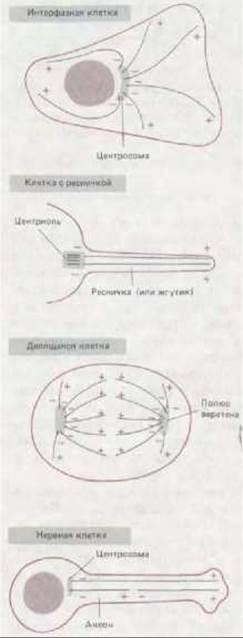
Figure 11-63. Microtubule minus ends are typically located at the microtubule-organizing center, whereas plus ends are frequently situated near The Plasma Membrane.
OBSERVED BEHAVIOR OF MICROTUBULES
Microtubules are dynamic structures, and their assembly and disassembly processes determine where and when they exist within the cell. Like actin filaments, they grow through the reversible addition of subunits, a process accompanied by conformational changes and nucleotide hydrolysis (see Scheme 11-1 in Sec. 11.2.10). However, microtubule polymerization possesses certain features that distinguish it from actin filament assembly.
In cells, the minus ends of microtubules are firmly anchored to microtubule-organizing centers, which prevents (or regulates) the assembly and disassembly of subunits at these ends (Section 11.4.4). Therefore, we will focus exclusively on the plus ends. In vitro, the plus end of an individual microtubule spontaneously switches between a phase of slow growth and a phase of rapid shortening, with each state lasting for many seconds. At any given moment, the overall microtubule population consists of Two Types of polymers, with transitions between them occurring relatively slowly:

This behavior, termed dynamic instability, can be explained by considering how the delayed hydrolysis of GTP affects polymerization.
STRUCTURAL BASIS OF DYNAMIC INSTABILITY
The conformational changes that tubulin molecules undergo upon polymerization are fundamentally similar to those of actin. Therefore, we will use the same notation as in the diagram:  will represent a monomer bearing GTP,
  — a subunit within the polymer that has already undergone a conformational change but still retains GTP (prior to its hydrolysis), and
  — a polymer subunit carrying GDP (after GTP hydrolysis). Because GTP hydrolysis occurs only after the subunit is incorporated into the polymer,
  -type subunits are typically found only at the growing end of a microtubule. This "cap" of
  -type subunits must be larger the faster the microtubule grows:
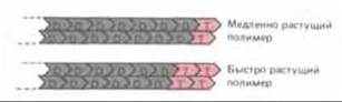
Since  -type subunits dissociate more readily than
  -type ones, any end that loses its
  cap will begin to lose subunits (depolymerize) at a much higher rate. (Microtubules lose subunits from an end terminated with
  roughly 100 times faster than from an end terminated with
  ). Consequently, once rapid depolymerization begins, it is difficult for a new
  cap to form. In addition, microtubules apparently undergo further structural alterations that render them unable to readily add new tubulin subunits.
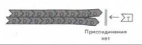
In the case shown here, the microtubule cap often fails to form altogether, and the microtubule continues to shorten until it completely disintegrates.
AN INDIVIDUAL MICROTUBULE LACKS A STEADY STATE
At steady state, the concentration of free subunits, [C], remains constant, such that
[fraction of shortening microtubules] × [depolymerization rate] =
= [fraction of growing microtubules] × [polymerization rate]
Because the depolymerization rate is much higher than the growth rate, at any given moment many microtubules are growing while only a small fraction are shortening. There is simply no critical concentration of free tubulin at which an individual microtubule maintains a constant length; instead, over a broad range of [C], we find a mixture of growing and shortening microtubules. Although the observed fraction of growing microtubules increases at higher [C] values, each individual microtubule (unlike an actin filament) never reaches a stable "steady state".
The Importance of POLYMER END "PROTECTION"
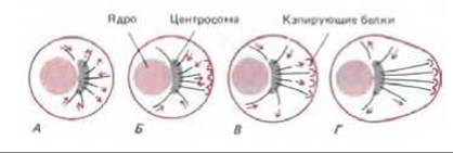
Due to its dynamic instability, a newly nucleated microtubule will survive only if both of its ends are protected from depolymerization. In cells, microtubule minus ends are typically protected by the microtubule-organizing center from which they grow, whereas certain plus ends are thought to be capped by specialized Proteins that regulate stability and, consequently, the spatial arrangement of microtubules within the cell. For example, in an unpolarized cell (A), new microtubules can grow and shorten with equal probability in all directions from the centrosome. Subsequently, some of them come into contact with cortical structures in a specific region of the cell that can cap the free microtubule plus ends (B). The selective stabilization of those microtubules that happen to encounter these capping structures results in a rapid redistribution of the entire microtubule network, transforming the cell into a polarized one (C and D).
Figure 11-2. Microtubule polymerization.
Generally speaking, one might expect actin filaments to exhibit dynamic instability as well. However, the ends of these filaments are structurally much simpler than those of microtubules, and the filaments themselves appear to transition between growth and shortening phases so rapidly that their behavior looks different—see Figure 11-1.
(Fig. 11-64). A probable explanation for this dynamic instability is illustrated in Scheme 11-2. This behavior is believed to result from the "delayed" hydrolysis of GTP that occurs during microtubule assembly. During rapid growth, the addition of tubulin molecules to the polymer outpaces the hydrolysis of their bound GTP molecules. As a result, a GTP "cap" forms at the growing end of the microtubule; because GTP-bearing tubulin molecules bind to one another with higher affinity than GDP-bearing ones, this "cap" promotes further microtubule growth. If a microtubule loses its GTP cap for any reason (e.g., due to slowed polymerization), it begins to shorten and tends to continue doing so. It should be noted, however, that microtubule shortening is also significantly facilitated by other factors, such as a permanent distortion in the tubulin lattice that typically develops at the microtubule end (see Scheme 11-2).
As we will see shortly, dynamic instability allows the cell to control the Spatial Organization of its microtubules through specialized cytoplasmic structures that bind to microtubule ends and stabilize them.
11-24
11.4.4. Most Microtubules in Animal Cells Grow from the Centrosome, Which Acts as a Microtubule-Organizing Center [44]
Microtubules in the cytoplasm of an interphase cell growing in vitro can be visualized by fixing the cell and staining it with fluorescent Antibodies against tubulin. They are densest around the nucleus, radiating outward toward the periphery as fine, intertwined threads (Fig. 11-65, A). THE ORIGIN OF microtubule growth within the cell becomes apparent when they are depolymerized with colchicine and then allowed to regrow (Fig. 11-65, B). Regenerating microtubules initially appear as a small, perinuclear, star-like structure called an aster. Its rays then elongate toward the cell periphery until their original distribution is restored. If the microtubules of a cultured cell are decorated with tubulin "hooks" (to determine polarity), one can see that their plus ends all point away from the microtubule-organizing center—the focal point of the aster. The primary microtubule-organizing center in almost all animal cells is the centrosome, or cell center. The centrosome is located adjacent to the nucleus and contains a pair of centrioles positioned at right angles to one another (in an L-shaped configuration, see Fig. 11-60). However, not all microtubule-organizing centers contain centrioles. For example, in mitotic cells of higher plants, microtubule ends are embedded in a poorly structured, electron-dense region devoid of centrioles. Centrioles are also absent from the meiotic spindle of mouse oocytes, although they appear later during embryonic development.
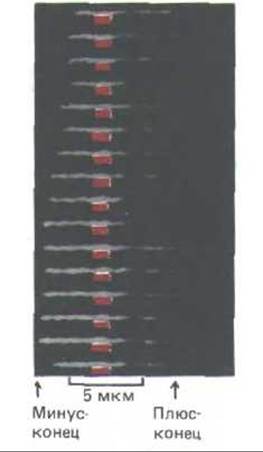
Fig. 11-64. Changes in the length of a single microtubule revealed by dark-field time-lapse video microscopy. Images were captured at 1–2 min intervals and arranged sequentially on a monitor screen. The two ends undergo cycles of elongation and shortening independently, with fluctuations being more pronounced at the plus end. (T. Horio and H. Hotani, Nature 321: 605-607, 1986.)

Fig. 11-65. Microtubule distribution in cultured cells (immunofluorescence micrographs, stained with anti-tubulin antibodies). A. Normal cell. B. Cells treated with colcemid for one hour to depolymerize their microtubules, then allowed to recover: microtubules initially appear as small asters and subsequently elongate toward the cell periphery. (A, courtesy of Eric Karsenti and Marc Kirschner; B, from M. Osborn, K. Weber, Proc. Natl. Acad. Sci. USA 73: 867-871, 1976.)
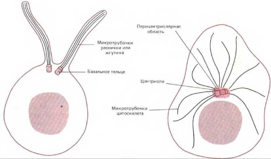
Fig. 11-66. Centrioles can act as organizing centers for entirely Different types of microtubule systems. The two centrioles in the cell on the left serve as basal bodies for ciliary axonemes, whereas in the cell on the right, two centrioles form part of the organizing center (centrosome) for microtubules radiating throughout the cytoplasm. Note that In the second case, microtubules do not grow directly from the centriole itself, but rather from the amorphous pericentriolar material.
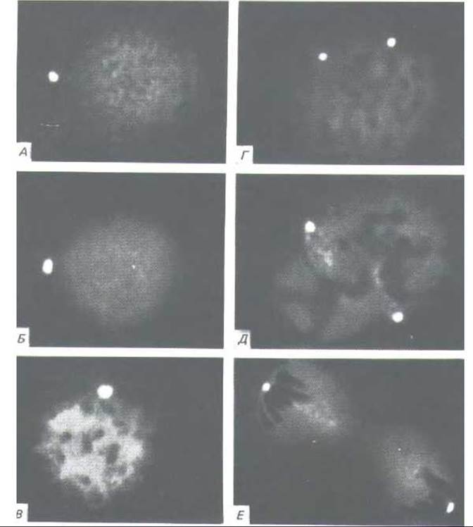
Fig. 11-67. Centrosomes in interphase and mitotic animal cells stained with fluorescent antibodies against a centrosomal protein. These micrographs illustrate the duplication and Separation of centrosomes during cell division. A and B — interphase; C and D — prophase; E — metaphase; F — telophase. For a description of mitotic stages, see Section 13.5.1. (S. L. Brenner, B.R. Brinkley, Cold Spring Harbor Symp. Quant. Biol. 46: 241-254, 1981. Courtesy of Bill Brinkley.)
Thus, unlike the ciliary axoneme, which grows directly from the edge of the centriole (Section 11.3.10), cytoplasmic microtubules do not grow straight from the centriole: their nucleation requires only the cloud of amorphous material surrounding the centriole (Fig. 11-66). Its exact composition remains unknown; however, antibodies against one of its protein components can stain the microtubule-organizing center in both PLANT AND ANIMAL cells, indicating that this protein is evolutionarily highly conserved (Fig. 11-67). If centrosomes (centrioles together with pericentriolar material) are isolated from a cell and mixed with purified tubulin, they will rapidly initiate microtubule assembly in vitro. In such microtubules, as in those formed in vivo, the minus ends are embedded in the pericentriolar material. Moreover, each isolated centrosome appears to give rise to a strictly defined number of microtubules, which is close to the number formed by the centrosome in the corresponding cell (e.g., about 250 in an interphase fibroblast). This suggests that the centrosome has a fixed number of nucleating sites.
Plants and Protozoa exhibit various types of microtubule-organizing centers. For example, the cytostome of Ciliates is equipped with a cyrtos — a complex structure functioning as a Pharynx and consisting of rows of microtubules radiating from the lower surface of a trilaminar sheet (Fig. 11-68). From the very beginning of the assembly process, these microtubules are arranged in a hexagonal lattice, suggesting that this organizing center contains a correspondingly ordered system of nucleation sites.
11-25
11.4.5. Microtubule dynamic instability can serve as one of the bases for cellular morphogenesis [45]
In animal cells, cytoplasmic microtubules radiate in all directions from the centrosome, to which their minus ends are anchored. However, most animal cells are polarized, and the assembly of tubulin molecules within them is regulated such that many microtubules are directed toward specific Regions of the cell. It is not yet entirely clear how this is achieved, but it seems likely that the mechanism relies on microtubule dynamic instability.
As we have already seen, in vitro individual microtubules most commonly exist in one of two states: a state of gradual growth or one of rapid, "catastrophic" depolymerization. In the cell, they likewise appear to exist in these two states. For instance, in cultured interphase fibroblasts, the average lifespan of a microtubule is very short, less than 10 min. This means that the entire array of microtubules radiating from the centrosome is continuously renewed — some microtubules depolymerize, while new ones grow in their place.
The inherent instability of microtubules helps explain how their growth can be regulated in specific required directions in a migrating cell (e.g., toward the leading edge) or in a dividing cell (toward condensed Chromosomes; Section 13.5.4). A microtubule with both ends "open" rapidly disappears in the cytoplasm. However, the organizing center continuously generates new tubules. They are oriented randomly, and their minus ends are protected from depolymerization because they are anchored in the organizing center. A microtubule growing from such a center can become stable provided that its plus end somehow becomes closed — "capped" — and thus prevented from dismantling. If a microtubule is capped by a structure located in a specific region of the cell, a fairly stable connection is established between that structure and the microtubule-organizing center. Thus, microtubules originating from their organizing center can be selectively stabilized by events occurring in other PARTS OF THE cell. It is believed that this is precisely how unknown structures or factors localized in specific areas of the cell cortex, which "capture" microtubule plus ends, influence cell polarity. The plane of cell division could similarly be determined in this manner (Section 13.5.13).
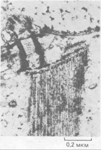
Fig. 11-68. A large and unusually ordered microtubule-organizing center in the region of the oral apparatus (cytopharynx) of the Ciliate Nassula. Microtubules grow in a regular hexagonal bundle from The surface of a flat trilaminar sheet that forms one of the elements of this cell's complex oral apparatus. (J. B. Tucker, J. Cell Sci. 6: 385-429, 1970.)
In many cells, the initial stabilization of microtubules by capping their plus ends is subsequently reinforced by other mechanisms that ensure more stable cell polarization. We now turn to the consideration of these mechanisms.
11.4.6. Microtubules gradually "mature" through post-translational modifications of their tubulin subunits [46]
Continuous formation and disappearance of microtubules is characteristic of cells undergoing significant internal reorganization, such as those dividing or migrating across a substrate. When cells become part of an established tissue, however, their microtubules transform into relatively permanent structures, particularly in cells that have differentiated and no longer divide (e.g., Neurons). This peculiar "maturation" of microtubules depends partly on the post-translational modification of tubulin molecules and partly on interactions with specific microtubule-associated proteins.
A number of Enzymes modify specific Amino Acids in tubulin. One of these is tubulin acetyltransferase, which acetylates a Lysine residue of $\alpha$-tubulin. In *Chlamydomonas*, this enzyme is located primarily in the flagellar axoneme; it appears to acetylate tubulin molecules after they have added to the distal end of the axoneme (Section 11.3.2). A specific deacetylase is contained in the cytoplasm and removes acetyl groups from unpolymerized tubulin. As a result of this localization of the two enzymes in *Chlamydomonas*, tubulin molecules in the axonemal microtubules are acetylated, whereas those in cytoplasmic microtubules, the majority of which rapidly turnover, are largely unacetylated. We will return to the potential consequences of tubulin Acetylation below.
A second, less common mode of tubulin modification is the removal of the C-terminal Tyrosine from $\alpha$-tubulin molecules incorporated into a microtubule. Tyrosine Cleavage is catalyzed by a specific enzyme present in the cytoplasm of many vertebrate cells. As with acetylation, there is also an enzyme that catalyzes the reverse reaction — the restoration of tyrosine to the C-terminus of depolymerized tubulin molecules. In cells with rapidly turning-over microtubules, most tubulin molecules remain in their original form (with tyrosine) simply because they do not have time to be detyrosinated before dissociating from the polymer. By contrast, increasingly "older" microtubules — those that have "managed to survive" the short turnover time — become enriched in detyrosinated tubulin. Thus, both acetylation and detyrosination serve as markers of the transition of microtubules from a temporarily stabilized form to a much more stable one (Fig. 11-69). If a fibroblast culture is treated with a drug that induces microtubule depolymerization (Section 11.4.1), the few surviving microtubules will be found to selectively bind antibodies that recognize the acetylated or detyrosinated form of tubulin.
Microtubules formed from acetylated and detyrosinated tubulin *in vitro* are no more stable than those formed from ordinary, unmodified tubulin; this indicates that the modifications apparently serve merely as a "signal" for binding specific proteins that subsequently stabilize the microtubules and alter their properties within cells.
11.4.7. The properties of cytoplasmic microtubules are altered by METABOLISM/18.html">The Influence of proteins associated with them [47]
Post-translational modifications of tubulin, firstly, mark microtubules as "mature" and, secondly, promote their stabilization. However, The most significant and important modifications experienced by microtubules result from the attachment of other proteins to them. These microtubule-associated proteins (MAPs) enhance microtubule resistance to depolymerization and mediate their interactions with other cellular components. Given how diverse microtubule functions are in cells, one would expect A wide variety of different types of MAPs to exist.
Two Major Groups of MAPs that can be co-isolated with microtubules from the Brain are high-molecular-weight proteins (with a molecular mass of about 200–300 kDa or even more) and tau proteins (with a molecular mass of 40–60 kDa). Proteins in both groups possess two domains, one of which is responsible for binding to microtubules; because this domain binds simultaneously to several unpolymerized tubulin molecules, MAPs accelerate microtubule nucleation *in vitro*. The other domain is believed to mediate the interaction of microtubules with other cellular components (Fig. 11-70). Both high-molecular-weight MAPs and tau proteins decorate cytoplasmic microtubules along their entire length, as demonstrated using antibodies against these proteins.
A considerable number of proteins that selectively bind to microtubules have already been isolated, though the functions of most remain unknown.
Some likely serve as structural components by stabilizing microtubules and anchoring them to other cellular structures (including other elements of the Cytoskeleton and the membranes of certain organelles), while others are responsible for moving organelles along the microtubules.
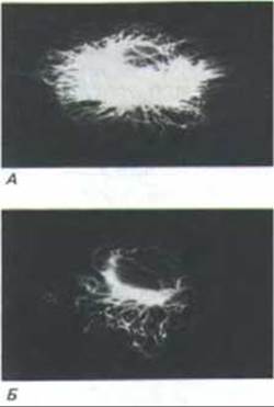
Figure 11-69. Immunofluorescence micrographs demonstrating that certain microtubules in the cytoplasm of a cultured cell are highly dynamic (A), whereas others are relatively stable (B). Cells were injected with tubulin covalently linked to a small molecule, biotin; an hour later, the cells were stained first with fluorescein-labeled antibodies against biotin, and then with antibodies against tubulin labeled with a different fluorescent dye, rhodamine. Rapidly turning-over microtubules incorporated biotinylated tubulin and thus reacted with the anti-biotin antibodies (A), which coated the microtubules and prevented the binding of anti-tubulin antibodies. Stable microtubules failed to incorporate biotinylated tubulin; consequently, they did not stain with anti-biotin antibodies but were instead labeled by the anti-tubulin antibodies (B). Stable Microtubules are also stained by antibodies that recognize detyrosinated or acetylated tubulin. (Courtesy of Eric Schulze and Marc Kirschner.)
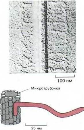
Figure 11-70. Regularly spaced "side arms" projecting from a microtubule, formed by a large microtubule-associated protein (MAP-2) from vertebrate brain. The electron micrograph shows a region of a microtubule associated with numerous MAP-2 molecules. Part of the MAP-2 molecule extends laterally from the microtubule, as illustrated in the diagram below. (Photographs kindly provided by William Voter and Harold Erickson.)
11-32
11.4.8. Microtubules often direct the intracellular movement of organelles [48]
Observing a living vertebrate cell under a phase-contrast microscope or a differential Interference contrast microscope (Section 4.1.5) reveals that its cytoplasm is in a state of constant motion. Mitochondria and smaller membrane-bound organelles change their positions within the cell within a matter of minutes via distinct periodic leaps that are far too orderly and directed to be mistaken for the incessant Brownian motion caused by the random thermal movement of molecules. Many of these Intracellular Movements occur in close association with microtubules. When a cell undergoing active organelle transport is rapidly fixed and sectioned for electron microscopy, the membranes of these organelles are frequently seen linked to cytoplasmic microtubules by thin, thread-like structures. This suggests that microtubules play a crucial role in such movement, although, as noted previously (Section 11.2.4), certain vesicle transports in the cytoplasm occur along actin filaments rather than microtubules. The most striking demonstration of the transport role of microtubules comes from studies of fast axonal transport in Nerve Cells, where membrane-bound vesicles move intensively in both directions along the axon—between the cell body and the nerve terminal.
11.4.9. Kinesin and cytoplasmic dynein drive the movement of vesicles along axonal microtubules in opposite directions using the energy of ATP hydrolysis [49]
The giant squid axon can be extracted from the animal and its cytoplasm extruded like toothpaste from a tube. When a droplet of this squeezed axoplasm is compressed under a cover slip and recorded on video through a microscope (Section 4.1.6), organelles can be observed moving along thin, filament-like "tracks." Immunofluorescence combined with electron microscopy reveals that these tracks are individual microtubules.
Isolated intracellular vesicles, and even synthetic particles such as polystyrene beads, can bind to microtubules in extruded axoplasm and move along them in much the same way as they do in a living cell. Because the extruded axoplasm is no longer enclosed by a plasma membrane, the concentrations of various ions and low-molecular-weight substances can be readily manipulated to investigate their effects on axonal transport. This approach demonstrated that AMPPNP—a nonhydrolyzable analogue of ATP—halts transport and locks organelles in a stationary, microtubule-bound state.
This AMPPNP-induced effect makes it possible to identify the components responsible for vesicle movement. By screening axoplasm extracts for proteins that bind to microtubules in the presence of AMPPNP but dissociate upon the addition of ATP, researchers isolated a large protein complex named kinesin. Kinesin proved to be an ATPase that utilizes the energy of ATP hydrolysis to drive the unidirectional movement of vesicles along microtubules (Section 3.4.11). This movement proceeds at a steady rate of 0.5 to 2 µm/s, lacking the pauses and sudden jumps characteristic of transport in the intact axon, which are thought to result from frequent collisions between the moving vesicle and various cytoplasmic elements.

Figure 11-71. Kinesin and cytoplasmic dynein are large microtubule-associated proteins (MAPs) capable of moving along microtubules in opposite directions by utilizing the energy of ATP hydrolysis (A). These proteins consist of complexes of two identical "heavy chains" and several smaller "light chains." Each heavy chain forms a globular HEAD through which the protein attaches to a microtubule in an ATP-dependent manner. Thus, similar to Myosin, kinesin (B, showing four molecules) and cytoplasmic dynein (C, two molecules) each have two heads. By contrast, ciliary dynein (D) possesses three heads (see Fig. 11-57). [Electron micrographs obtained by John Heuser using the quick-freeze, deep-etch technique; proteins isolated by Trina Schroer, Jeff Gelles, Michael Scheetz (B); Eric Steuer (C); Ursula Goodenough (D).]
The direction of movement generated by kinesin was determined using polystyrene beads driven along microtubules polymerized in vitro from centrosomes. While crude axoplasm extracts transport particles in both directions, purified kinesin Supports transport exclusively in a "rard-out" direction—toward the plus ends of microtubules (recall that ciliary dynein moves in the opposite direction; see Section 11.3.10). Because axonal microtubules are known to be oriented with their plus ends pointing away from the cell body (see Fig. 11-63), kinesin-mediated vesicle transport must be directed from the cell body toward the axon terminal. It seems likely that transport in the reverse direction is mediated by a dynein-like protein. Indeed, a high-molecular-weight MAP with these exact properties has recently been isolated from several sources, including mammalian brain (Fig. 11-71). This protein was named cytoplasmic dynein.
11.4.10. Microtubules determine the intracellular positioning of the Endoplasmic reticulum and the Golgi apparatus [50]
Motor proteins similar to kinesin and dynein are not restricted to neurons; they appear to be present in all cells that contain microtubules, and several vital organizational functions of microtubules are presumably executed through these proteins. Recent in vitro studies have shown that kinesin binds to The endoplasmic reticulum membrane and can stretch it along oriented microtubules, transforming this organelle into its characteristic complex network. Experiments on intact cells similarly indicate that the endoplasmic reticulum is pulled outward along microtubules away from the centrosome, exactly as expected if the process is driven by kinesin. Finally, immunofluorescence Methods reveal that the delicate margins of the reticulum cisternae in the peripheral regions of cells frequently align with microtubules (Fig. 11-72A and B).
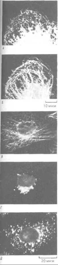
Figure 11-72. Distribution of endoplasmic reticulum (ER) membranes and microtubules in cultured cells. A.
Immunofluorescent staining of ER proteins reveals ER cisternae as a lace-like network at the cell periphery. B. Arrangement of microtubules in the same cell. C, D, and E. The Effect of microtubules on the Golgi apparatus. When a cultured cell is double-labeled with fluorescent antibodies against both microtubules (C) and Golgi membranes (D), the latter appear as clusters surrounding the centrosome. However, when the cell is treated with nocodazole to depolymerize microtubules, the Golgi membranes disperse throughout the cytoplasm (E), much as they do during mitosis. [Courtesy of Mark Terasaki and Lan Bo Chen (A, B); Viki Allan and Thomas Kreis (C, D, E).]
While endoplasmic reticulum membranes actively move outward away from the centrosome along microtubule "tracks" (remaining continuous at the opposite end with the nuclear envelope), Golgi cisternae appear to be transported in the reverse direction, as though linked to dynein-like proteins, resulting in the positioning of the Golgi apparatus near the centrosome (Fig. 11-72C and D). Both the Golgi apparatus and the endoplasmic reticulum undergo extensive fragmentation during mitosis (Section 13.5.16), and when microtubules subsequently re-form in the cytoplasm, their orientation presumably guides the reassembly of these organelles from small vesicles and membrane fragments (Fig. 11-72E).
Summary
Microtubules are formed by the polymerization of tubulin molecules, which subsequently hydrolyze their tightly bound GTP (a process that lags slightly behind polymerization). Microtubules grow slowly, are inherently unstable, and are prone to rapid, "catastrophic" depolymerization; however, they can be stabilized through association with other structures that cap their ends. Microtubule-organizing centers, such as centrosomes, continuously nucleate the growth of new microtubules that extend in random directions. Any microtubule that encounters a structure capable of capping its free plus end is selectively stabilized, whereas uncapped microtubules eventually depolymerize. This dynamic process is thought to be the primary mechanism determining the polarity and spatial arrangement of microtubule arrays within the cell.
In microtubules that form in appropriate locations, tubulin subunits undergo post-translational modifications—acetylation and detyrosination. These modifications serve as "markers" for mature microtubules and create binding sites for specialized microtubule-associated proteins (MAPs) that further enhance microtubule resistance to depolymerization and adapt them for specific cellular functions. A distinct group of such proteins utilizes the energy of ATP hydrolysis to drive unidirectional movement along microtubules, thereby powering the directed cytoplasmic transport of cellular organelles and maintaining their proper spatial organization.
Last update: 12/08/2026
Editorial and Educational Adaptation: This material has been compiled based on the primary/original source text. The project team performed an editorial review, corrected technical inaccuracies, structured sections, and adapted the content for an educational format.
What was processed:
- elimination of formatting defects (OCR errors, structural breaks, corrupted characters);
- editorial organization of content;
- standardization of terminology in accordance with academic sources;
- verification of factual statements against the original source text.
All mentions of the author, publication year, and origin of the primary text have been preserved in accordance with the source.