Biochemistry and Molecular Biology - Belyasova, N.A. 2002
Structure and Functions of Cellular Components
Biomembranes
Organization and Functions of Membrane Proteins
Lipids in Cell membranes are primarily credited with structural properties — they form a bilayer, or matrix, that houses the active Components of the membrane, namely Proteins. It is the proteins that impart uniqueness to diverse membranes and provide their specific properties. Numerous Membrane Proteins perform the following core Functions: they mediate the Transport of substances across membranes (transport functions), catalyze biochemical reactions, support photo- and Oxidative Phosphorylation, METABOLISM/36.html">DNA Replication, protein Translation and modification, signal reception, and Nerve Impulse transmission, among others.
Membrane proteins are conventionally divided into two groups: integral (intrinsic) and peripheral (extrinsic). The criterion for this Classification is the strength of the protein's association with the membrane and, consequently, the harshness of the Treatment required to extract the protein from it. For instance, peripheral Proteins can be released into solution simply by washing membranes with low-ionic-strength Buffer solutions and low pH values in the presence of chelating agents, such as ethylenediaminetetraacetic acid (EDTA), which bind divalent cations. Peripheral proteins are extracted under such mild conditions because they are bound to lipid headgroups or other membrane proteins via weak Electrostatic Interactions, or to lipid tails via hydrophobic interactions. Conversely, integral proteins are amphiphilic molecules featuring large hydrophobic domains on their surfaces and reside within the interior of the membrane; therefore, their extraction requires disrupting the bilayer. Detergents or organic Solvents are most commonly used for this purpose. The ways in which proteins attach to the membrane vary quite widely (Fig. 4.8).
Transport proteins. The Lipid Bilayer serves as an impermeable barrier for most Water-soluble molecules and ions, and their transit across Biomolecules relies on The activity of transport proteins. Two MAIN TYPES OF these proteins can be distinguished: channels (pores) and carriers. Channels are tunnels spanning the membrane in which the binding sites for transported substances are accessible from both surfaces of the membrane simultaneously. Channels do not undergo any conformational changes during the transport process; their conformation shifts only during opening and closing. Carriers, on the other hand, alter their conformation while transporting substances across the membrane. Furthermore, at any given moment, the binding site for the transported substance on a carrier is accessible from only one surface of the membrane.
Channels, in turn, can be divided into two main groups: voltage-gated and Ligand-gated (chemically regulated). An example of a voltage-gated channel is the Na-channel, the operation of which is regulated by Changes in the electric field voltage. In other words, these channels open and close in response to changes in the transmembrane potential. Ligand-gated channels open and close in response to the binding of specific chemical agents. For instance, the nicotinic Acetylcholine Receptor transitions into an open conformation upon binding a neurotransmitter and allows monovalent cations to pass through (subsection 4.7 of this chapter). The terms "pore" and "channel" are generally interchangeable, but a pore is more often understood to mean a non-selective Structure that distinguishes substances primarily by size and permits the passage of all sufficiently small molecules. Channels are more commonly understood as Ion Channels. The rate of transport through an open channel reaches 106—108 ions per second.
Class="center">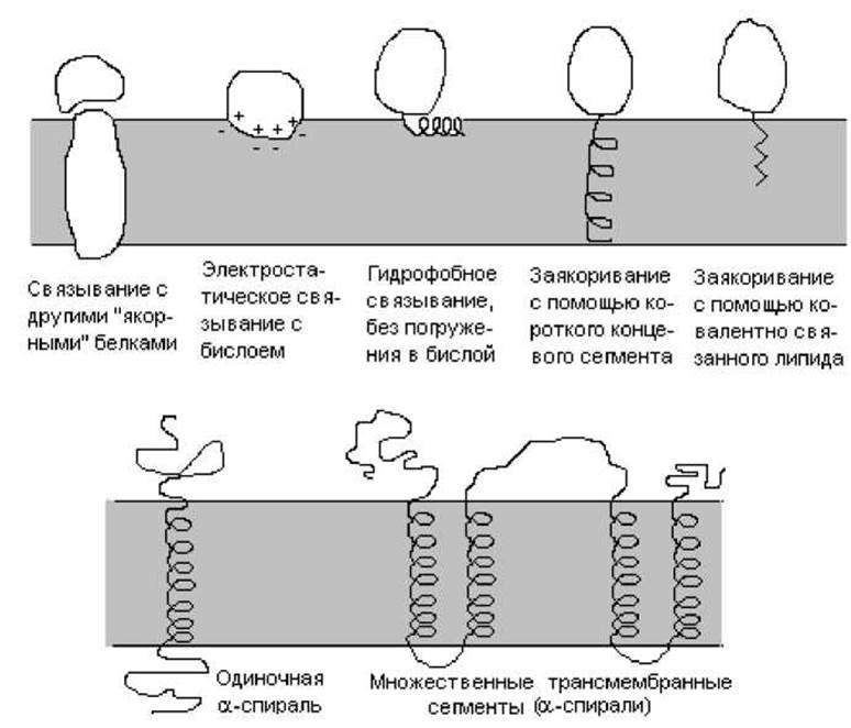
Fig. 4.8. Various Modes of Protein attachment to the membrane
Carriers can also be divided into two groups: passive and active. Passive carriers facilitate The transport of a single type of substance across the membrane. Passive carriers are involved in Facilitated Diffusion and merely increase the flux of a substance moving down its electrochemical gradient (e.g., glucose transport across Erythrocyte membranes). Active carriers transport substances across the membrane with an expenditure of energy. These transport proteins accumulate substances on one side of the membrane by moving them against their electrochemical gradient. The rate of carrier-mediated transport depends heavily on the specific type and ranges from 30 to 105 s-1. The terms "permease" and "translocase" are often used to designate individual carriers and can be considered synonyms for the term "carrier".
Enzymatic functions of membrane proteins. Cell membranes host a vast array of diverse Enzymes. Some are localized within the membrane, finding a suitable environment there for The conversion of hydrophobic compounds; others, owing to the Structural Organization of membranes, are arranged in strict succession, catalyzing Sequential Stages of vital processes; still others require the assistance of lipids to stabilize their conformation and maintain activity. Biomembranes contain enzymes representing all known classes. They may span the membrane entirely, exist within it in a dissolved form, or, acting as peripheral proteins, bind to membrane surfaces in response to a specific signal. The following characteristic Types of Membrane Enzymes can be distinguished:
1) transmembrane enzymes that catalyze coupled reactions on opposite sides of the membrane. As a rule, these enzymes possess multiple active sites located on opposing sides of the membrane. Typical Examples of such enzymes include components of the Respiratory Chain or photosynthetic redox centers, which catalyze oxidation-reduction processes coupled with Electron Transport and the generation of ion gradients across the membrane;
2) transmembrane enzymes involved in the transport of substances. Transport proteins that couple substance transport with ATP Hydrolysis, for instance, possess catalytic activity;
3) enzymes that catalyze the conversion of membrane-bound substrates. These enzymes participate in the metabolism of membrane components: Phospholipids, Glycolipids, Steroids, etc.
4) enzymes involved in the conversion of water-soluble substrates. By means of membranes—most frequently in an anchored state—enzymes can be concentrated in Regions of the membrane where the concentration of their substrates is highest. For example, enzymes that hydrolyze proteins and starch are attached to the microvillar membranes of the intestine, which helps increase the rate at which these substrates are broken down.
Cytoskeletal proteins. The Cytoskeleton is a complex network of various types of protein filaments found exclusively in Eukaryotic Cells. The cytoskeleton provides mechanical support for The Plasma Membrane, determines cell shape, and dictates the positioning of Organelles as well as their movement during mitosis. Cellular processes vital to cell function, such as endocytosis, exocytosis, phagocytosis, and amoeboid movement, also rely on the cytoskeleton. Thus, the cytoskeleton serves as the dynamic framework of The Cell and governs its mechanics.
The cytoskeleton is formed by Three types of filaments:
1) microfilaments (~ 6 nm in diameter). These are thread-like organelles composed of polymers of the globular protein Actin and other associated proteins;
2) Intermediate filaments (8–10 nm in diameter). These are formed by Keratins and related proteins;
3) microtubules (~ 23 nm in diameter) — long tubular structures. They consist of the globular protein tubulin, whose subunits form a hollow cylinder. The length of microtubules can reach several micrometers in the Cell Cytoplasm and several millimeters in nerve axons.
These cytoskeletal structures permeate the cell in various directions and are tightly linked to the membrane, anchoring to it at specific points. These membrane regions play a crucial role in intercellular contacts, allowing cells to attach to a substrate. They also play an essential role in the transmembrane Distribution of lipids and proteins within membranes.
Last update: 06/08/2026
Editorial and Educational Adaptation: This material has been compiled based on the primary/original source text. The project team performed an editorial review, corrected technical inaccuracies, structured sections, and adapted the content for an educational format.
What was processed:
- elimination of formatting defects (OCR errors, structural breaks, corrupted characters);
- editorial organization of content;
- standardization of terminology in accordance with academic sources;
- verification of factual statements against the original source text.
All mentions of the author, publication year, and origin of the primary text have been preserved in accordance with the source.