Biological Membranes - A. N. Ogurtsov 2012
Structure and Functions of Biomembranes
Passive Transmembrane Transport
Facilitated Diffusion
Facilitated diffusion across Introduction/36.html">Biological Membranes occurs with the participation of carrier molecules. A distinction is made between facilitated diffusion mediated by mobile carrier molecules and by stationary carrier molecules fixed in a specific orientation across the membrane.
An example of a mobile carrier molecule is valinomycin [L-lactate - L-valine - D-oxy-isovaleric acid - 0-valine]3, a mobile carrier for potassium ions. The valinomycin molecule has the shape of a cuff, lined on the inside with polar groups and on the outside with nonpolar groups (Figure 53).
Class="center">
Figure 53 - Structure OF THE valinomycin molecule: (a) chemical formula, (b) Van der Waals model of the valinomycin-potassium ion complex
Due to its chemical structure, valinomycin, first, is capable of forming a complex with potassium ions entering the interior of the cuff-like molecule (the potassium ion is held in the center via ion-dipole interactions involving the peptide carbonyl groups) and, second, valinomycin is soluble in the lipid phase of the membrane because its exterior is nonpolar. Valinomycin molecules located at the membrane surface can capture potassium ions from the surrounding solution. By diffusing within the membrane, the molecules transport potassium across it, and some of them release the ions into the solution on the other side of the membrane.
The transport of potassium by valinomycin can occur in both directions across the membrane. Therefore, if the potassium concentrations on both sides of the membrane are equal, the potassium flux in one direction will be equal to that in the other, resulting in no net potassium Transport Across the membrane.
However, if the concentration on one side is higher than on the other, the potassium flux down the concentration gradient will exceed the flux in the opposite direction.
Facilitated diffusion thus proceeds from regions of higher concentration of the transported substance to regions of lower concentration.
Facilitated diffusion also accounts for the Transport of Amino acids, sugars, and other biologically vital substances across biological membranes.
Four key features distinguish facilitated diffusion from simple diffusion.
1. Substance transport involving a carrier occurs significantly faster.
2. Facilitated diffusion exhibits saturation kinetics (Figure 54)—as the concentration on one side of the membrane increases, the flux density of the substance rises only up to a certain limit where all carrier molecules become fully occupied.
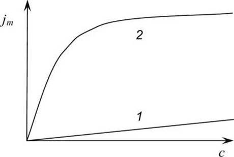
Figure 54 - Dependence of the flux density jm of substances into The Cell through a biological membrane on their concentrations c in the extracellular medium: 1 - for simple diffusion; 2 - for facilitated diffusion
3. Facilitated diffusion demonstrates competitive inhibition among transported substances when the carrier transports multiple compounds; certain substances are transported more efficiently than others, and The addition of some substances hinders the transport of others. For instance, among sugars, glucose is transported better than fructose, fructose better than xylose, xylose better than arabinose, and so on.
4. Certain substances can block facilitated diffusion by forming stable complexes with carrier molecules; for example, phloridzin inhibits the transport of sugars across biological membranes.
If the transport of a given substance across a biological membrane exhibits these characteristics, one can assume that facilitated diffusion is taking place.
A variation of facilitated diffusion is transport mediated by stationary carrier molecules fixed in a specific orientation across the membrane.
Transmembrane Transport of glucose. Facilitated diffusion of glucose and other small hydrophilic molecules across the membrane is mediated by uniporter Proteins.
One of the most thoroughly studied uniporters is the glucose transporter protein GLUT1. GLUT1 proteins were isolated from Erythrocyte membranes. Most mammalian Cells utilize GLUT1 proteins to deliver Blood-borne glucose into the Cytosol by expressing the corresponding GLUT1 Gene.
Like other uniporters, GLUT1 can adopt two conformational states: in one state, the glucose-binding site is exposed externally on the exoplasmic side of the membrane, while In the second state, it is exposed internally on the cytosolic side. Figure 55 illustrates the operational cycle of such a glucose uniporter. The GLUT1 uniporter can also mediate glucose transport out of the cytosol if the cytosolic glucose concentration exceeds that of the extracellular space.
The Human Genome contains twelve genes encoding homologous uniporters, GLUT1–GLUT12, whose transmembrane domains are highly similar; all are structurally assembled in a comparable manner from twelve transmembrane a-helices composed predominantly of hydrophobic Amino Acids. However, some a-helices incorporate Serine, asparagine, Threonine, and glutamine residues, which form Hydrogen Bonds with the hydroxyl groups of glucose, thereby establishing the glucose-binding sites within the uniporters.

Figure 55 - Operational cycle of the glucose transporter uniporter. The triangle indicates the glucose concentration gradient
Glucose uniporter genes are expressed in various cells across different Tissues, and the synthesized uniporters differ in their efficiency of transmembrane glucose transport.
For example, GLUT2 is expressed in Liver cells (hepatocytes) and Insulin-secreting pancreatic ß-cells, and the efficiency of glucose transport via the GLUT2 uniporter is twice as high as that of GLUT1 (which operates in erythrocyte membranes). In hepatocytes, this "excess" glucose is converted into Glycogen, whereas in ß-cells, the surplus of glucose triggers the secretion of the hormone insulin into the bloodstream. Insulin further reduces blood glucose concentration by stimulating glucose transport mechanisms into the cytosol of Muscle cells and inhibiting hepatic glucose synthesis.
Another glucose uniporter isoform, the GLUT4 protein, is expressed exclusively in adipose and Muscle Tissues, while the GLUT5 uniporter transports fructose instead of glucose.
The functioning of transport proteins can only be studied within the context of a membrane. Most cellular membranes contain too many different integral proteins to allow the observation of a single one in isolation. Two approaches are employed to investigate transport proteins.
1. Genetic Engineering techniques are used to significantly increase the expression level of the target transport protein, after which The rate of transmembrane transport of the corresponding metabolite is compared between the wild-type and genetically modified systems.
2. The protein of interest is extracted and purified, and then integrated into a pure lipid bilayer, such as a liposome membrane (Figure 56).

Figure 56 - Transfer of the GLUT1 transport protein from a biomembrane into a liposome
Last update: 13/08/2026
Editorial and Educational Adaptation: This material has been compiled based on the primary/original source text. The project team performed an editorial review, corrected technical inaccuracies, structured sections, and adapted the content for an educational format.
What was processed:
- elimination of formatting defects (OCR errors, structural breaks, corrupted characters);
- editorial organization of content;
- standardization of terminology in accordance with academic sources;
- verification of factual statements against the original source text.
All mentions of the author, publication year, and origin of the primary text have been preserved in accordance with the source.