Review of Medical Physiology - William F. Ganong 2002
Introduction
General Principles and Cellular Basis of Human Physiology
Transport Across the Cell Membrane
Transport Across Cell Membranes occurs primarily via exocytosis, endocytosis, movement through Ion Channels, and Primary and secondary Active Transport.
Exocytosis
Proteins secreted by Cells move from The Endoplasmic reticulum to the Golgi apparatus, and from the trans-Golgi network they are extruded into secretory granules or vesicles (see Figs. 1-24). These granules and vesicles move toward The Cell membrane, with which their membranes subsequently fuse (Figs. 1-25), and the fusion site breaks down. As a result, the Contents of the granules or vesicles are released outside the cell, while the cell membrane remains intact. The process of extruding substances outside the cell is called exocytosis. It requires Ca2 and energy, as well as acceptor proteins (see below and Chapter 4).
Note that There are two pathways for cellular secretion (see Figs. 1-24). In the non-constitutive (regulated) pathway, proteins from the Golgi apparatus enter secretory granules, where prohormones undergo Processing into mature Hormones prior to exocytosis. The other pathway—the constitutive pathway—involves the rapid transport of proteins to the cell membrane in vesicles with little or no processing or storage. The non-constitutive pathway is sometimes referred to as the regulated pathway, although this term is a misnomer, as the release of proteins via the constitutive pathway is also regulated.
Endocytosis
The process of endocytosis is the reverse of exocytosis and occurs in several forms. Phagocytosis ("cell eating") is the process by which Bacteria, dead tissue, or other microscopic particles of matter are engulfed by cells, such as polymorphonuclear leukocytes in the Blood. The material comes into contact with the cell membrane, which invaginates. The invagination then pinches off, trapping the engulfed material within a membrane-bound vacuole while leaving the cell membrane intact. Pinocytosis ("cell drinking") is similar to phagocytosis, with the sole difference that the ingested particles are in solution and therefore not visible microscopically.
Each type of endocytosis can be either constitutive or clathrin-mediated. Constitutive endocytosis is a non-specific process, whereas clathrin-mediated endocytosis occurs almost entirely through clathrin-coated pits on the cell membrane. Clathrin molecules are triskelion-shaped, with three legs radiating from a central hub (Fig. 1-26). As endocytosis proceeds, clathrin molecules assemble into a geometric lattice that surrounds the budding vesicle. The neck of the vesicle contains the protein dynamin (a guanosine triphosphatase), which directly or indirectly pinches off the vesicle (hence this protein is often referred to as a "pinchase"). Once the vesicle is fully formed, the clathrin coat disassembles, and the triskelion proteins are recycled to form other vesicles. These vesicles subsequently uncoat and deliver their contents to early endosomes (see Figs. 1-24). From the early endosome, a new vesicle buds off and returns to the cell membrane (see Figs. 4-5). Alternatively, an early endosome can mature into a late endosome. Late endosomes fuse with Lysosomes (see Figs. 1-24), and their contents are degraded by lysosomal Enzymes.
Clathrin-mediated endocytosis can occur more rapidly than constitutive endocytosis and is more specific in the sense that the molecules stimulating it are concentrated on their receptors within coated pits and, consequently, coated vesicles. This type of endocytosis is responsible for the internalization of many Ligand-bound receptors, such as nerve growth factor and low-density Lipoproteins—essential elements of cellular Cholesterol METABOLISM (see Chapter 17). It also plays a crucial role in synaptic function (see Chapter 4).
The cell membrane contains small dimples called caveolae, which are coated with the protein caveolin rather than clathrin. These serve as endocytic sites that facilitate the uptake of various Vitamins and Peptides.
Naturally, exocytosis increases the total surface area of the membrane surrounding the cell, whereas endocytosis decreases it. Therefore, the coupled processes of exocytosis and endocytosis maintain the cell surface area within its normal limits.
Class="center">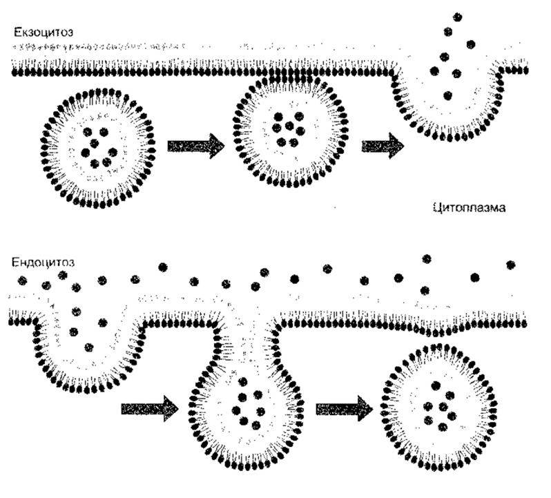
Fig. 1-25. Exocytosis and endocytosis. Note that in exocytosis, the cytoplasmic faces of the two membranes fuse, whereas in endocytosis, the non-cytoplasmic faces fuse (reproduced with permission from Alberts B et al. Molecular Biology of the Cell, 2nd ed. Garland, 1989).
Mechanisms Involved in Vesicular Transport
Significant progress has been made in elucidating the BIOCHEMICAL BASIS OF vesicle formation, transport, and docking within cells. Intracellular Transport is best analyzed alongside exocytosis and endocytosis, as these processes share very similar mechanisms.

Fig. 1-26. A clathrin molecule On the surface of an endocytic vesicle. Note the characteristic triskelion shape of clathrin and its role in forming a supportive lattice for the vesicle alongside other molecules.
It is now established that all transport vesicles possess protein coats. Four MAIN TYPES OF coats are recognized: AP-1 clathrin, AP-2 clathrin, COPI, and COPII. Vesicles transporting proteins from the trans-Golgi network to lysosomes are coated with AP-1 clathrin, whereas endocytic vesicles transporting proteins to endosomes are coated with AP-2 clathrin. Vesicles mediating transport between the Endoplasmic reticulum and the Golgi apparatus have COPI and COPII coats. Specific Amino acid sequences or attached chemical groups on the transported proteins direct them to their appropriate destinations. For instance, the Amino Acid Sequence Asn-Pro-X-Tyr directs Transport from the cell surface to endosomes, whereas attached mannose-6-phosphate groups direct transport from the Golgi apparatus to endosomes. Vesicles diffuse over short distances within the cell and move along microtubules over long distances. Once a vesicle reaches its target, it is docked when a v-SNARE (vesicle membrane protein) locks with a t-SNARE (target membrane protein). Each target has a unique set of t-SNAREs, ensuring that it only accepts vesicles with the matching set of v-SNAREs. Target and receptor proteins are discussed in greater detail in Chapter 4 in the context of synaptic transmission.
Various small Rab GTP (guanosine 5'-triphosphate)-binding proteins are associated with the Different types of vesicles (see below).
Distribution of Ions and Other Substances Across the Cell Membrane
Differences in the composition of intracellular and interstitial fluid depend on the specific properties of cell membranes. Typical concentrations for a mammalian tissue are listed in Table 1-2. Average values for humans are shown in Fig. 1-27, and the distribution of Na and K in various body compartments is given in Table 1-6.
Membrane Permeability and Membrane Protein Transport
The cell membrane is virtually impermeable to intracellular proteins and other organic anions, which constitute the majority of all intracellular anions, conventionally denoted by the symbol A. When analyzing membrane permeability to smaller molecules, It is important to clearly distinguish between The Lipid Bilayer itself and the numerous diverse transport proteins embedded within it. Transport proteins are transmembrane proteins that selectively facilitate the movement of a few, or often just a single, substance across the membrane. The lipid bilayer is primarily permeable to Water. Its permeability to other substances depends on their size (Table 1-7), solubility, and charge. Nonpolar, hydrophobic molecules such as O2 and N2 dissolve in the bilayer and cross it easily. Small uncharged polar (hydrophilic) molecules, such as CO2, also diffuse rapidly through lipid bilayers, whereas the diffusion of large uncharged polar molecules (such as glucose) and charged particles (ions) is extremely slow. However, in vivo, ions, glucose, urea, and many other small molecules utilize transport proteins to cross the cell membrane, while water diffusion is facilitated by water channels.
Our understanding of transport proteins has been greatly expanded by the patch-clamp technique. In this method, the tip of a micropipette is placed in tight contact with the cell membrane. A tiny patch of membrane beneath the pipette tip containing only a few transport proteins can then be studied in detail (Fig. 1-28). There are several variants of this technique: the cell can be left intact (cell-attached patch); the membrane patch can be detached from the cell (excised patch); or the patch within the micropipette can be ruptured while remaining continuous with the rest of the cell membrane, providing direct access to the cell interior (whole-cell recording).
Some transport proteins function as simple aqueous ion channels. Some of these channels are constantly open, whereas others have gates that open and close. In some cases, gating is regulated by Changes in membrane potential (voltage-gated channels), while in others, channels open or close upon binding a ligand (ligand-gated channels). Often the ligand is extracellular (e.g., a neurotransmitter or hormone), but it can also be intracellular. Specifically, intracellular Ca2+, cAMP, or one of the G proteins produced within the cell (see below) can bind directly to channels and activate them. Some channels open in response to mechanical stretch. A classic example of a voltage-gated channel is the Na+ channel (see below), and a classic example of a ligand-gated channel is the Acetylcholine Receptor (see Chapter 4).
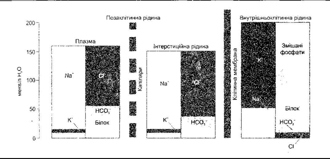
Fig. 1-27. Electrolyte Composition of human Body Fluids. Note that values are expressed per liter of water rather than body fluid (reprinted with permission from Jonson LR [editor] Essential Medical Physiology. Raven Press, 1992).
Table 1-6. Distribution of sodium and potassium in The Human Body
Component |
Content, % |
|
Na+ |
К+ |
|
Entirely intracellular |
9,0 |
89,6 |
Entirely extracellular |
91,0 |
10,4 |
Plasma |
11,2 |
0,4 |
Interstitial fluid |
29,0 |
1,0 |
Cytology/practical/45.html">Dense Connective Tissue and Cartilage |
11,7 |
0,4 |
Bone |
36,51 |
7,6 |
Intercellular Location |
2,6 |
1,0 |
1 Of these, 11.0 are exchangeable and 25.5 are non-exchangeable.
Other transport proteins are carriers that bind ions and other molecules and then undergo a conformational change to transport the bound molecule from one side of the cell membrane to the other. Molecules move from areas of high concentration to areas of low concentration (along their chemical gradient), cations move toward negatively charged areas, and anions toward positively charged areas (along their electrical gradient). The process by which carrier proteins transport substances along their chemical or electrical gradient does not require energy and is termed facilitated transport (or Facilitated Diffusion). A classic example is The transport of glucose by glucose transporters, which move it down its concentration gradient from the ECF into the Cell Cytoplasm (see Chapter 19). Other carriers transport substances against their Electrical and Chemical gradients. This form of transport requires energy and is called active transport. In animal cells, energy is overwhelmingly provided by ATP Hydrolysis (see above and Chapter 17).
Table 1-7. Size of hydrated ions and other biologically important substances1
Substance |
Atomic or molecular mass |
Radius, nm |
Cl- |
35 |
0,12 |
К+ |
39 |
0,12 |
Н2O |
18 |
0,12 |
Са2+ |
40 |
0,15 |
Na+ |
23 |
0,18 |
Urea |
60 |
0,23 |
Li+ |
7 |
0,24 |
Glucose |
180 |
0,38 |
Sucrose |
342 |
0,48 |
Inulin |
5 000 |
0,75 |
Albumin |
69 000 |
7,50 |
1 Data from Moore EW: Physiology of Intestinal Water and Electrolyte Absorption, American Gastroenterological Association, 1976.
Therefore, it is not surprising that carrier molecules are ATPases—enzymes that catalyze the hydrolysis of ATP. One such ATPase is sodium-potassium-activated adenosine triphosphatase
(Na+-K+-ATPase), also known as the Na+-K+ pump. There are also H+-K+-ATPases in the gastric mucosa (see Chapter 26) and renal tubules (see Chapter 38). Specifically, V-ATPases are proton ATPases that acidify many intracellular Organelles, including PARTS OF THE Golgi apparatus and lysosomes; F-ATPases are found in Mitochondria and synthesize ATP from ADP. Some membranes contain ATPases that transport Ca2+.

Fig. 1-28. Types of patch configurations used to study ion channel activity across the cell membrane. A and B also show membrane changes over time (modified from Ackerman MJ, Clapman DE: Ion channels: Basic science and clinical disease. N Engl J Med 1997;336:1575).

Fig. 1-29. Two families of K+ channels. Left: Inward rectifier K+ channel. Top: Association with cell membranes. Bottom: Formation of an aqueous pore by four subunits. Right: Voltage-gated "gates" (reprinted with permission from Kubo Y et al: Primary Structure and Functional expression of a mouse inward rectifier potassium channel. Nature 1993;362:127)
Some transport proteins are called uniporters because they transport only a single substance. Others are termed symporters, as this mode of transport involves the binding of more than one substance to the transport protein, which are then transported across the membrane together. An example is the symport in the intestinal mucosa responsible for the coupled transport of Na+ and glucose from the intestinal lumen into mucosal cells during facilitated diffusion (see Chapter 25). Other transporters are called antiporters because they exchange one substance for another. A classic antiporter is the aforementioned Na+-K+-ATPase: for every three Na+ ions it moves out of the cell, two K+ ions are transported into the cell.
Ion Channels
Ion channels are known for K+, Na+, Ca2+, and Cl-. Each of these exists in numerous forms with diverse properties. The inward rectifier K+ channels were likely among the first to evolve, named so because they facilitate K+ influx while having almost no effect on efflux. These channels lack voltage-gated gates, and each consists of four subunits containing two transmembrane domains (M1 and M2; Fig. 1-29). These subunits surround an aqueous pore. Another family of K+ channels probably evolved later and possesses four additional transmembrane domains in the outer shell. They contain voltage-gated gates, and the amino terminals of their subunits are shaped like a ball and chain. Upon activation, the ball undergoes thermal motion and enters the pore, causing rapid channel inactivation despite ongoing depolarization. Following repolarization, the conformational changes of the channel return to their normal resting state (Fig. 1-30).
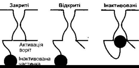
Fig. 1-30. Rapid inactivation of voltage-gated K+ channels. Depolarization opens the activation gates, and subsequently the amino terminal of the subunits swings toward the channel, arresting conduction despite ongoing depolarization. Following membrane repolarization, the protein conformational changes reset to the resting state (reprinted with permission from Antz C, Fakler B: Fast inactivation of voltage-gated K+ channels: from cartoon to structure. News Physiol Sci 1998;13:177)

Fig. 1-31. SCHEMATIC STRUCTURE OF the principal subunits of two ion channels. Some Na+ and Ca2+ channels have additional subunits. Arabic numerals denote the alpha-helical domains spanning the cell membrane. The H5 domain is not shown (after Catterall WA. Modified and reprinted from Hall ZW: An Introduction to Molecular Neurology. Sinauer, 1992).
Over 40 different K+ channels have been described in mammals. All of them are tetramers, and each subunit type is encoded by a different Gene.
Ca2+ channels and voltage-gated Na+ channels also consist of four homologous domains, each containing six transmembrane domains surrounding an aqueous pore with a diameter greater than 0.5 nm. However, they differ from K+ channels in that each is the product of a single gene, with linking peptide chains connecting the domains (Fig. 1-31). Over 30 different voltage-gated or cyclic nucleotide-gated Na+ and Ca2+ channels have been described. Some of these have non-transporting auxiliary subunits associated with them in vivo. The toxins tetrodotoxin (TTX) and saxitoxin (STX) bind to Na+ channels and block them. The number and distribution of Na+ channels can be determined by labeling TTX or STX and analyzing the distribution of the label.
The epithelial Na+ channel (ENaC) family, which has a distinct structure, is found in the apical membranes of epithelial cells in the Kidney, colon, lung, and Brain. Each epithelial sodium channel consists of three subunits encoded by three different genes. Each subunit likely spans the membrane twice, with both the amino and carboxy terminals located inside the cell; the alpha-subunit transports Na+, whereas the beta- and gamma-subunits do not. The precise stoichiometry of ENaC has not been definitively established, meaning there may be more than one of each of the three subunits in the functional channel in vivo. These channels are inhibited by the diuretic amiloride, which binds to the alpha-subunit. They are frequently referred to as amiloride-sensitive Na+ channels. In the kidney, they play a crucial role in regulating ECF volume via aldosterone (see Chapter 38). Mutant mice lacking functional ENaCs are born alive but die rapidly because their bodies are unable to pump out Na+ and consequently water from the Lungs.
There are also numerous Cl- channels that play major roles in cell Volume Regulation, transepithelial ion transport, and presumably the Regulation of Muscle and renal function. For example, the GABA receptor and Glycine receptor are Cl- channels (see Chapter 4). Cl- channels are present in large quantities in Skeletal Muscle. The normal physiological function of these muscle channels remains unclear, but Mutations in the gene encoding them cause myotonia congenita, a disease characterized by muscle hyperexcitability.
Nа+-К+-ATPase
As noted above, the Na+-K+-ATPase catalyzes the hydrolysis of ATP to ADP, utilizing the energy to extrude three Na+ ions from the cell and import two K+ ions into the cell for each mole of hydrolyzed ATP. Consequently, this electrogenic pump has a coupling ratio of 3:2, as it moves three positive charges out of the cell for every two moving in. Such pumps are found throughout the body. Their activity is inhibited by ouabain and similar digitalis Glycosides, which are used to treat Heart Failure. It is a heterodimer composed of an α-subunit with a molecular mass of approximately 100,000 and a β-subunit with a molecular mass of approximately 55,000. Both span the cell membrane (Fig. 1-32). Dissociation of the subunits results in the cessation of activity. However, the β-subunit is a glycoprotein, whereas Na+ and K+ transport occurs via the α-subunit. The β-subunit has a single transmembrane domain and three extracellular glycosylation sites, each with attached carbohydrate residues. These residues account for one-third of its molecular mass. The α-subunit spans the membrane ten times, with its NH2 and COOH termini located intracellularly. It possesses intracellular Na+- and ATP-binding sites, a phosphorylation site, and an extracellular binding site for K+ and ouabain. When Na+ binds to the α-subunit, ATP also binds and is converted to ADP, while the phosphate is transferred to Asp 376 at the phosphorylation site. This induces a conformational change in the protein, extruding Na+ into the ECF. Subsequently, K+ binds extracellularly, dephosphorylating the α-subunit, which then returns to its original conformation and releases K+ into the cytoplasm. Notably, the α- and β-subunits are heterogeneous, comprising α1, α2, and α3, as well as β1, β2, and β3 subunits; the α1 isoform is found in the membranes of most cells, whereas α2 is present in muscle, heart, adipose tissue, and the brain, and α3 in The Heart and brain. The β1 subunit is widespread, although it is absent in certain astrocytes, vestibular Cells of the Inner ear, and fast-twitch glycolytic Muscles; these muscles contain exclusively the β2 subunit. The diverse structures of the α- and β-subunits of the Na+-K+-ATPase across various Tissues clearly reflect the specialization of specific tissue Functions.

Fig. 1-32. Na+-K+-ATPase. The intracellular portion of the α-subunit contains the Na+-binding site (1), phosphorylation site (4), and ATP-binding site (5). The extracellular portion contains the K+- (2) and ouabain-binding sites (3) (from Horisberger J-D et al. Structure-function relationship of Na-K-ATPase. Annu Rev Physiol 1991;53:565. Reprinted with permission from Annual Review of Physiology, vol. 53. Annual Reviews, 1991).
Regulation of Na+-K+-ATPase
Naturally, The amount of Na+ entering the cell is not constant. For example, if intracellular Na+ increases, pump activity is upregulated, thereby increasing the amount of Na+ extruded from the cell. Pump activity is influenced by intracellular second messengers, notably cAMP, diacylglycerol (DAG), and arachidonic acid derivatives (see below). The magnitude and direction of these effects vary depending on experimental conditions. THYROID HORMONES increase pump activity via genomic action by accelerating the synthesis of Na+-K+-ATPase molecules. Aldosterone also increases the number of pumps, although this effect is secondary (see Chapters 20 and 38). Dopamine inhibits the pump in the kidney by phosphorylating it, leading to natriuresis. Insulin enhances pump activity through various mechanisms. Finally, the Na+-K+-ATPase is linked to the membrane Cytoskeleton, and interestingly, G-Actin (see above) also increases pump activity.
Secondary Active Transport
In many cases, active transport of Na+ is coupled with the transport of other substances (secondary active transport). For example, the luminal membranes of small intestinal mucosal cells contain a symporter that transports glucose into the cell only when Na+ simultaneously binds to the protein and is transported down its electrochemical gradient.

Fig. 1-33. Composite diagram of the major secondary effects of active Na+ and K+ transport; the Na+-K+-ATPase converts the chemical energy of ATP hydrolysis to maintain an inward gradient for Na+ and an outward gradient for K+. The energy of these gradients is used for countertransport, cotransport, and Maintenance of the Membrane Potential (reprinted with permission from Skou JC. The Na-K pump. News Physiol Sci 1992;7:95)
The electrochemical gradient of Na+ is maintained by active transport of Na+ from the mucosal cells into the ISF (see Chapter 25). Other Examples are shown in Fig. 1-33. In the heart, the Na+-K+-ATPase indirectly affects Ca2+ transport. The antiporter in cardiac muscle cell membranes normally exchanges intracellular Ca2+ for extracellular Na+. The stoichiometry of this exchange is proportional to the Na+ concentration gradient across the cell membrane. If The activity of the Na+-K+-ATPase is inhibited (e.g., by ouabain), intracellular Na+ concentration increases, the transmembrane Na+ gradient decreases, and Ca2+ extrusion is impaired. Consequently, the resulting increase in intracellular Ca2+ enhances myocardial contraction (positive inotropic effect; see Chapter 3).
Active transport of Na+ and K+ is one of the most energy-demanding processes in the human body. On average, it accounts for about 24% of the energy produced by cells, and up to 70% in Neurons. Consequently, it is responsible for a significant portion of the basal metabolic rate.
Last update: 10/08/2026
Editorial and Educational Adaptation: This material has been compiled based on the primary/original source text. The project team performed an editorial review, corrected technical inaccuracies, structured sections, and adapted the content for an educational format.
What was processed:
- elimination of formatting defects (OCR errors, structural breaks, corrupted characters);
- editorial organization of content;
- standardization of terminology in accordance with academic sources;
- verification of factual statements against the original source text.
All mentions of the author, publication year, and origin of the primary text have been preserved in accordance with the source.