Review of Medical Physiology - William F. Ganong 2002
Introduction
General Principles and Cellular Foundations of Human Physiology
Structure and Functions of DNA and RNA
Genome
DNA is found in Bacteria, the nuclei of Eukaryotic Cells, and Cell/35.html">Mitochondria. It consists of two extremely long nucleotide chains containing the bases adenine (A), guanine (G), thymine (T), and cytosine (C) (Fig. 1-14). The Chemical Structure of these purine and pyrimidine bases is described in Chapter 17. The chains are held together by Hydrogen Bonds between the complementary bases, with adenine pairing with thymine, and guanine with cytosine. The ultimate double-helix STRUCTURE OF THE molecule is shown in Fig. 1-15. Evidence for The complexity of this molecule is that in the human haploid genome (the complete genetic text), DNA comprises 3x10⁹ Base Pairs.
DNA is a component of Chromosomes, which contain Genetic information regarding all the hereditary CHARACTERISTICS OF THE cell and its precursors. Each chromosome contains a segment of the DNA double helix. Genetic information is encoded by The sequence of purine and pyrimidine bases in the nucleotide chains. The reading of genetic information is reflected in the order in which Amino Acids are arranged in Proteins synthesized by The Cell. This information is carried by RNA to Ribosomes, the sites of Protein Synthesis in the Cytoplasm. RNA differs from DNA in that it has a single-stranded structure, contains uracil instead of thymine, and its sugar moiety is ribose rather than 2'-deoxyribose (see Chapter 17). Proteins formed from the DNA template include all the Enzymes that, accordingly, control cellular METABOLISM. A Gene is defined as the Amount of Information required to form a single protein molecule. However, a protein encoded by a single gene may subsequently be cleaved into multiple proteins with different physiological activities. Genes also contain promoters, which are DNA sequences that facilitate RNA formation. Mutations occur when The base sequence within a DNA molecule is altered under The Influence of X-rays, cosmic rays, or other mutagenic agents. It has been estimated that The Human Genome consists of 50,000 to 100,000 genes or three billion base pairs.
DNA Polymorphism
Class="center">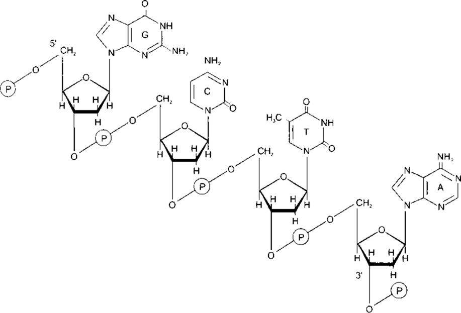
Fig. 1-14. Segment of the DNA molecular structure, in which the purine and pyrimidine bases adenine (A), thymine (T), cytosine (C), and guanine (G) are linked by a backbone formed of phosphodiester bridges between 2'-deoxyribose molecules, which are attached to the nucleotide bases via N-glycosidic bonds. Note that the backbone has polarity (5' and 3' directions) (reprinted with permission from Murray RK et al., Harper’s Biochemistry, 25th ed., McGraw-Hill, 2000).
Protein-coding genes (exons) account for only 3% of the human genome; the remaining 97% consists of introns (see below) and Other types of DNA with undetermined or unknown Functions. Sometimes this 97% is referred to as junk DNA. A characteristic feature of human DNA is its structural Variability among individuals. Most variations occur in non-coding regions, although they can also occur in coding regions; they may be silent or expressed as identifiable changes in proteins. Predominantly, these variations manifest as variable numbers of tandem repeats (VNTRs) ranging from one to hundreds of times. This variation alters the length of the DNA chain between the points where it is cleaved by various restriction enzymes. Consequently, restriction fragment length polymorphisms (RFLPs) can be observed in the DNA fragments of different individuals. RFLP analysis among the population yields samples that are essentially a characteristic DNA map or fingerprint. The Importance of DNA fingerprinting has been demonstrated using additional specialized techniques. The probability of obtaining identical DNA profiles using these Methods in individuals who are not monozygotic twins varies According to the number of enzymes applied, the familial relationships of the individuals, and other factors. Given this, the question of which statistical data are most appropriate for analysis has been fiercely debated. Still, the probability of an accidental RFLP match has been estimated to range from 1 in 1,000,000 to 1 in 100,000. RFLPs can be analyzed from samples of semen, Blood, or other Tissues. Numerous copies of DNA samples are obtained using the Polymerase Chain Reaction (PCR), an ingenious method for DNA Amplification. DNA fingerprinting is of paramount importance for solving crimes and establishing paternity, although reliable and proven methods must be used and the results interpreted with caution. RFLP analysis is also valuable in studying animal and Human Evolution and in identifying the chromosomal mapping of genes that cause Hereditary diseases.
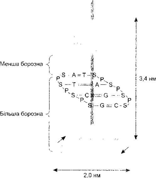
Fig. 1-15. The double-helix structure of DNA, showing adenine (A) pairing with thymine (T), and cytosine (C) pairing with guanine (G) (reprinted with permission from Murray RK et al.: Harper’s Biochemistry, 25th ed., McGraw-Hill, 2000).
Mitosis
During each somatic Cell Division (mitosis), the two DNA strands separate, and each serves as a template for the synthesis of a new complementary strand. This reaction is catalyzed by DNA polymerase. One of the newly formed Double helices goes to one daughter cell and the second to the other, so the total amount of DNA in each daughter cell is identical to that in the parent cell.
Telomerases
Cellular Replication involves not only DNA polymerase but also a specialized Reverse Transcriptase that synthesizes the short DNA repeats characteristic of chromosome ends (telomeres). Cells lacking this transcriptase, also known as telomerase, progressively lose DNA at their telomeres, which can lead to the cessation of cell division and cellular Aging. Conversely, cells—including most Cancer cells—in which telomerase exhibits high activity, continue to divide. It is unsurprising that there is intense interest in The Mechanism of telomerase action regarding aging and oncogenesis. It is now evident that the mechanism of chromosome end replication is complex, and further research is required to fully elucidate its therapeutic potential.
In Germ Cells, reduction division (meiosis) occurs during their maturation. As a result, one chromosome from each pair ends up in each germ cell; therefore, each mature germ cell contains half The amount of chromosomal material found in somatic cells. Accordingly, when a spermatozoon fuses with an ovum, the newly formed zygote possesses a full Complement of DNA, with half coming from the paternal and half from the maternal cell. The behavior of chromosomes during Fertilization is detailed in Chapter 23. The term "ploidy" is sometimes used to refer to the number of chromosomes in a cell. Normal resting diploid cells are euploid and become tetraploid just prior to division. Aneuploidy is a condition in which a cell contains an increased or decreased number of chromosomes that is not a multiple of the haploid set; this condition is typical of cancer cells.
Clearly, mitosis and normal cell division are initiated in accordance with a sequence of events commonly referred to as the cell cycle. A diagram of these events is shown in Fig. 1-16. There is considerable interest in the biochemical mechanisms driving mitosis due to its link to oncological diseases. Mitosis in cells is initiated by cyclins and cyclin-dependent protein Kinases, which phosphorylate other proteins. However, it should be noted that Cell cycle regulation is a complex process that will not be analyzed in detail here.
The strands of the DNA double helix not only undergo replication but also serve as a template, through the alignment of complementary bases, for the formation in The Nucleus of Messenger RNA (mRNA), Transfer RNA (tRNA), ribosomal RNA (rRNA), and others. The formation of mRNA is called transcription (Fig. 1-17) and is catalyzed by various RNA polymerases. Typically, following post-transcriptional Processing (see below), mRNA directs the formation of a polypeptide chain—a protein (translation). This process takes place in the ribosomes, where tRNA links amino acids to mRNA. mRNA molecules are smaller than DNA molecules, and each is a transcript of a small segment of the DNA chain. A tRNA molecule contains only 70 to 80 nitrogenous bases (compared with hundreds in mRNA and 3 billion in DNA).
It is worth noting that DNA is responsible for the preservation of species; it is passed down from generation to generation through the germ cells. In contrast, RNA is responsible for the reproduction of the individual; it transcribes the information encoded in DNA and shapes the mortal human being. This process has been termed "the budding of the germ line from accumulated haploid cells."
Genes
Information regarding Gene Structure and its regulation is accumulating at a rapid pace. The structure of a typical eukaryotic gene is illustrated in Fig. 1-18. This gene consists of a DNA chain containing both coding and non-coding regions. In eukaryotes, unlike prokaryotes, the portions of genes responsible for protein synthesis are typically split into several segments (exons) and separated from non-translated segments (introns). Pre-mRNA is synthesized from DNA, after which post-transcriptional processing in the nucleus eliminates introns and certain exons. Consequently, the mature mRNA that enters the cytoplasm consists of exons (Fig. 1-19). Introns are excised, and exons are joined together through several diverse pathways. The removal of introns in some genes is mediated by spliceosomes—complexes composed of small RNAs and proteins. Other introns are removed via self-splicing RNA, which is driven by two distinct mechanisms. RNA can also catalyze other reactions, making the catalytic activity of RNA a subject of significant current interest.
Due to the presence of introns and Alternative Splicing, more than one mRNA can be produced from a single gene. The difference between these mRNAs lies in the inclusion of different exons. Other physiological functions of introns have not yet been definitively discovered, but it is likely that they contribute to variations in genetic information and, consequently, to evolution.
Near the transcription start site lies the promoter, the site where RNA polymerase binds along with its Cofactors. It frequently includes a TATA sequence (the TATA box), which ensures that transcription initiates at the correct Location. Further upstream, at the 5'-end, are regulatory elements that encompass enhancer and silencer sequences. It has been found that, on average, There are five regulatory sites per gene. Regulatory sequences are occasionally located at the 3'-end. There is evidence that sequences at this end may also influence the functions of other genes.
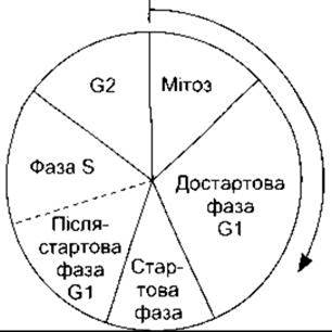
Fig. 1-16. Sequence of events during the cell cycle.
Introduction/30.html">Regulation of Gene Expression
Every somatic cell in the body contains the complete genetic information; however, among the diverse types of adult cells, there is significant differentiation and specialization of functions, with only a small fraction of the genetic information being actively transcribed under normal conditions. Consequently, genetic information is typically in a repressed state. Nevertheless, genes are controlled both spatially and temporally. What activates The genes of this specific cell rather than others? What activates genes in a cell at this particular stage of development rather than at inappropriate stages? What maintains orderly cell proliferation and prevents uncontrolled growth, known as cancer? Naturally, DNA sequences such as the TATA box facilitate the correct Transcription of the gene of which they are a part (cis-regulation). Yet, the master key to selective Gene Expression lies in proteins that bind to regulatory Regions of the gene, enhancing or repressing its activity. These transcription factors are products of other genes and thus act as intermediaries in trans-regulation. They are extremely numerous, encompassing activated steroid Hormone Receptors and many other factors. Stimuli, such as Neurotransmitters binding to The cell membrane, generally initiate chemical cascades that activate immediate-early genes. These, in turn, produce transcription factors that act on other genes. The best-characterized immediate-early genes are c-fos and c-jun. The proteins encoded by these genes—c-Fos, c-Jun, and several related proteins—form homo- and heterodimers that bind to a regulatory DNA sequence known as the AP-1 site (Fig. 1-20). Some dimers enhance transcription, while others repress it. The presence of c-Fos, c-Jun, and certain related proteins is a hallmark of cellular activation, and immunocytochemical methods targeting them or measuring their mRNAs make it possible to determine which cells in The Nervous system or any other tissue are activated by specific stimuli.
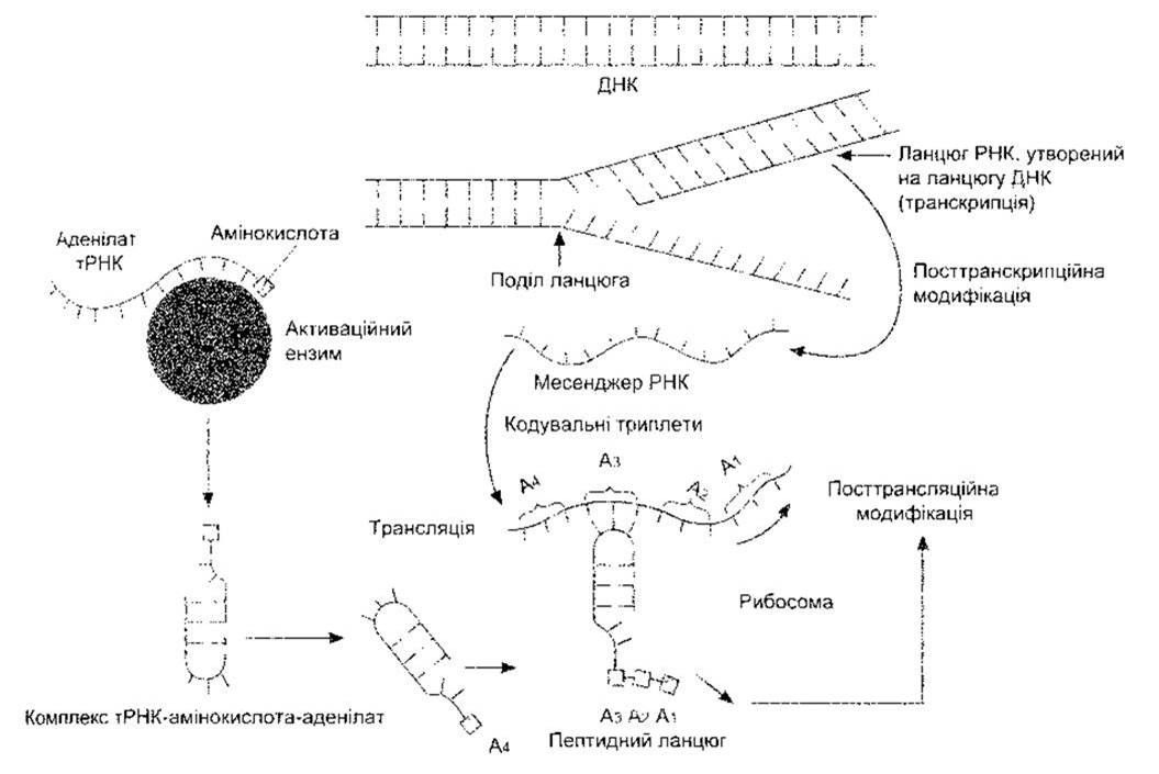
Fig. 1-17. Schematic diagram of protein synthesis. Nucleic Acids are shown as lines with numerous short projections representing individual bases.
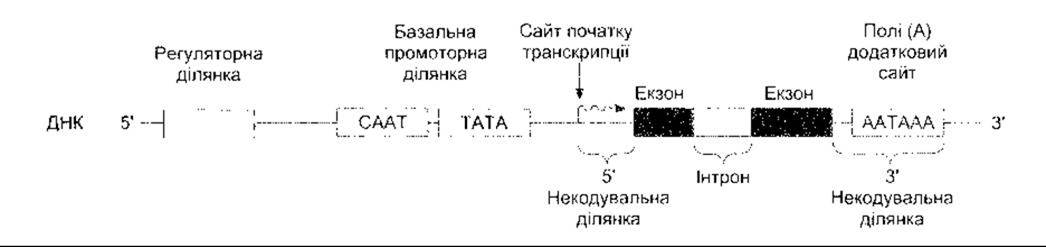
Fig. 1-18. Schematic representation of the components of a typical eukaryotic gene. The coding region consists of introns and exons, flanked on either side by non-coding regions. The 5’ flanking region contains DNA fragments that interact with proteins to promote or inhibit transcription, and the 3’ region contains a poly(A) addition site (reproduced with permission from Murray RK et al., Harper’s Biochemistry, 25th ed. McGraw-Hill, 2000).
Over 80% of known transcription factors feature one of four DNA-binding motifs. The most common is the zinc finger, in which characteristic structural complexes are formed by the coordination of a Zn2+ ion with two Cysteine and two Histidine residues, or four cysteine residues. Various transcription factors contain from 2 to 37 such zinc fingers, which mediate DNA binding. Another motif is the leucine zipper, in which the $\alpha$-helical domains of dimers contain regularly spaced leucine residues that interact with one another to form a coiled-coil structure. The dimer extensions beyond the joining region are rich in Arginine and Lysine, which bind to DNA. In addition, helix-turn-helix and helix-loop-helix structures also bind to DNA.
Today, molecular biology techniques allow researchers to upregulate specific gene functions, transfer human genes into animals, and disrupt the function of individual genes (gene knockout). The gene knockout method is widely used in numerous experimental studies.
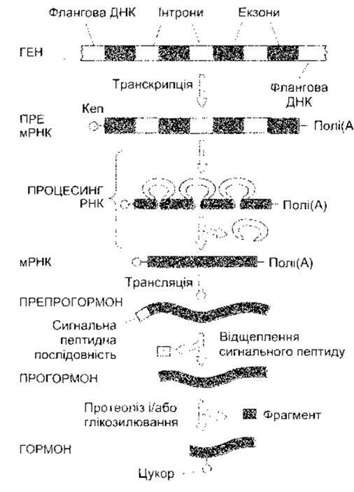
Fig. 1-19. Transcription, post-transcriptional mRNA modification, ribosomal translation, and post-translational processing during the formation of Hormones and other proteins. Cap denotes the cap site (modified and reproduced with permission from Baxter JD. Principles of Endocrinology. In: Cecil Textbook of Medicine, 16th ed. Wyngaarden JB, Smith LH Jr [editors]. Saunders, 1982).
Protein Synthesis
The process of Protein synthesis is both complex and fascinating. As noted, it consists of four stages: transcription, post-transcriptional modification, translation, and post-translational modification. These stages are illustrated and briefly characterized in Fig. 1-19. Once sufficient activation is achieved, gene transcription begins at the so-called cap site (see Fig. 1-19) and terminates approximately 20 bases downstream from the AATAAA sequence. The RNA transcript is capped in the nucleus by The addition of 7-methylguanosine triphosphate to the 5'-end; this cap is essential for proper ribosomal attachment (see below). A poly(A) tail consisting of over 100 bases is attached to the untranslated segment at the 3'-end. The exact function of the poly(A) tail remains unclear, but it is thought to help maintain mRNA stability. During pre-mRNA Processing, the capped and polyadenylated transcript undergoes intron elimination (see Fig. 1-19), and upon completion of this post-transcriptional modification, the mature mRNA is transported to the cytoplasm. Post-transcriptional modification of pre-mRNA is a regulated process, and as mentioned above, alternative splicing of a single pre-mRNA can yield more than one mRNA.
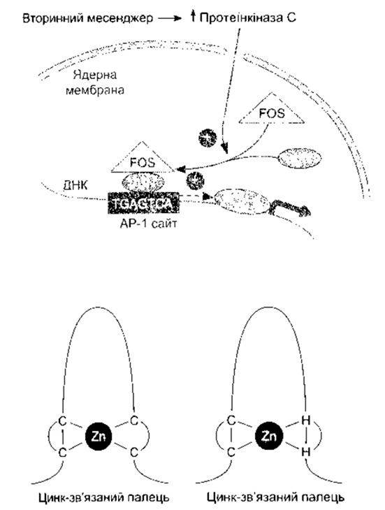
Fig. 1-20. Top: Activation of genes by secondary messengers. The action of immediate-early genes increases the amount of protein kinase C, leading to the formation of c-Fos and c-Jun. The c-Fos–c-Jun heterodimer binds to the AP-1 site, activating the transcription of other genes. Bottom: Zinc fingers. Curved lines represent polypeptide chains binding to DNA, while straight lines indicate the binding of zinc to cysteines (C) or to cysteines and histidines (H) (Reproduced with permission from Murray RK et al.: Harper's Biochemistry, 25th ed. McGraw-Hill, 2000).
Once the mature mRNA reaches a ribosome in the cytoplasm, it directs the assembly of a polypeptide chain. Amino Acid Activation in the cytoplasm occurs by combination with an enzyme and adenosine monophosphate (adenylate), after which each activated amino acid binds to a specific tRNA molecule. For each of the 20 unmodified amino acids found in Abundance in animal body proteins, there is at least one tRNA (see Chapter 17), and for Some amino acids, more than one. The tRNA-amino acid-adenylate complex attaches to the mRNA template. This process takes place within ribosomes and is diagrammed in Fig. 1-17. A tRNA molecule "recognizes" the corresponding attachment site on the mRNA template because its active end bears a triplet of bases complementary to a specific three-base sequence in the mRNA chain. The Genetic Code consists of these triplets, or codons—sequences of three purine and/or pyrimidine bases—with each triplet specifying a particular amino acid.
Translation begins in the ribosomes at an AUG codon (transcribed from ATG in the gene), which encodes Methionine. An amino acid residue is then added, lengthening the chain by one amino acid. Messenger RNA attaches to the 40S ribosomal subunit during protein synthesis; the nascent polypeptide chain binds to the 60S subunit, while tRNA interacts with both. Because amino acids are added in the sequence dictated by the triplet code, the ribosome moves along the mRNA molecule like a bead on a string. The translation process terminates at one of three stop or nonsense codons—UGA, UAA, or UAG—and the polypeptide chain is released. tRNA molecules are recycled, whereas mRNA molecules are reused approximately ten times before being replaced.
Typically, more than one ribosome is associated with each mRNA strand. The mRNA chain and its cluster of ribosomes can be visualized under an Electron microscope as aggregates known as polyribosomes (Polysomes).
At least in theory, the synthesis of certain Proteins can be halted using antisense oligonucleotides—short synthetic base fragments complementary to segments of a protein's mRNA. These fragments bind to the mRNA, blocking translation. Initial attempts using this technique were unsuccessful due to non-specific binding and immune reactions; however, research is ongoing, and it is hoped that such agents will aid in the Treatment of various diseases, including cancer.
Post-translational Modification
The Modification of the nascent polypeptide chain into a functional protein involves a combination of reactions, notably hydroxylation, carboxylation, glycosylation, or phosphorylation of amino acid residues; peptide bond Cleavage, which converts a large polypeptide into a smaller form; as well as protein folding, packaging, and the acquisition of a final, often complex conformation. It has been estimated that a typical Eukaryotic Cell synthesizes roughly 10,000 different proteins during its lifespan. How do these proteins reach their correct cellular destinations? Synthesis begins on free ribosomes. As noted, most proteins destined for secretion or incorporation into Organelles, as well as all transmembrane proteins, possess an N-terminal signal peptide (leader sequence) that directs them to The Endoplasmic reticulum. This peptide consists of 15–30 predominantly hydrophobic amino acid residues. The synthesized signal peptide binds to the signal recognition particle (SRP)—a complex macromolecule comprising six Polypeptides and 7S RNA, a small RNA species. The SRP halts translation until it binds to the translocon, a channel-like structure in the endoplasmic reticulum membrane formed by a heterotrimer of Sec61 proteins. The ribosome also binds, and the signal peptide guides the growing peptide chain into the lumen of the endoplasmic reticulum (Fig. 1-21). Subsequently, the signal peptide is cleaved from the rest of the peptide by a signal peptidase while Synthesis of the remainder of the peptide chain proceeds.
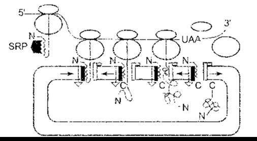
Fig. 1-21. Protein translation into the endoplasmic reticulum according to the signal hypothesis. Protein-synthesizing ribosomes move along the mRNA from the 5'- to the 3'-end. When the signal peptide of a protein destined for secretion, the cell membrane, or Lysosomes emerges from the large ribosomal subunit, it binds to the signal recognition particle (SRP), arresting further translation until it binds to the translocon on the endoplasmic reticulum. This releases the SRP, which is recycled in the cytoplasm. Binding to the ribosomal receptor also occurs, opening a channel that allows the growing protein chain to enter the endoplasmic reticulum. The signal peptide is cleaved by a signal peptidase. Upon completion of protein synthesis, the two ribosomal subunits dissociate, and the C-terminal end enters the endoplasmic reticulum; N, amino-terminal end of the protein; C, carboxy-terminal end of the protein. (Reproduced with permission from Walter P, Lingappa VR: Transport of proteins into and across the endoplasmic reticulum membrane. In: Protein Transfer and Organelle Biogenesis. Das RC, Robbins PW [editors]. Academic Press, 1988.)
Signals directing Newly synthesized proteins to their appropriate cellular compartments are generated in the Golgi apparatus (see below) and involve specific modifications of the carbohydrate residues on Glycoproteins.
Secreted Proteins
Most proteins secreted by cells are synthesized as large proteins, and polypeptide sequences are cleaved from them during maturation. In the case of hormones, these larger forms are called prehormones and prohormones (see Figs. 1-19 and 1-22). An example is parathyroid hormone (see Chapter 21). It is synthesized as a molecule containing 115 amino acid residues (preproparathyroid hormone). The 25-amino-acid leader sequence at the N-terminus is rapidly removed, thus generating proparathyroid hormone. Before secretion, six additional amino acids are cleaved from the N-terminus to form the secreted molecule. The function of this six-amino-acid peptide remains unknown.
Although most secreted polypeptides and proteins possess a leader sequence that directs them to the Endoplasmic reticulum and are secreted via exocytosis (see below), an increasing number of such proteins lack this sequence. In humans, these include the cytokines interleukin-1α (IL-1α) and IL-1β, three growth factors, and various factors involved in hemostasis. Secretion apparently occurs via ATP-dependent membrane transporters. There is a large family of these ATP-binding cassette (ABC) transport proteins that transport ions and other substances, as well as proteins, between organelles and across the cell membrane. Typically, they consist of two cytoplasmic ATP-binding domains and two membrane-spanning domains, each of which anchors into the membrane and contains six long α-helical sequences (Fig. 1-23). The cystic fibrosis transmembrane conductance regulator (CFTR) is one such ABC transporter, partially regulated by cAMP. It transports Cl- and is defective in individuals with cystic fibrosis (see Chapter 37).
Protein Folding
Protein folding is an additional post-translational modification. This complex process is driven primarily by the Amino Acid Sequence within the polypeptide chain. However, in certain instances, newly formed proteins associate with other proteins called chaperones. They prevent improper interactions with other proteins and ensure that the protein achieves its correct final conformation. Misfolded proteins and other proteins "flagged" for degradation are conjugated with ubiquitin and degraded in organelles known as 26S proteasomes (see Chapter 17).
Apoptosis
Not only cell division and growth but also cell death and clearance are under Genetic control. This process is known as programmed cell death (PCD) or apoptosis. The word apoptosis stems from the Greek term for leaves falling from a tree. This process can also be described as "cellular suicide," since the cell's own genes are largely responsible for its demise. It must be distinguished from necrosis (cell death resulting from injury), in which the destruction of healthy cells is caused by external factors such as inflammation. Apoptosis is a normal process during development and maturity. Notably, A large number of Neurons are formed in the Central Nervous System that ultimately die during remodeling occurring during development and synapse formation (see Chapter 4). In The Immune System, inappropriate clones of immunocytes are eliminated via apoptosis (see Chapter 27); it accounts for the lytic effects of glucocorticoids on lymphocytes (see Chapter 20). Apoptosis is also a vital factor in processes such as the removal of webbing between fingers in the fetus and the regression of fetal tubular systems during sexual maturation (see Chapter 23). In adults, apoptosis drives the cyclic breakdown of the endometrium leading to menstruation (see Chapter 23) and accounts for the shedding of enterocytes sloughed off from the tips of intestinal villi (see Chapter 26). Abnormal apoptosis is thought to occur in autoimmune diseases, neurodegenerative disorders, and cancer.
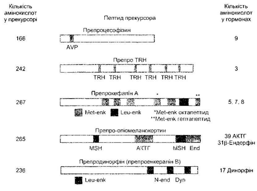
Fig. 1-22. Examples of large precursors (preprohormones) for small Peptide Hormones. See also Figs. 14-12. TRH, thyrotropin-releasing hormone; AVP, arginine vasopressin; Met-enk, Met-enkephalin; Leu-enk, Leu-enkephalin; MSH, melanocyte-stimulating hormone; ACTH, adrenocorticotropic hormone; End, β-endorphin; Dyn, dynorphin; N-end, neoendorphin.
A variety of extracellular ligands can activate receptors that trigger apoptosis. One of these is Fas, a transmembrane protein protruding from lymphocytes, natural killer (NK) cells, and T lymphocytes (see Chapter 27), which exist in various forms and circulate in the blood. Apoptosis is conveniently characterized in terms of activation, Intracellular Signaling, and execution. The two main extracellular factors that activate the process are Fas and TNF. When Fas binds to its receptor, a cascade of intracellular enzymatic reactions is triggered, leading to cell destruction. The apoptosis-inducing cascade is initiated by the activation of caspases, members of a large family of cysteine proteases. To date, thirteen mammalian caspases have been identified and characterized.
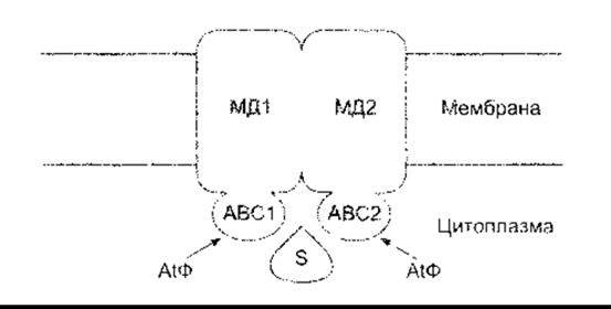
Fig. 1-23. General structure of eukaryotic ABC transport proteins that move ions, other substances, and proteins across membranes. ABC1 and ABC2, ATP-binding domains; MD1 and MD2, membrane domains; S, substrate (modified from Kuchler K, Thorner J. Secretion of Peptides and Proteins lacking hydrophobic signal sequences: The Role of adenosine triphosphate-driven membrane translocators. Endocr Rev 1992; 13:499).
Tumor necrosis factor can also mimic apoptosis. This initiates the action of activated apoptotic genes, causing DNA fragmentation, cytoplasmic and Chromatin Condensation, and ultimately membrane blebbing, cell fragmentation, and the removal of cellular debris via phagocytosis.
One of the main mechanisms by which the product of the tumor suppressor gene *p53* (see below) performs its anticancer function is the activation of apoptosis.
Molecular Medicine
Fundamental research into the molecular aspects of genetics, The regulation of gene expression, and protein synthesis is increasingly essential in clinical medicine.
Among the earliest achievements in this field was the understanding of the Mechanisms of action of Antibiotics (Table 1-5). Many of these agents act by inhibiting protein synthesis at one of the levels described above. Antiviral drugs act similarly; for example, acyclovir and ganciclovir inhibit DNA polymerases. Some of these drugs primarily affect bacteria, whereas others inhibit protein synthesis in animal cells, including those of mammals. This highlights the importance of antibiotics both for research and for the treatment of infections.
Table 1-5. Mechanisms of inhibition of protein synthesis by antibiotics and Other Compounds
Agent |
Effect |
Chloramphenicol |
Prevents normal binding of mRNA to ribosomes |
Streptomycin, neomycin, kanamycin |
Cause misreading of the genetic code |
Cycloheximide, tetracycline |
Inhibit The transfer of the tRNA-amino acid complex to the polypeptide |
Puromycin |
The puromycin-amino acid complex substitutes for the tRNA-amino acid complex, preventing the addition of amino acids to the polypeptide |
Mithramycin, mitomycin C, dactinomycin (actinomycin D) |
Bind to DNA, preventing RNA polymerization on DNA templates |
Chloroquine, colchicine, novobiocin |
Inhibit DNA polymerase |
Nitrogen mustards, e.g., mechlorethamine (mustargen) |
Bind to guanine in base pairs |
Diphtheria toxin |
Prevents the movement of the ribosome along mRNA |
Single genetic abnormalities causing over 600 human diseases have now been identified. Many of these disorders are rare, but a significant group are well-known, and some lead to severe, occasionally fatal consequences. Examples include a defectively regulated Cl- channel in cystic fibrosis (see above and also Chapter 34); unstable trinucleotide repeats in various regions of The Genome causing Huntington's disease; fragile X syndrome; and several other neurological disorders (see Chapter 12). Abnormalities in Mitochondrial DNA can also cause various human diseases, notably Leber's hereditary optic neuropathy and certain forms of cardiomyopathy. Unsurprisingly, the greatest attention is focused on the GENETIC ASPECTS OF cancer. Some cancers are driven by oncogenes—genes carried within the genomes of cancer cells that are responsible for their malignant phenotype. Oncogenes arise via somatic mutations from closely related Proto-oncogenes. More than 100 oncogenes have already been described.
Another group of genes produces proteins that suppress tumors; more than 10 such tumor suppressor genes have been described. Among them, the best studied is the *TP53* gene on human chromosome 17. The p53 protein produced by this gene induces apoptosis. It also acts as a nuclear transcription factor that upregulates The production of a 21 kDa protein, which inhibits cell cycle enzymes, thus slowing the cycle and allowing time for the repair of mutations and other DNA defects. The *TP53* gene is mutated in nearly 50% of cancer patients, impairing the production of p53 proteins that attenuate cell cycle arrest and prevent the accumulation of further DNA mutations. The accumulated mutations ultimately lead to cancer.
Gene Therapy is still in its early Selection/3.html">Stages of development; nonetheless, various ingenious approaches are being developed to introduce genes into cells. One such approach is already undergoing clinical trials for the treatment of certain diseases and involves isolating cells from an affected patient, transfecting these cells with normal genes *in vitro*, and reinfusing them as autografts. Another method involves inserting the appropriate gene into relatively safe Viruses, which are then administered to patients to deliver the genes to the cells they infect.
The Golgi Apparatus and Intracellular Vesicular Transport
The Golgi apparatus is a set of membrane-enclosed sacs (cisternae) stacked like plates on a dining table (see Fig. 1-4). There are usually about six such sacs, though occasionally more. One or more Golgi apparatuses are present in all eukaryotic cells, typically located near the nucleus. The Golgi apparatus is a polarized structure with *cis* and *trans* faces (Fig. 1-24). Membrane vesicles containing newly synthesized proteins bud off from the rough endoplasmic reticulum and fuse with the cisternae at the *cis* face of the apparatus. Proteins then travel via vesicles through the medial cisternae and finally to the *cisterna* at the *trans* face, from which vesicles branch out into the cytoplasm. From the *trans* face, vesicles traffic to lysosomes and out of the cell via constitutive and non-constitutive pathways, both of which involve exocytosis (see below). In the reverse direction, vesicles bud inward from The Plasma Membrane via endocytosis (see below), enter endosomes, and ultimately reach lysosomes. From the lysosomes, certain proteins traffic back to the *trans* Golgi network. The terminal glycosylation of proteins occurs via the addition of preformed Oligosaccharides in the endoplasmic reticulum, but these oligosaccharides are modified into A wide variety of diverse carbohydrate constituents within the Golgi apparatus.
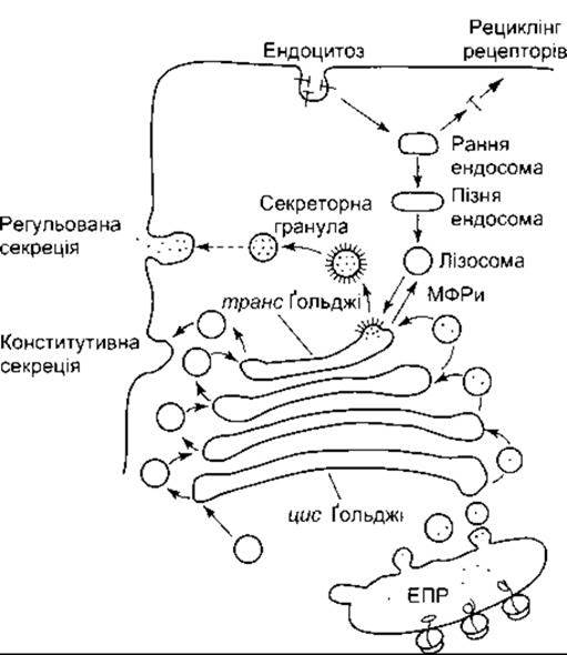
Fig. 1-24. Pathways involved in protein processing in the Golgi apparatus, secretion via exocytosis, and membrane regeneration via endocytosis. RER, rough endoplasmic reticulum. Note the different pathways for regulated (non-constitutive) and constitutive secretion via exocytosis. M6PR, mannose-6-phosphate receptors.
Quality Control
The processes involved in protein synthesis, folding, and migration to other PARTS OF THE cell reflect a complex network of diverse checkpoints designed to prevent errors and abnormalities. A corresponding "quality control" mechanism operates at every level. Damaged DNA is detected and repaired. Various RNAs are also monitored during the translation process. Finally, as protein chains reside in the endoplasmic reticulum or the Golgi apparatus, defective structures are identified, and abnormal proteins are degraded in lysosomes and proteasomes. The ultimate result is precise monitoring of protein production, which is essential for the normal functioning of the Organism.
Last update: 10/08/2026
Editorial and Educational Adaptation: This material has been compiled based on the primary/original source text. The project team performed an editorial review, corrected technical inaccuracies, structured sections, and adapted the content for an educational format.
What was processed:
- elimination of formatting defects (OCR errors, structural breaks, corrupted characters);
- editorial organization of content;
- standardization of terminology in accordance with academic sources;
- verification of factual statements against the original source text.
All mentions of the author, publication year, and origin of the primary text have been preserved in accordance with the source.