Molecular Biology of the Cell - Volume 2 - Alberts B., Bray D., Lewis J., Raff M., Roberts K., Watson J. 1993
Intracellular macromolecule sorting and maintenance of cellular compartments
Golgi apparatus
The Golgi Apparatus (also known as the Golgi complex) is typically located near the Cell Nucleus, and in animal Cells, it is often found in the vicinity of the centrosome, or cell center. It consists of a set of membrane-enclosed, flattened cisternae resembling a stack of plates. Each Golgi stack (referred to as a dictyosome in plants) usually contains four to six cisternae, generally with a diameter of about 1 µm (Fig. 8-59). The number of Golgi stacks in a cell largely depends on its type: some cells contain one large stack, whereas others have hundreds of very small stacks.
A multitude of small (approximately 60 nm in diameter) membrane-bounded vesicles are constantly associated with Golgi stacks. They cluster on the face adjacent to the ER as well as along the periphery of the stack near the expanded margins of each cisterna (see Fig. 8-59). It is believed that these vesicles (Golgi vesicles) transfer Proteins and Lipids into the Golgi apparatus, transport them out of it, and mediate traffic between the remaining cisternae. Many of these vesicles are coated and covered with clathrin or another specific protein. Such coated vesicles can frequently be observed budding off from Golgi cisternae.
Class="center">
Fig. 8-59. A, Three-dimensional Structure OF THE Golgi apparatus reconstructed from Cytology/cytology/93.html">ELECTRON MICROGRAPHS OF an animal secretory cell. The stacks of flattened Golgi cisternae feature expanded edges from which small vesicles apparently bud off. Large secretory vesicles are formed from the trans-compartment of the Golgi apparatus. B, Transmission electron micrograph of a cross-section of the Golgi apparatus in a plant cell (the green alga Chlamydomonas). The Golgi apparatus in plant cells is typically more pronounced and more clearly segregated from other intracellular membranes compared to animal cells. (A, after R. V. Krstic, ULTRASTRUCTURE OF THE Mammalian Cell. New York: Springer-Verlag, 1979; B, courtesy of George Palade.)
The Golgi apparatus has two distinct faces: the forming, or cis face, and the mature, or trans face. The cis face is closely associated with transitional elements of the ER (see Section 8.1.3); the trans face expands to form a tubular reticulum known as the trans-Golgi network. Proteins and lipids packaged in small vesicles enter the Golgi stack from the cis face and leave it en route to various compartments via vesicles budding off from the trans face. As they move from one Golgi stack to another, these molecules undergo a sequential series of modifications.
The Golgi apparatus is prominently developed in secretory cells, such as the goblet Cells of the intestinal epithelium, which secrete large amounts of mucus. Unusually large vesicles form on the trans face of the Golgi apparatus, oriented toward the region of The Plasma Membrane where secretion takes place (Fig. 8-60).
8-33
8.7.1. Modification of Oligosaccharide Chains Takes Place in the Golgi Apparatus [48]
As noted above, an N-linked oligosaccharide is added to proteins in the ER. This oligosaccharide undergoes initial modifications while still in the ER (see Section 8.6.12), with further alterations subsequently occurring in the Golgi apparatus.
The Oligosaccharides attached to asparagine residues in mature Glycoproteins fall into two broad classes: complex oligosaccharides and high-mannose oligosaccharides (Fig. 8-61). Sometimes oligosaccharides of both types are attached (at different sites) to the same polypeptide chain. High-mannose oligosaccharides do not acquire new sugars within the Golgi apparatus. They contain two N-acetylglucosamines and numerous mannose residues—often nearly as many as were present in the lipid-linked oligosaccharide precursor in the ER. Complex oligosaccharides may possess more than two N-acetylglucosamines, along with variable numbers of galactose, sialic acid, and (in some cases) fucose residues. Sialic acid is of particular interest as the only carbohydrate residue in glycoproteins that carries a negative charge (see Section 6.1.6). Complex oligosaccharides are generated by the "trimming" of oligosaccharides added in the ER and the subsequent addition of extra sugars. Thus, each complex oligosaccharide consists of a core region derived from the initial N-linked oligosaccharide (typically containing two N-acetylglucosamines and three mannose residues) and a terminal region composed of a variable number of trisaccharides (N-acetylglucosamine-galactose-sialic acid) linked to the core mannose residues. Occasionally, the terminal region appears truncated, containing only N-acetylglucosamine and galactose, or even just N-acetylglucosamine. In some instances, the presence of fucose attached to the core N-acetylglucosamine is also optional. All these sugars making up the terminal domain are added in the trans-Golgi by a battery of Glycosyltransferases operating in a strictly defined sequence (Fig. 8-62). Specific activated nucleotide sugars serve as substrates for these Enzymes, being transported into the lumen of the Golgi apparatus by a set of membrane-bound carriers. These same carriers export the nucleotide byproducts resulting from glycosylation. One of the characterized glycosyltransferases (galactosyltransferase) is commonly used as a marker to identify vesicles originating from the Golgi apparatus in membrane fractions purified by differential centrifugation.

Fig. 8-60. Goblet cell of the Small Intestine. This cell is specialized for the secretion of mucus, which is a mixture of Glycoproteins and Proteoglycans synthesized in the ER and the Golgi apparatus. The Golgi apparatus is highly polarized, facilitating the release of mucus via exocytosis from the apical surface of The Cell. (After R. Krstic, Illustrated Encyclopedia of Human Histology. New York: Springer-Verlag, 1984.)

Fig. 8-61. Examples of the two principal classes of asparagine-linked (N-linked) oligosaccharides found in mature glycoproteins: "complex" oligosaccharides (A) and "high-mannose" oligosaccharides (B). Numerous structural variations exist. For instance, the complex oligosaccharide shown here has three terminal branches, but structures with two or four terminal branches are also common, depending on the glycoprotein and the cell type in which it is produced. "Hybrid" oligosaccharides, featuring one mannose-rich branch and one GlcNAc-Gal branch, are also encountered. The three highlighted Amino Acids represent the recognition sequence for the enzyme that transfers the initial oligosaccharide to the protein. Abbreviations: Asn, asparagine; Man, mannose; GlcNAc, N-acetylglucosamine; NANA, N-acetylneuraminic acid (sialic acid); Gal, galactose; X, any amino acid.
The reactions leading to The formation of complex oligosaccharides proceed via the strictly ordered pathway illustrated in Fig. 8.63.

Fig. 8-62. Scheme of sequential sugar residue addition. These reactions take place within the Golgi apparatus and result in the formation of complex oligosaccharides. Each step is mediated by one of three distinct glycosyltransferases that utilize nucleotide-activated sugars as substrates. Glycosylation proceeds on the luminal surface of the membrane. The chemical structures of the nucleotide sugars UDP-N-acetylglucosamine, UDP-galactose, and CMP-N-acetylneuraminic acid are depicted at the top. For abbreviations, see the legend to Fig. 8-61.

Fig. 8-63. Oligosaccharide Processing in the ER and the Golgi apparatus. Processing is highly ordered, with each stage shown depending on the preceding reaction in the sequence. Processing begins in the ER with the removal of a glucose residue from the oligosaccharide initially transferred to the protein. All three glucose residues may be removed even before Protein Synthesis is complete. Subsequently, a mannosidase residing in the ER membrane removes a specific mannose residue. In the Golgi stack, mannosidase I removes three additional mannose residues, and N-acetylglucosaminyltransferase I adds a GlcNAc residue that prevents mannosidase II from removing two further mannose residues. Ultimately, this yields a core of three mannose residues characteristic of "complex-type" oligosaccharides. At this stage, the linkage between the two GlcNAc residues in the core becomes resistant to attack by the highly specific endoglycosidase H (Endo H). Because all subsequent processing steps are also Endo H-resistant, Treatment with this enzyme is widely used to distinguish between complex and high-mannose oligosaccharides. Finally, additional GlcNAc, galactose, and sialic acid residues are added. The extent of Processing of the initial oligosaccharide depends on the protein type and the local conformation around the asparagine residue to which the oligosaccharide is attached. The processing of some oligosaccharides halts within the Golgi apparatus, whereas others undergo the modifications shown here to varying degrees. For abbreviations, see the legend to Fig. 8-61.
The details of this pathway were elucidated through The Use of various drugs and Antibiotics that inhibit specific Stages of the process (Table 8-4). Whether a given oligosaccharide remains high-mannose or undergoes further modification is largely determined by the conformation of the protein to which it is attached: if the oligosaccharide remains sterically accessible to the modifying Enzymes of the Golgi apparatus after attachment, it will likely be converted into the complex form; otherwise, it will remain high-mannose.
Table 8-4. Inhibitors of Various Stages of N-Glycosylation
|
Drug(s) |
Inhibited Step |
|
Tunicamycin |
Dolichol-P → dol-P-P-GlcNAc |
|
Castanospermine and N-methyldeoxynojirimycin |
Glucose3-Man9-GlcNAc2-Asn → glucose2-Man9-GlcNAc2-Asn |
|
Bromoconduritol |
Glucose2-Man9-GlcNAc2-Asn → Man9-GlcNAc2-Asn |
|
Deoxymannojirimycin |
Man8-GlcNAc2-Asn → Man5-GlcNAc2-Asn |
|
Swainsonine |
GlcNAc-Man5-GlcNAc2-Asn → GlcNAc-Man3-GlcNAc2-Asn |
8.7.2. CARBOHYDRATES of Cell Membranes Face the Membrane Surface Topologically Equivalent to the Extracellular Space
Because oligosaccharide chains are added on the luminal side of the ER and the Golgi apparatus, the distribution of carbohydrates on Membrane Proteins and lipids is asymmetric. Like the Asymmetry of The Lipid Bilayer itself, this asymmetric orientation of glycosylated molecules is maintained during transport to the plasma membrane, secretory vesicles, or Lysosomes. As a result, the oligosaccharides of all glycoproteins and Glycolipids in these cellular membranes face the lumen of the Organelles and, in the plasma membrane, the extracellular space (Fig. 8-64).
8.7.3. What is the Biological Significance of N-Glycosylation? [49]
There is an important distinction between the synthesis of oligosaccharide molecules and other macromolecules such as DNA, RNA, and proteins. Nucleic Acids and Proteins are copied from a template through the repeated execution of identical steps, using the same enzyme (or enzymes). Complex carbohydrates, however, require different enzymes at various stages of synthesis, with the product of each reaction being recognized as the substrate for the next. Given The complexity of the biochemical mechanisms that evolved for oligosaccharide synthesis, it is reasonable to assume that these compounds serve vital Functions, although most of these functions remain unknown.

Fig. 8-64. The orientation of a transmembrane protein in the ER membrane is preserved during its transport to other membranes. The black circles at the end of each glycoprotein molecule indicate an N-linked oligosaccharide, which is attached to proteins within the lumen (cavity) of the ER. Note that these sugar residues are located exclusively in the lumen of internal organelles and become exposed to the extracellular space only after a transport vesicle fuses with the plasma membrane.
For example, N-linked glycosylation predominates in all eukaryotes, including Yeast, but is absent in eubacteria. Since most proteins transported through the ER and the Golgi apparatus bear one or more N-linked oligosaccharides (and this transport process is specific to Eukaryotic cells), it was initially hypothesized that these oligosaccharides are involved in sorting and transport. However, it turned out that agents blocking certain stages of glycosylation (Table 8-4) generally do not affect transport (with one notable exception—transport to lysosomes, discussed below in Section 8.8). Mutant cultured cells in which Golgi glycosylation is blocked at various stages remain viable, and protein transport proceeds normally in them. It has been established that certain proteins cannot fold properly without their cognate oligosaccharides, causing them to precipitate in the ER and rendering them transport-incompetent; nevertheless, the majority of proteins retain normal activity even without glycosylation.
Because sugar chains have limited flexibility, even a small N-linked oligosaccharide protrudes well above The surface of the glycoprotein (Fig. 8-65) and can thus restrict the approach of other macromolecules to that surface. Consequently, the presence of the oligosaccharide sometimes confers relative resistance to proteases upon the glycoprotein. It is possible that oligosaccharides provided the ancestral Introduction/5.html">Eukaryotic Cell with a protective coat that, unlike the rigid Bacterial Cell wall, allowed the cell to change shape and move. Subsequently, these oligosaccharides could have been modified to perform other functions as well.
8.7.4. Proteoglycans Are Assembled in the Golgi Apparatus [50]
During the transit of proteins from the ER to their final destinations through the Golgi apparatus, N-linked oligosaccharides are not the only structures to be altered; many proteins are modified in other ways as well. For example, as noted above, in some proteins sugars are added to the side chains of specific Serine or Threonine residues. Such O-linked glycosylation, like the elongation of N-linked oligosaccharide chains, is catalyzed by glycosyltransferases. These enzymes add sugars to the protein one by one, utilizing nucleotide sugars present within the lumen of the Golgi apparatus as substrates. Typically, N-acetylgalactosamine is added first, followed by a variable number of additional sugar residues, ranging from a few up to 10 or more.
Glycosylation is most extensive in proteoglycan core proteins, which are modified in the Golgi apparatus to yield proteoglycans. This process involves the polymerization of one or more glycosaminoglycan chains (long, unbranched polymers composed of repeating disaccharide units) onto serine residues of the core protein. The Fate of proteoglycans varies: some are secreted as Components of the Extracellular matrix, while others remain anchored in the plasma membrane. In addition, Proteoglycans form the backbone of mucus, which provides a protective coating for numerous epithelia.
The sugars comprising glycosaminoglycans are heavily sulfated immediately upon polymerization in the Golgi apparatus, imparting a strong negative charge to proteoglycans. The sulfate is transferred from an activated sulfate donor, 3'-phosphoadenosine-5'-phosphosulfate (PAPS), which is imported from the Cytosol into the Golgi. The most subtle modification occurring in this organelle is The transfer of a sulfate group from PAPS to the hydroxyl group of specific Tyrosine residues in proteins. Sulfated tyrosines are characteristic of secreted proteins and are occasionally found in plasma membrane proteins (within their extracellular domains).

Fig. 8-65. Three-dimensional structure of a small N-linked oligosaccharide, determined by X-ray crystallography of a glycoprotein. This oligosaccharide contains only 6 sugar residues, whereas the N-linked oligosaccharide initially attached to the protein in the ER contains 14 sugar residues (see Fig. 8-52). (A) Ball-and-stick model showing all atoms except hydrogens; (B) space-filling model, with dark atoms representing the asparagine residue. (Courtesy of Richard Feldman.)
8.7.5. Proteins Often Undergo Proteolysis During Secretory Vesicle Formation [51]
The most radical modification to which proteins are subjected prior to secretion occurs last. Many polypeptide Hormones and Neuropeptides are synthesized as inactive precursor proteins, which are subsequently cleaved by proteolysis to generate the active molecule. This Cleavage is thought to begin in the trans Golgi network and continue within secretory vesicles. Initially, a membrane-bound protease cleaves the protein at bonds adjacent to pairs of basic amino acids (Lys-Arg, Lys-Lys, Arg-Lys, or Arg-Arg), after which the secreted product undergoes its final maturation (Fig. 8-66). In the simplest case, a polypeptide often contains only a single N-terminal pro-region, which is cleaved off to yield the mature protein shortly before secretion. Consequently, such proteins are synthesized as pre-pro-proteins, in which the pre-region is an ER signal peptide that is removed in the rough ER. In a more complex scenario, peptide molecules are synthesized as polyproteins containing multiple copies of the same Amino Acid Sequence (see Fig. 8-66). Finally, some cells contain Peptides that act as precursors for numerous distinct end products. These end products are cleaved one by one from the initial polypeptide chain. In different cell types, the same polyproteins can be cleaved in various ways, thereby increasing The Diversity of molecules involved in intercellular chemical signaling.
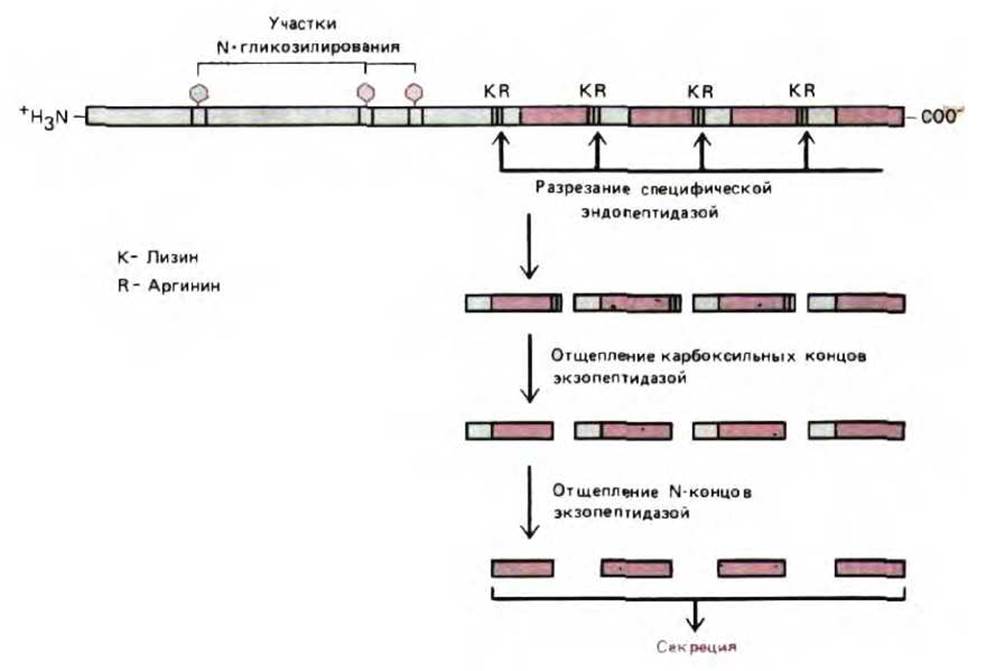
Fig. 8-66. An example of a polyprotein that is cleaved to generate multiple copies of the same signaling peptide molecule. Processing typically begins with cleavage at pairs of basic amino acids (here, Lys-Arg pairs) catalyzed by a specific membrane-bound protease located in secretory vesicles or the trans Golgi network. The processing mechanism shown yields the 13-amino-acid peptide $\alpha$-factor of the yeast Saccharomyces cerevisiae, a secreted peptide that regulates the mating process in yeast. (From R. Fuller, A. Brake, and J. Thorner, in Microbiology 1986 [L. Lieve, ed.], pp. 273–278. Washington, D. C.: American Society for Microbiology, 1986.)
Why is such "delayed" proteolysis characteristic of so many Polypeptides? Perhaps many of them, such as enkephalins (five-amino-acid neuropeptides), are simply too short to be synthesized efficiently on Ribosomes, given that even longer peptides sometimes lose the signals required for packaging into secretory vesicles. Furthermore, delaying the Formation of the active product until it reaches a secretory vesicle can prevent the product from acting prematurely within the cell.
8.7.6. Golgi Cisternae Are Assembled into Successive Compartments in Which Product Processing Occurs [52]
Processing within the Golgi stack is highly ordered. Each cisterna constitutes a distinct compartment with its own unique set of enzymes, and the stack as a whole thus functions as a multi-stage processing assembly line. Proteins are modified sequentially as they move from cisterna to cisterna.
Proteins entering from the ER arrive at the first Golgi cisterna (the cis compartment), move to the next compartment (the medial compartment), and finally reach the trans compartment (represented by the terminal cisterna of the stack), where glycosylation is completed. From the trans compartment, proteins enter the trans Golgi network (TGN); within this tubular reticulum, they are sorted into various transport vesicles and dispatched to their final destinations—the plasma membrane, lysosomes, or secretory vesicles.
The functional differences among the cis, medial, and trans compartments of the Golgi stack were first discovered through studies of the enzymes involved in N-linked oligosaccharide processing. These experiments employed both physical organelle fractionation and immunoelectron Microscopy. This approach demonstrated, for instance, that the removal of mannose residues and The addition of N-acetylglucosamine occur in the medial compartment, whereas the addition of galactose and sialic acid takes place in the trans compartment (Figs. 8-67 and 8-68).
Proteins entering the Golgi apparatus from the ER (except for those destined to remain resident in a particular Golgi cisterna) "flow" through the stack from the cis to the medial and then to the trans compartment, undergoing stepwise processing along the way. The exact mechanism by which proteins and lipids are transferred from one cisterna to the next is not yet fully understood, but it is thought to involve coated vesicles that bud off from the dilated margins of the cisternae. Because proteins entering the Golgi apparatus have been shown to move through the stack sequentially—from the cis compartment through the medial to the trans compartment—without taking direct shortcuts or skipping steps, each vesicle must be able to fuse exclusively with the membrane of the next cisterna in line. Although only three functionally distinct Regions of the Golgi apparatus have been identified to date, each of these regions is sometimes represented by two or more sequentially arranged cisternae, and even finer distinctions between them may yet be uncovered. Alternatively, there may indeed be only three fundamentally distinct compartments, with some cisternae within them simply representing redundant copies of the same functional unit.

Fig. 8-67. Histochemical staining demonstrates that the Golgi apparatus is biochemically polarized. A. Unstained preparation. B. Osmium stains primarily the cisternae of the cis compartment. C. The enzyme nucleoside diphosphatase (see Fig. 8-62) is localized in the trans Golgi cisternae; this enzyme was formerly known as "thiamine pyrophosphatase." D. The enzyme acid phosphatase serves as a marker for the trans Golgi network. (Courtesy of Daniel S. Friend.)
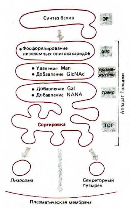
Fig. 8-68. Compartmentalization of the Golgi apparatus. As they progress through the closely packed cisternae of the Golgi stack, proteins undergo sequential covalent modifications. The trans Golgi network (TGN) is a tubular reticulum that functions primarily as a sorting station. The localization of each processing step depicted here was determined by combining various techniques, including membrane subfractionation of the Golgi apparatus and Electron microscopy following immunogold labeling for specific processing enzymes.
The sites of many other reactions remain to be established.
Proteins enter the Golgi apparatus from the ER and are subsequently directed to the plasma membrane, lysosomes, and secretory vesicles. The Golgi apparatus is a polarized structure consisting of one or more stacks of flattened cisternae surrounded by numerous small vesicles. These cisternae are organized into at least three distinct compartments (cis, medial, and trans compartments) of the Golgi apparatus. Proteins from the ER lumen and membrane are transported to the cis face of the Golgi stack via transport vesicles. Proteins destined for secretory vesicles, the plasma membrane, and lysosomes move sequentially from one cisterna to the next. Finally, they reach the trans-Golgi network, from which each protein is dispatched in specialized vesicles to its proper destination.
Unlike the ER, the Golgi apparatus contains a high concentration of nucleotide sugars. Various glycosyltransferases utilize these as substrates in the glycosylation reactions of proteins and lipids passing through the Golgi apparatus. For example, mannose residues are cleaved from N-linked oligosaccharides, and additional sugars—such as N-acetylglucosamine, galactose, and sialic acid residues—are added. Furthermore, O-glycosylation and The conversion of proteoglycan core proteins into proteoglycans occur within the Golgi apparatus. The sulfation of sugars in proteoglycans and of certain tyrosine residues in proteins also takes place specifically in the Golgi apparatus.
Last update: 12/08/2026
Editorial and Educational Adaptation: This material has been compiled based on the primary/original source text. The project team performed an editorial review, corrected technical inaccuracies, structured sections, and adapted the content for an educational format.
What was processed:
- elimination of formatting defects (OCR errors, structural breaks, corrupted characters);
- editorial organization of content;
- standardization of terminology in accordance with academic sources;
- verification of factual statements against the original source text.
All mentions of the author, publication year, and origin of the primary text have been preserved in accordance with the source.