Human Biochemistry, Volume 2 - Murray R. 1993
Special Topics
Glycoproteins and Proteoglycans
Proteoglycans and Glycosaminoglycans — Functions of Glycosaminoglycans and Proteoglycans
The binding of glycosaminoglycans to other extracellular macromolecules makes a significant contribution to the Structural Organization of the Extracellular matrix. Glycosaminoglycans can interact with extracellular macromolecules, Plasma Proteins, Cell surface components, and intracellular macromolecules.
The binding of glycosaminoglycans is typically electrostatic in nature due to their prominent polyanionic character, although some binding reactions are more specific. In general, glycosaminoglycans containing IdUA, such as dermatan sulfate and heparan sulfate, bind proteins with higher affinity than those containing GlcUA as their sole uronic acid.
Interaction with Extracellular Macromolecules
All glycosaminoglycans, with the exception of those lacking sulfate (hyaluronan) or carboxyl groups (keratan sulfates), bind electrostatically to Collagen at neutral pH. The presence of IdUA promotes tighter binding, allowing Proteoglycans to interact with collagen more strongly than the corresponding free glycosaminoglycans. Between 2 and 5 polysaccharide chains bind to each collagen monomer. All soluble collagens (types I, II, and III) bind chondroitin sulfate proteoglycans.
Chondroitin sulfate and heparan sulfate specifically bind to Elastin.
As noted above, chondroitin sulfate and keratan sulfate chains within their respective proteoglycans form aggregates with hyaluronic acid through the mediation of link proteins. Up to 100 proteoglycan molecules can bind to a single hyaluronan molecule.
Interactions with Plasma Proteins
The arterial wall contains proteoglycans that include hyaluronan, chondroitin sulfate, dermatan sulfate, and heparan sulfate. Among these, dermatan sulfate forms bonds with plasma Lipoproteins. Furthermore, dermatan sulfate appears to be the primary glycosaminoglycan synthesized by arterial smooth Muscle Cells. Because these specific cells proliferate during atherosclerotic lesions in Arteries, dermatan sulfate may play a substantial role in The formation of atherosclerotic plaques.
Although heparin is synthesized and stored in mast cells, it is always closely associated with Blood Vessels. Owing to its high negative charge (conferred by IdUA and sulfate residues), heparin strongly interacts with several plasma components. It specifically binds Blood Coagulation factors IX and XI. More importantly for heparin's anticoagulant activity is its ability to interact with an a2-plasma glycoprotein known as antithrombin III. Stoichiometric binding to heparin (1:1) significantly enhances the inactivating effect of antithrombin III on Serine proteases, particularly Thrombin. The binding of heparin to Lys residues of antithrombin III appears to induce conformational changes that facilitate the interaction of the latter with serine proteases. These processes are illustrated schematically in Fig. 54.13.
Commercially available heparin contains two components—high-affinity and low-affinity—which likely bind to the same region of the antithrombin III molecules. However, high-affinity heparin exhibits 10 times greater anticoagulant activity and a higher binding constant than low-affinity heparin. N-Desulfation or Modification of the IdUA residues of heparin reduces its anticoagulant activity.
Heparan sulfate, which is structurally similar to heparin, also has The ability to accelerate the action of antithrombin III, though its effect is considerably weaker than that of heparin.
Heparin can specifically bind to lipoprotein lipase present in the Capillary Wall and trigger the release of this enzyme into the bloodstream. Hepatic lipase binds to heparin and enters the bloodstream in a similar manner, albeit with a lower affinity than lipoprotein lipase. Partially N-desulfated heparin binds to lipoprotein lipase more intensely than to antithrombin III.
Class="center">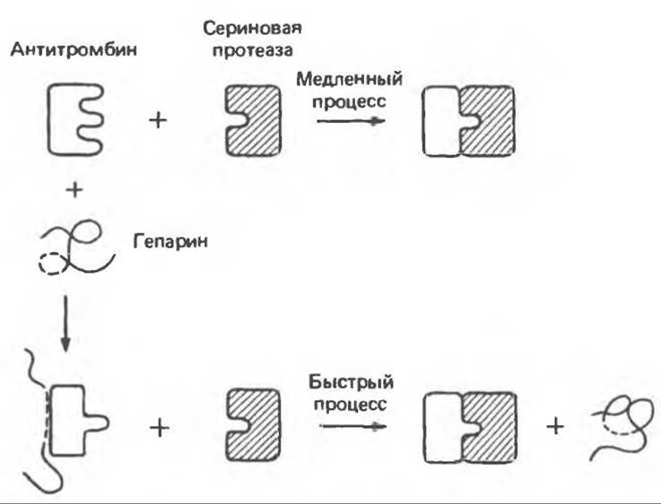
Fig. 54.13. Schematic representation of the inactivation by antithrombin of serine proteases (such as thrombin) involved in blood coagulation. Heparin appears to accelerate inactivation by binding to antithrombin and inducing Conformational Changes in it that facilitate the interaction between antithrombin and thrombin. (Heparin may also bind directly to thrombin.) This interaction requires the presence of a specific binding site (- - - - - - ) within the polysaccharide chain. (Reproduced, with permission, from Lennarz W. J. The Biochemistry of Glycoproteins and Proteoglycans. Plenum Press, 1980.)
Glycosaminoglycans and Cell Surface Molecules
Heparin is capable of binding to many cell types, including platelets, arterial endothelial cells, and Liver cells. Chondroitin sulfate, dermatan sulfate, and heparan sulfate bind to distinct Regions of the cell surface, such as those of fibroblasts. It is precisely within these regions that Glycosaminoglycans and Proteoglycans undergo degradation.
Hyaluronan is deposited by cells growing on plastic substrates. In addition, hyaluronan appears to participate in cell-Cell Adhesion processes, which play a vital role in the GROWTH AND DEVELOPMENT of Multicellular Organisms.
Certain proteoglycans likely serve as receptors and carriers for macromolecules, including lipoproteins, lipases, and antithrombin. Proteoglycans may also take part in the Regulation of Cell growth, intercellular interactions, and the protection of cell surface receptors.
Glycosaminoglycans and Intracellular Macromolecules
In addition to interacting with Enzymes involved in their Biosynthesis AND DEGRADATION, proteoglycans and their glycosaminoglycan components influence Protein Synthesis AND intranuclear functions. Specifically, heparin can affect Chromatin Structure and activate DNA polymerase in vitro. The extent to which these effects are physiological remains unclear. Glycosaminoglycans are present in significant quantities in the nuclei of various cell types, and existing evidence Supports a role for heparan sulfate in sea urchin embryo development.
Chondroitin sulfates, dermatan sulfates, and heparin can either activate or inhibit lysosomal acid Hydrolases. These enzymes are capable of forming natural complexes with glycosaminoglycans, yielding protected or inactive forms.
Numerous storage or secretory granules, such as chromaffin granules of The adrenal medulla, prolactin secretory granules of the Pituitary Gland, and basophilic granules of mast cells, contain sulfated glycosaminoglycans. The glycosaminoglycan-peptide complexes present within these granules may play a role in the release of biogenic amines.
Buckwalter J. A., Rosenberg L. С. Electron microscopic studies of Cartilage proteoglycans, J. Biol. Chem., 1982, 257, 9830. DiNatale P., Neufeld E. F. The biochemical Diagnosis of Mucopolysaccharidoses, mucolipidosis and related disorders. In: Perspectives in Inherited Metabolic Diseases, Vol. 2, Barra B. et al. (eds.), Editiones Ermes (Milan), 1979.
Höök M. et al. Cell-surface glycosaminoglycans, Annu. Rev. Biochem., 1984, 53, 847.
Hubbard S. C., Ivatt R. J. Synthesis and Processing of asparagine-linked Oligosaccharides, Annu. Rev. Biochem., 1981, 50, 555.
Jagues L. B. Heparin: an old drug with a new paradigm. Science, 1979, 206, 528.
Kornfeld R., Kornfeld S. Assembly of asparagine-linked oligosaccharides, Annu. Rev. Biochem., 1985, 54, 631. Kornfeld S., Sly W. S. Lysosomal storage defects, Hosp. Pract. (Aug), 1985, 20, 71.
Lennarz W. J. The Biochemistry of Glycoproteins and Proteoglycans, Plenum Press, 1980.
Poole A. R. et al. Proteaglycans from bovine nasal cartilage: Immunochemical studies of link protein, J. Biol. Chem., 1980, 255, 9295.
Reitman M. L. et al. Fibroblasts from patients with I-cell disease and pseudo-Hurler polydystrophy are deficient in uridine 5'-diphosphate-N-acetylglucosamine: glycoprotein N- acetylglucosaminylphosphotranspherase activity, J. Clin. Invest., 1981, 67, 1574.
Schächter H. Biosynthetic controls that determine the branching and heterogeneity of protein-bound oligosaccharides, Biochem. Cell Biol., 1986, 64, 163.
Snider M. D., Rogers О. C. Transmembrane Movement of oligosaccharide-Lipids during glycoprotein synthesis, Cell, 1984, 36, 753.
Last update: 06/08/2026
Editorial and Educational Adaptation: This material has been compiled based on the primary/original source text. The project team performed an editorial review, corrected technical inaccuracies, structured sections, and adapted the content for an educational format.
What was processed:
- elimination of formatting defects (OCR errors, structural breaks, corrupted characters);
- editorial organization of content;
- standardization of terminology in accordance with academic sources;
- verification of factual statements against the original source text.
All mentions of the author, publication year, and origin of the primary text have been preserved in accordance with the source.