Principles of Biochemistry, Volume 1 - A. Lehninger 1985
Biomolecules
Carbohydrates: Structure and Biological Functions
Glycosaminoglycans and Proteoglycans: Important Components of Connective Tissue
Another group of structural and protective Polysaccharides comprises the glycosaminoglycans, or acidic mucopolysaccharides. These are typically attached to Proteins to form Proteoglycans (Fig. 11-24), i.e., polysaccharide-Structure/178.html">Protein Complexes in which the polysaccharide accounts for the major portion of the molecule—usually more than 95%. Unlike proteoglycans, Glycoproteins also consist of polysaccharides and proteins, but the protein moiety constitutes the greater part of the molecule.
Glycosaminoglycans are composed of repeating disaccharide units, each of which is a derivative of an amino hexose—usually D-glucosamine or D-galactosamine. At least one of the two sugars in the repeating disaccharide unit of a glycosaminoglycan contains an acidic group that carries a negative charge at pH 7; as a rule, this is a carboxyl or sulfate group. An example of an acidic hexose is D-glucuronate, formed from D-glucose by oxidation at the sixth carbon atom (Fig. 11-25). Glycosaminoglycans are therefore classified as Heteropolysaccharides because they consist of alternating monosaccharide residues of two different types (Table 11-2). The name "mucopolysaccharides" reflects the fact that these polysaccharides were originally isolated from mucin, a slippery, lubricating proteoglycan found in secreted mucus. Subsequently, the term "acidic mucopolysaccharides" came to be used for the acidic polysaccharides of various vertebrate Tissues. Proteoglycans are present in the gel-like ground substance ("intercellular cement") that fills the extracellular space in most tissues. In addition, they are found in Cartilage, tendons, and Skin, as well as in synovial fluid, which acts as a lubricant in joints.
Hyaluronic acid is a glycosaminoglycan of the extracellular ground substance of animal tissues, consisting of repeatedly alternating residues of D-glucuronic acid and N-acetyl-D-glucosamine (Fig. 11-26). Hyaluronic acid forms highly viscous, gel-like solutions. It frequently occurs in association with other glycosaminoglycans. The enzyme hyaluronidase, secreted by certain pathogenic Bacteria, is capable of hydrolyzing the glycosidic bonds of hyaluronic acid, thereby facilitating bacterial penetration into tissues. During Fertilization, vertebrate hyaluronidase hydrolyzes the glycosaminoglycan-rich outer coat of the ovum, making it permeable to the spermatozoon. Chondroitin, the principal polysaccharide of cartilage proteoglycans, contains alternating residues of D-glucuronic acid and N-acetyl-D-galactosamine. Dermatan sulfate of the skin also contains alternating residues of two different sugars (Table 11-2).
Class="center">Table 11-2. Monosaccharide components of acidic polysaccharides characteristic of Connective Tissue
|
Polysaccharide |
Components |
|
Hyaluronic acid |
D-glucuronate + N-acetyl-D-glucosamine |
|
Chondroitin |
D-glucuronate + N-acetyl-D-galactosamine |
|
Dermatan sulfate |
D-iduronate + N-acetyl-D-galactosamine-4-sulfate |
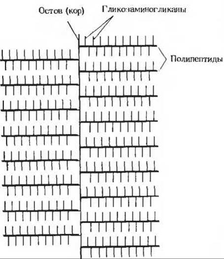
Fig. 11-24. Schematic representation of a proteoglycan, consisting of a branched protein backbone to which numerous glycosaminoglycan side chains are attached. The entire structure is highly hydrated.
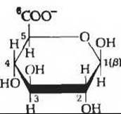
Fig. 11-25. D-glucuronate. The carboxyl group at C-6, which dissociates near pH 7, is highlighted in red.
Another important glycosaminoglycan is heparin; it is synthesized only in specific Cells located primarily in the arterial walls. Heparin contains repeating units of six-sugar sequences, each consisting of alternating residues of sulfated derivatives of N-acetyl-D-glucosamine and D-iduronate. Heparin is a potent anticoagulant; it prevents Blood clot formation in the Circulation. Isolated from lung tissue, heparin is used medically to prevent the clotting of donor blood and to inhibit intravascular thrombosis in various pathological conditions, such as following angina attacks.
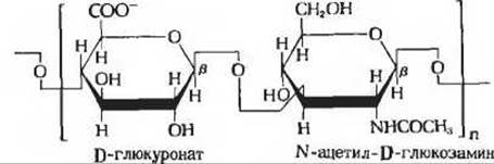
Fig. 11-26. Repeating unit of hyaluronate, a linear polymer containing thousands of such disaccharide units. At pH 7, the carboxyl group of the D-glucuronate residue is fully ionized.
In cartilage proteoglycans, the glycosaminoglycan molecules are covalently linked to protein. A typical cartilage proteoglycan contains about 150 polysaccharide chains, each with a Molecular Weight of 20,000, covalently attached as side chains to a core polypeptide. Such proteoglycans are highly hydrated structures (Fig. 11-24).
Last update: 06/08/2026
Editorial and Educational Adaptation: This material has been compiled based on the primary/original source text. The project team performed an editorial review, corrected technical inaccuracies, structured sections, and adapted the content for an educational format.
What was processed:
- elimination of formatting defects (OCR errors, structural breaks, corrupted characters);
- editorial organization of content;
- standardization of terminology in accordance with academic sources;
- verification of factual statements against the original source text.
All mentions of the author, publication year, and origin of the primary text have been preserved in accordance with the source.