Genetics - A. V. Sivolob 2008
The Nature of Genetic Material
Structural Organization of DNA in Cells
Chromatin Structure
At the first level of Chromatin Organization, DNA forms elementary structures known as nucleosomes through interaction with Proteins. The protein component of the nucleosome (the core) consists of eight core histone molecules—H2A, H2B, H3, and H4—with two molecules of each type (see The Structure of the H2A-H2B dimer, a core component, in Fig. 1.7, a). The octameric histone complex features a specific track of positively charged amino acid residues on its surface, which serves to bind 145 Base Pairs of nucleosomal DNA: the DNA wraps around the octamer surface forming ~1.7 turns of a left-handed superhelix (Fig. 1.11).
Class="center">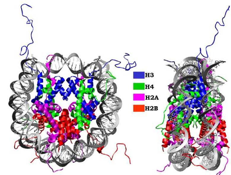
Fig. 1.11. Nucleosome structure in two projections.
Images generated using UCSF Chimera, Protein Data Bank structure code 1KX5
In chromatin, all DNA forms nucleosomes with an average density of one nucleosome per 200 base pairs, with adjacent nucleosomes connected by internucleosomal linker regions. The nucleosomal DNA together with the linker region constitutes the so-called nucleosome repeat, the length of which varies both along the polynucleotide chain and depending on the functional state, Cell type, etc. The distribution pattern of nucleosomes along genomic DNA is of great functional importance: naturally, linker DNA is more accessible to external regulatory influences.
As shown in Fig. 1.11, the disordered terminal regions of Histones (tails) extend beyond the nucleosome boundary. Due to their structural lability, they participate in organizing chromatin at the supranucleosomal level and also serve as a crucial platform for binding various proteins. Such Protein Interactions have significant functional consequences for Gene activity regulation and depend on post-translational modifications of the tails—the attachment of specific chemical groups to particular amino acid residues, such as Acetylation, phosphorylation, methylation, and others. The relationship between the modification pattern and the set of proteins that recognize a specific distribution of modified groups on the tails is referred to as the histone code.
The linker regions connecting adjacent nucleosomes continue the trajectory of the nucleosomal DNA in a straight line, resulting in a zig-zag arrangement of nucleosomes within the polynucleotide fiber (Fig. 1.12). Through interaction with DNA, the disordered tails of core molecules and the fifth histone—histone H1 (one molecule per nucleosome)—cause the polynucleotide zig-zag to condense into a 30 nm fiber, representing the second level of chromatin compaction.
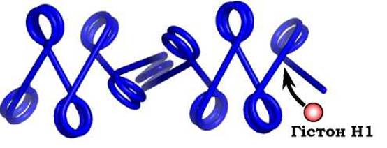
Fig. 1.12. Zig-zag configuration of the polynucleosomal fiber
The 30 nm fiber is the primary form of chromatin during interphase, the period between cell divisions. However, chromatin exhibits significant heterogeneity in its degree of Condensation. On the one hand, the activation of specific chromatin regions requires fiber decondensation. On the other hand, in repressed regions, the chromatin fiber may either be additionally stabilized in a compact state or undergo higher-order compaction. The fraction of chromatin that maintains an increased state of compaction throughout interphase is termed heterochromatin (the remaining chromatin, where METABOLISM/31.html">Transcription activation can potentially occur, is designated as euchromatin). Heterochromatin formation occurs primarily in regions containing repeats—at centromeres, telomeres, adjacent pericentric and subtelomeric regions, and zones concentrated with mobile elements.
At the next level of structural organization within the Cell Nucleus, the chromatin fiber forms loops whose ends are rigidly anchored to the skeletal protein structures of The Nucleus, known as the nuclear matrix (Fig. 1.13). A single loop containing between 20 and 200 kb of DNA (encompassing one or several genes) is often viewed as an essential element in The regulation of Transcription and Replication processes. DNA segments ranging from 300 to 1,000 base pairs—matrix-associated regions (MARs)—interact with the matrix proteins.
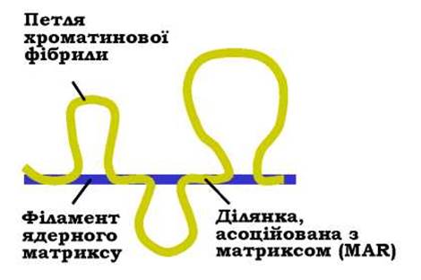
Fig. 1.13. Schematic representation of the loop organization of chromatin
The nuclear matrix is a system of protein filaments that forms the structural framework of the nucleus. At the nuclear periphery lies a specialized portion of the matrix associated with the inner nuclear membrane, known as the nuclear lamina. Filaments of the internal nuclear matrix extend from the lamina into the nuclear interior. A substantial portion of heterochromatin, including chromosome centromeres and telomeres, interacts with the lamina. The euchromatic portion of the chromosome "hangs" into the nuclear interior, where chromatin loops are anchored to the internal matrix. As a result, each chromosome occupies a distinct region within the nuclear volume, known as a chromosome territory.
Last update: 11/08/2026
Editorial and Educational Adaptation: This material has been compiled based on the primary/original source text. The project team performed an editorial review, corrected technical inaccuracies, structured sections, and adapted the content for an educational format.
What was processed:
- elimination of formatting defects (OCR errors, structural breaks, corrupted characters);
- editorial organization of content;
- standardization of terminology in accordance with academic sources;
- verification of factual statements against the original source text.
All mentions of the author, publication year, and origin of the primary text have been preserved in accordance with the source.