Molecular Biology: Protein Structure and Function - Stepanov V.M. 2005
Post-translational protein modification
Lipoproteins
Strictly speaking, lipoproteins are defined as Proteins containing covalently bound Lipids. Traditionally, however, the term also encompasses proteins involved in lipid transport that form non-covalent complexes with lipids, such as Blood lipoproteins. Here, we will focus exclusively on lipoproteins that fit the strict definition.
11.7.1. Lipoproteins with C-Terminal Glycolipids
Roughly 30 proteins are known to anchor into animal Cell membranes via hydrophobic interactions between Membrane Lipids and a uniquely structured C-terminal glycolipid domain (often referred to as "PIG-tailed" proteins, derived from Phospho-Inositol-Glycan). Examples include alkaline phosphatase, the globular form of acetylcholinesterase, the highly variable surface glycoprotein of trypanosomes, and neural Cell Adhesion molecules.
Following The Biosynthesis of the polypeptide chain, the C-terminal segment of such a protein—typically comprising 17 to 31 predominantly hydrophobic amino acid residues—is cleaved off and replaced by the following Structure:
Class="center">protein—СО—NH—СН2СН2О—glycan—glucosamine—phosphoinositol—diacylglycerol ethanolamine
The glycolipid moiety varies somewhat among different proteins in The structure of its glycan (which contains galactose) and in the COMPOSITION OF THE Fatty acids within its diacylglycerol unit, with palmitic and myristic acids among those identified.
In certain cases, the entire glycolipid moiety can be cleaved off by Treatment with proteinase. The bond between glucosamine and phosphoinositol is cleaved by nitrous acid. Particularly characteristic is the release of the lipid by the action of phospholipases D or C, which hydrolyze the phosphodiester bonds between inositol and phosphatidic acid, or between phosphoinositol and diacylglycerol, respectively, thereby detaching the protein from the membrane. Evidence suggests that phospholipase C plays a role in The Mechanism of Insulin action by cleaving the glycolipid fragment from lipoprotein lipase. As a result, the enzyme loses its attachment to the outer membrane of fat Cells, which directly affects triglyceride Catabolism. In any case, it is clear that the glycosylphosphatidylinositol anchor in these proteins is far from being an inert tether.
11.7.2. Lipoproteins with N-Terminal Lipid Moieties
Two main pathways are known for attaching a lipid moiety to the amino terminus of a protein. The simplest is N-myristoylation, which involves The transfer of a myristic acid residue—–СН3(СН2)12СО— from myristoyl-CoA to the α-amino group of the N-terminal Glycine residue of the protein. This hydrophobic myristyl group helps anchor the protein within The Lipid Bilayer or potentially binds to a specialized receptor protein.
Certain microbial proteins, such as the β-lactamase of Bacillus cereus, interact with membrane lipids via a distinct lipid anchor. This anchor is formed at the amino terminus of the polypeptide chain after the Cleavage of a signal peptide that initially guides the translocation of the protein across the membrane. In these proteins, the amino-terminal position is invariably occupied by a Cysteine residue. A diacylglycerol moiety is attached to its sulfhydryl group via a thioether bond, and an additional fatty acid residue subsequently acylates the α-amino group of the cysteine, yielding the following structure:

The concentration of three fatty acid residues at the N-terminal cysteine renders this region of the polypeptide chain highly hydrophobic, which enables its interaction with membrane lipids. This entire assembly securely anchors the protein to the membrane, while the protein globule itself remains fully immersed in the aqueous environment adjacent to the membrane, such as the bacterial periplasmic space.
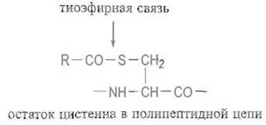
These are not the only structural types of lipoproteins. Notably, the Rhesus factor protein (Rh protein) located in the human erythrocyte membrane appears to contain several (up to four) palmitic acid residues linked via thioether bonds to cysteine sulfhydryl groups, rendering it exceptionally hydrophobic:
11.7.3. Prenylated Proteins
S-prenylation involves the attachment of Isoprenoids to the sulfur atom of a cysteine residue located near the C-terminus of a polypeptide chain. This modification affects about 100 proteins, notably the Ras protein (see Chapter 12). The primary precursor for prenyl groups is isopentenyl pyrophosphate, which isomerizes via a double-bond shift into dimethylallyl pyrophosphate:
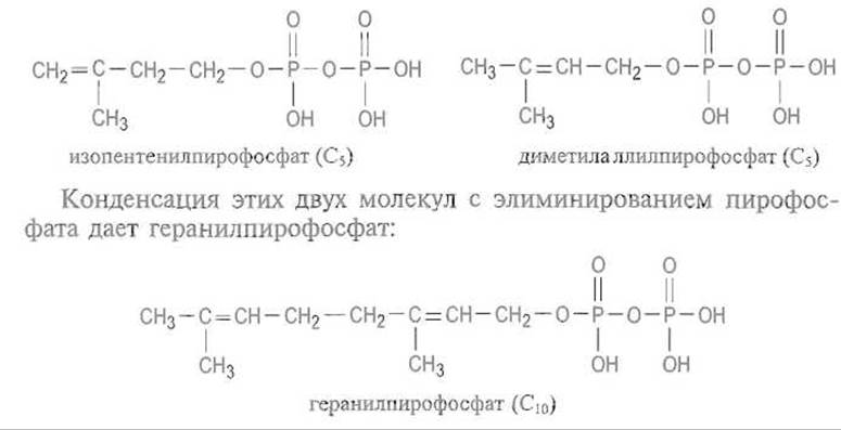
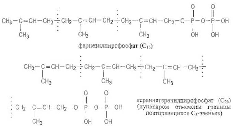
The carbon Skeleton is then extended through the sequential addition of isopentenyl pyrophosphate via the same mechanism, incorporating a C5 unit at each step. This process yields farnesyl pyrophosphate (C15)—which also serves as a precursor for Steroids and carotenoids—and geranylgeranyl pyrophosphate (C20):
The farnesyl or geranylgeranyl groups are transferred (with the release of pyrophosphate) to the sulfur atom of a cysteine residue located near the C-terminus within a C—A—A—X motif (the so-called CAAX box), where C represents cysteine, A denotes aliphatic amino acid residues, and X is the C-terminal amino acid that dictates which of the two prenylation pathways takes place. If X is Serine, Methionine, or glutamine, a farnesyl group (C15) is attached; if X is leucine, a geranylgeranyl group (C20) is added. Protein prenylation proceeds through a specific sequence of reactions. Prenyltransferase transfers the prenyl moiety from the corresponding prenyl pyrophosphate to the SH group of the cysteine residue within the CAAX box at the protein's C-terminus. Next, a specific peptidase cleaves off the now-obsolete A—A—X signaling sequence. Finally, a methyltransferase transfers a methyl group from S-adenosylmethionine to the carboxyl group of the prenylated cysteine, converting it into a methyl ester:
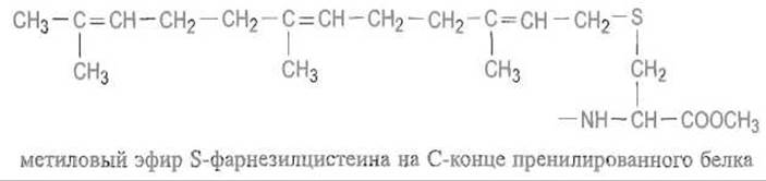
Clearly, attaching a bulky hydrophobic prenyl group to a protein promotes its integration into the cytoplasmic membrane. Furthermore, the type of prenyl group dictates the protein's orientation within the membrane: farnesylated proteins typically insert into the outer leaflet, whereas geranylgeranylated proteins embed into the inner leaflet facing the Cytoplasm. Naturally, this rule can have exceptions, and other structural elements likely assist in targeting prenylated proteins. Thus, prenylation is a key process governing protein localization within biological structures—their topogenesis.
Last update: 13/08/2026
Editorial and Educational Adaptation: This material has been compiled based on the primary/original source text. The project team performed an editorial review, corrected technical inaccuracies, structured sections, and adapted the content for an educational format.
What was processed:
- elimination of formatting defects (OCR errors, structural breaks, corrupted characters);
- editorial organization of content;
- standardization of terminology in accordance with academic sources;
- verification of factual statements against the original source text.
All mentions of the author, publication year, and origin of the primary text have been preserved in accordance with the source.