LEHNINGER PRINCIPLES OF BIOCHEMISTRY - VOL. 2. BIOENERGETICS AND METABOLISM - 2014
PART II. BIOENERGETICS AND METABOLISM
22. BIOSYNTHESIS OF AMINO ACIDS, NUCLEOTIDES, AND RELATED MOLECULES
22.4. Nucleotide Biosynthesis and Degradation
As described in Chapter 8 (Vol. 1), NUCLEOTIDES play a variety of crucial roles in The Cell. They serve as precursors for DNA and RNA. They are essential for energy transfer—primarily as ATP, and to some extent GTP. They are components of Cofactors such as NAD, FAD, S-adenosylmethionine, and coenzyme A, as well as activated biosynthetic intermediates like UDP-glucose and CDP-diacylglycerol. Some nucleotides, such as cAMP and cGMP, function as second messengers.
Cells obtain nucleotides via two pathways: de novo synthesis and the salvage pathway. De novo nucleotide synthesis begins with metabolic precursors: Amino Acids, ribose 5-phosphate, CO2, and NH3. The salvage pathway reuses free bases and nucleotides derived from The breakdown of Nucleic Acids. Both biosynthetic pathways are vital for cellular METABOLISM and are discussed later in this chapter.
The de novo Synthesis of Purines and Pyrimidines is remarkably similar across virtually All living organisms. Notably, guanine, adenine, thymine, cytidine, and uracil are not intermediates of this pathway; that is, the cell does not synthesize the bases first and then attach them to ribose, as one might expect. Instead, the purine ring is "built" directly by adding one or several atoms at a time to the ribose moiety throughout the biosynthetic cycle. The pyrimidine ring is synthesized as orotate, which is then attached to ribose phosphate and subsequently converted into the common pyrimidine nucleotides required for nucleic acid synthesis. Free bases do not participate in de novo synthesis; they appear only as intermediates in certain Salvage Pathways.
Several key precursors are involved in the de novo synthesis of both purines and pyrimidines. Phosphoribosyl pyrophosphate (PRPP) plays a crucial role in the synthesis of both types of nucleotides, and unlike The Biosynthesis of Tryptophan and Histidine, the ribose molecule is ultimately retained in the nucleotide, as shown previously. In nucleotide synthesis, the primary amino acid Donors are Glycine for purines and aspartate for pyrimidines. Glutamine serves as the most important source of amino groups, acting at five different steps in the de novo pathways. Aspartate is also utilized as an amino group donor in two reactions during purine synthesis.
Two features of nucleotide synthesis are particularly noteworthy. First, cellular Enzymes—especially those involved in de novo purine synthesis—are organized into large multienzyme complexes, a recurring theme we will frequently encounter in metabolism. Second, the cellular pool of nucleotides (all except ATP) is quite small, accounting for probably ~1% or less of the amount required for cellular DNA Synthesis. Consequently, cells must continue to synthesize nucleotides even while nucleic acids are being formed, and in some cases, nucleotide synthesis can be the rate-limiting factor for DNA Replication and Transcription. Because these biological processes are critical for dividing cells, agents that inhibit nucleotide synthesis have earned a well-established place in modern medicine.
Here we will examine the biosynthetic pathways of purines and pyrimidines, their regulation, The formation of deoxynucleotides, and the degradation of purines and pyrimidines into uric acid and urea. We will conclude our Structure/133.html">Discussion by examining chemotherapeutic agents that interfere with nucleotide synthesis.
De Novo Nucleotide Synthesis Begins with PRPP
The two main purine nucleotides incorporated into nucleic acids, adenosine 5'-monophosphate (AMP; adenylate) and guanosine 5'-monophosphate (GMP; guanylate), contain the purine bases adenine and guanine. Figure 22-32 illustrates the Water/144.html">Origin of the carbon and nitrogen atoms in the purine ring, as determined by John Buchanan in avian experiments using isotopic tracers. The detailed pathway of Purine Biosynthesis was elucidated primarily by Buchanan and J. Robert Greenberg in the 1950s.
Class="center">John Buchanan

Figure 22-32. Origins of the atoms of the purine ring. Data were obtained from experiments using precursors radiolabeled with 14C or 15N. Formate is provided as N10-formyltetrahydrofolate.
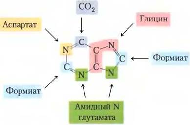
In the first step of this pathway, an amino group from glutamate is transferred to the C-1 atom of PRPP (Fig. 22-33). The resulting 5-phosphoribosylamine is highly unstable, with a half-life of 30 s at pH 7.5. The purine ring is subsequently built upon this structure. The pathway described here is identical in all organisms, except for a single step that differs in higher eukaryotes, as discussed below.
The second step involves The addition of the three carbons of glycine (Fig. 22-33, step (2)). This Condensation reaction requires ATP to activate the carboxyl group of glycine (forming an acyl phosphate). Next, a formyl group from N10-formyltetrahydrofolate is added to the amino group of glycine (step (3)), followed by the addition of a nitrogen atom from glutamine (step (4)). A dehydration reaction and ring closure then yield the five-membered imidazole ring of the purine Nucleus, forming 5-aminoimidazole ribonucleotide (AIR; step (5)).
Figure 22-33. De novo Synthesis of purine nucleotides: Formation of the purine ring of inosinate (IMP). Each atom newly incorporated into the purine ring is color-coded to match Figure 22-32. After step (2), R represents the 5-phospho-D-ribosyl group upon which the purine ring is constructed. The formation of 5-phosphoribosylamine (step (1)) is the committed step of the purine synthesis pathway. Note that the AICAR product of step (9) also appears in histidine biosynthesis (see Fig. 22-20, step (5)). To simplify Enzyme Nomenclature, many intermediates are referred to by Abbreviations. Step (6a) occurs in higher eukaryotes.

At this stage, three of the six atoms required to build the second ring of the purine structure are in place. To complete this process, a carboxyl group is added first (step (6)). This carboxylation is somewhat unusual because it utilizes bicarbonate—which is present in sufficient concentrations in the aqueous cellular environment—rather than biotin. Through a rearrangement, the carboxylate is transferred from the exocyclic amino group to the fourth atom of the imidazole ring (step (7)). Steps (6) and (7) occur in Bacteria and Fungi. In higher eukaryotes, including humans, the 5-aminoimidazole ribonucleotide formed in step (5) is directly carboxylated to yield carboxyaminoimidazole ribonucleotide in a single step rather than two (step (6a)). The enzyme catalyzing this reaction is aminoimidazole ribonucleotide carboxylase (AIR carboxylase).
In the two subsequent steps ((8) and (9)), aspartate serves as an amino group donor: an amide bond is formed, accompanied by the removal of the carbon Skeleton of aspartate (as fumarate). Recall that aspartate plays an analogous role in two steps of The Urea Cycle (see Fig. 18-10). The final carbon atom is contributed by N10-formyltetrahydrofolate (step (10)), and ring closure of the second ring completes the adjacent second ring of the purine nucleus (step (11)). This produces the first intermediate, inosinate (IMP), which features a fully assembled purine ring.
As in the tryptophan and histidine biosynthetic pathways, the enzymes involved in IMP synthesis are organized within the cell into a massive multienzyme complex. This is further evidenced by the existence of single multifunctional Polypeptides that catalyze non-sequential steps of the pathway. In Eukaryotic cells, ranging from Yeast to fruit flies and chickens, steps (1), (3), and (5) (Fig. 22-33) are catalyzed by a multifunctional protein. Additionally, another multifunctional protein catalyzes steps (10) and (11). In humans, a multifunctional enzyme combines AIR synthase and SAICAR synthetase activities (steps (6a) and (8)). In bacteria, these activities reside in separate Proteins, but their cells can form a massive non-covalently linked complex. The channeling of reaction intermediates directly from one enzyme to another, enabled by such a complex, is likely especially important for unstable intermediates such as 5-phosphoribosylamine.
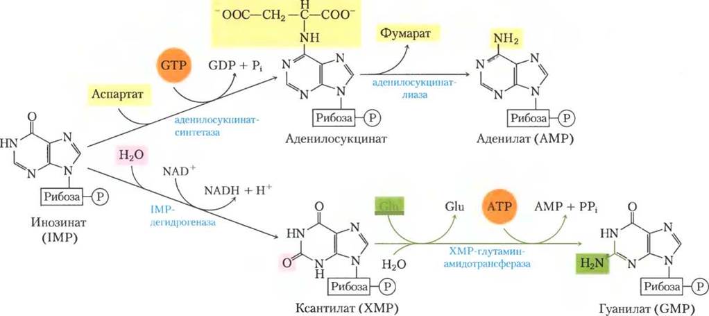
The conversion of inosinate into adenylate requires the incorporation of an amino group derived from aspartate (Fig. 22-34); this proceeds in two steps analogous to those involved in incorporating the N-1 atom into the purine ring (Fig. 22-33, steps (8) and (9)). The main difference is that GTP, rather than ATP, is used as the high-energy phosphate source for the synthesis of adenylosuccinate. Guanylate is formed via an NAD+-dependent oxidation of inosinate, accompanied by the Addition of an amino group derived from glutamine. In the final step, ATP is cleaved into AMP and PPi (Fig. 22-34).
Figure 22-34. Biosynthesis of AMP and GMP from IMP.
Nucleotide biosynthesis is regulated by negative feedback
The overall rate of de novo purine nucleotide synthesis and The ratio of the two end products, adenylate and guanylate, are coregulated by three main control mechanisms (Fig. 22-35). The first mechanism regulates the initial reaction unique to purine biosynthesis—The transfer of an amino group to PRPP to yield 5-phosphoribosylamine. This reaction is catalyzed by the allosteric enzyme glutamine-PRPP amidotransferase, which is inhibited by the end products IMP, AMP, and GMP. In this concerted inhibition, AMP and GMP act synergistically, enhancing each other's effect. Thus, whichever product accumulates in excess—AMP or GMP—the first step of biosynthesis from PRPP will always be partially inhibited.
Fig. 22-35. Regulation of adenine and guanine nucleotide biosynthesis in E. coli. Regulation of these pathways differs in other organisms.

The second control mechanism, operating at a later stage, relies on the fact that an excess of cellular GMP inhibits the conversion of xanthylate to inosinate by IMP dehydrogenase without affecting AMP formation (Fig. 22-35). Conversely, the accumulation of adenylate inhibits the synthesis of adenylosuccinate by adenylosuccinate synthetase without affecting GMP formation. The third mechanism relies on the requirement for GTP during the conversion of IMP to AMP (Fig. 22-34), whereas ATP is required for the conversion of IMP to GMP—a reciprocal regulation that maintains balance in the Synthesis of the two ribonucleotides.
The final control mechanism is the inhibition of PRPP synthesis via the Allosteric Regulation of ribose phosphate pyrophosphokinase. In addition to metabolites from other pathways originating from PRPP, this enzyme is inhibited by ADP and GDP.
Pyrimidine nucleotides are formed from aspartate, PRPP, and carbamoyl phosphate
Common pyrimidine nucleotides include cytidine 5'-monophosphate (CMP; cytidylate) and uridine 5'-monophosphate (UMP; uridylate), which contain the pyrimidines cytosine and uracil. The de novo BIOSYNTHESIS OF PYRIMIDINE nucleotides (Fig. 22-36) differs somewhat from that of purine nucleotides; the six-membered pyrimidine ring is assembled before being attached to ribose-5-phosphate. This reaction requires carbamoyl phosphate, another intermediate of the urea cycle (see Fig. 18-10). However, as noted in Chapter 18, the carbamoyl phosphate required for the urea cycle in animals is synthesized by carbamoyl phosphate synthetase I in the Mitochondria, whereas the carbamoyl phosphate involved in pyrimidine biosynthesis is produced in the Cytosol by a different enzyme form, carbamoyl phosphate synthetase II. In bacteria, a single enzyme provides the pool of carbamoyl phosphate for both Arginine and pyrimidine synthesis. The bacterial enzyme has three distinct active sites connected by a channel about 100 Å long (Fig. 22-37). Bacterial carbamoyl phosphate synthetase serves as a striking example of the channeling of unstable reaction intermediates between active sites.
Fig. 22-36. De novo synthesis of pyrimidine nucleotides: biosynthesis of UTP and CTP via orotidylic acid. The pyrimidine ring is formed from carbamoyl phosphate and aspartate. Orotate phosphoribosyltransferase then adds ribose-5-phosphate to the completed pyrimidine ring. The first step of this pathway (not shown here; see Fig. 18-11a) is the synthesis of carbamoyl phosphate from CO2 and NH4+, catalyzed in eukaryotes by carbamoyl phosphate synthetase II.
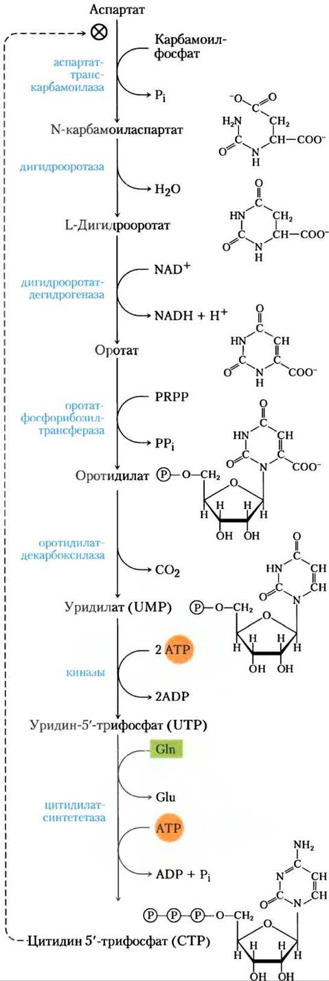
In the first committed step of the pyrimidine biosynthetic pathway, carbamoyl phosphate condenses with aspartate to form N-carbamoylaspartate (Fig. 22-36). This reaction is catalyzed by aspartate transcarbamoylase. In bacteria, this step is tightly regulated, and bacterial aspartate transcarbamoylase is one of the most thoroughly studied allosteric enzymes (see below). Dehydration of N-carbamoylaspartate, catalyzed by dihydroorotase, closes the pyrimidine ring to yield L-dihydroorotate. This compound is oxidized to the pyrimidine derivative orotate in a reaction where NAD+ serves as the ultimate electron acceptor. In eukaryotes, the first three enzymes of this pathway—carbamoyl phosphate synthetase II, aspartate transcarbamoylase, and dihydroorotase—are combined in a single multifunctional protein, CAD. This protein contains three identical polypeptide chains (each with an Mr of 230,000), each bearing active sites for all three reactions. Consequently, large multienzyme complexes may be quite common in this pathway.
Fig. 22-37. Intermediate channeling in bacterial carbamoyl phosphate synthetase (based on PDB ID 1M6V). The reaction catalyzed by this enzyme is shown in Fig. 18-11a. The large and small subunits are colored gray and blue, respectively; the channel between active sites (nearly 100 Å long) is represented by a yellow mesh. A glutamine molecule (green) binds to the small subunit, and in the glutamine amidotransferase reaction, its amide nitrogen is cleaved off as NH4+. The NH4+ enters the channel leading to the second Active Site, where it combines with bicarbonate in an ATP-dependent reaction (bound ADP is shown in blue). The carbamate then re-enters the channel to reach the third active site, where it is phosphorylated to form carbamoyl phosphate (bound ADP is shown in red).

Following the formation of orotate, a ribose-5-phosphate side chain, again donated by PRPP, is attached to it (Fig. 22-36). The resulting orotidylate is then decarboxylated to uridylate, which is subsequently phosphorylated to UTP. CTP is synthesized from UTP by the action of cytidylate synthetase via an acyl phosphate intermediate (consuming one molecule of ATP). Glutamine typically serves as the nitrogen donor, although in many cases cytidylate synthetases can utilize NH4+ directly.
Pyrimidine Nucleotide Biosynthesis is regulated by negative feedback
The rate of pyrimidine nucleotide synthesis in bacteria is regulated primarily via aspartate transcarbamoylase, which catalyzes the first step of the pathway and is inhibited by CTP, the end product of this reaction sequence (Fig. 22-36). The bacterial aspartate transcarbamoylase molecule consists of six catalytic subunits (see Fig. 6-32, vol. 1). The catalytic subunits bind substrate molecules, whereas the Regulatory Subunits bind the allosteric inhibitor CTP. Like its subunits, the aspartate transcarbamoylase molecule exists in two distinct Conformations: active and inactive. When CTP is not bound to the regulatory subunits, the enzyme is maximally active. As CTP accumulates, it binds to the regulatory subunits and induces a conformational change. This change is transmitted to the catalytic subunits, which then also adopt the inactive conformation. ATP counteracts these CTP-induced changes. Figure 22-38 illustrates The Effect of Allosteric regulators on aspartate transcarbamoylase activity.
Fig. 22-38. Allosteric regulation of aspartate transcarbamoylase by CTP and ATP. The addition of 0.8 mM CTP, an allosteric inhibitor of aspartate transcarbamoylase, increases the K0.5 for aspartate (lower curve) and the rate of aspartate conversion to N-carbamoylaspartate. The Introduction of 0.6 mM ATP almost completely reverses this effect (middle curve).

Nucleoside monophosphates are converted to nucleoside triphosphates
In nucleotide biosynthesis, monophosphates are generally converted to nucleoside triphosphates via pathways common to all cells. The phosphorylation of AMP to ADP is carried out by adenylate kinase via the reaction
ATP + AMP ⇄ 2ADP
The resulting ADP is then phosphorylated to ATP by glycolytic enzymes during Oxidative Phosphorylation.
ATP also participates in the Formation of other nucleoside diphosphates through the action of nucleoside monophosphate Kinases. This class of enzymes is generally specific for a particular base but relatively nonspecific with respect to the sugar moiety (ribose or deoxyribose), catalyzing the reaction:
ATP + NMP ⇄ ADP + NDP
Efficient systems for the rephosphorylation of ADP to ATP in the cell shift the equilibrium of this reaction toward product formation.
Nucleoside diphosphates are converted into triphosphates by the action of the widespread enzyme nucleoside diphosphate kinase, which catalyzes the reaction
NTPD + NDPA ⇄ NDPD + NTPA
This enzyme is unusual in that it lacks Specificity for either the base (purine or pyrimidine) or the sugar (ribose or deoxyribose). This lack of specificity also applies to both the phosphate acceptor (A) and donor (D), although ATP almost invariably serves as the donor (NTPD) because under aerobic conditions its concentration in the cell is much higher than that of other nucleoside triphosphates.
Ribonucleotides serve as precursors of deoxyribonucleotides
Deoxyribonucleotides, the Building Blocks of DNA, are formed from the corresponding ribonucleotides by the direct reduction of the 2'-carbon atom of D-ribose to yield the 2'-deoxy derivative. For example, adenosine diphosphate (ADP) is reduced to 2'-deoxyadenosine diphosphate (dADP), and GDP to dGDP. This reaction is somewhat unusual because the carbon atom is not activated prior to reduction; no other analogous reactions are currently known. The reaction is catalyzed by Ribonucleotide reductase, which is best characterized in E. coli; its substrates are nucleoside diphosphates.
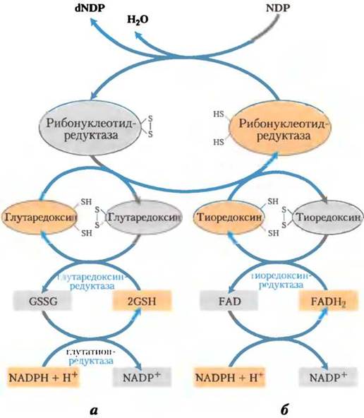
The reduction of certain D-ribose residues in ribonucleoside diphosphate to 2'-deoxy-D-ribose involves two hydrogen atoms supplied by NADPH via an intermediate hydrogen-carrier protein, thioredoxin. This widespread protein performs a similar function in oxidation-reduction reactions during Photosynthesis (see Fig. 20-19) and other processes. Thioredoxin contains paired -SH groups that transfer hydrogen atoms from NADPH to the ribonucleoside diphosphate. Its oxidized (disulfide) form is reduced by NADPH in a reaction catalyzed by thioredoxin reductase (Fig. 22-39), and is then used by ribonucleotide reductase to reduce nucleoside diphosphates (NDP) to deoxyribonucleoside diphosphates (dNDP). Another source of reducing equivalents for ribonucleotide reductase is Glutathione (GSH). Glutathione acts as a reductant for glutaredoxin, a protein related to thioredoxin, which further passes the reducing potential on to ribonucleotide reductase (Fig. 22-39).
Fig. 22-39. Reduction of ribonucleotides to deoxyribonucleotides by ribonucleotide reductase. Electrons are transferred (blue arrows) to the enzyme from NADPH via (a) glutaredoxin or (b) thioredoxin. The sulfide groups of glutaredoxin reductase belong to bound glutathione (GSH; GSSG is oxidized glutathione). Note that thioredoxin reductase is a flavoenzyme in which FAD serves as the prosthetic group.
Ribonucleotide reductase is remarkable because its catalytic mechanism has been described in detail as an example of a free-radical reaction in biochemical processes—an event once considered quite rare in living systems. In E. coli and most eukaryotes, this enzyme is a dimer whose subunits are designated R1 and R2 (Fig. 22-40). The R1 subunit contains Two Types of regulatory sites, as described below. The two active sites of the enzyme are formed by the interaction of the R1 and R2 subunits. In each active site, the two sulfhydryl groups required for enzyme activity belong to the R1 subunit, whereas the stable Tyrosine radical belongs to the R2 subunit. The R2 subunit also contains a cofactor with two Fe3+ iron ions that help generate and stabilize the tyrosyl radicals (Fig. 22-40). The tyrosine radical is located too far from the active site to interact with it directly; however, it induces the formation of another radical at the active site, which actually Functions in catalysis. The most likely mechanism of the ribonucleotide reductase reaction is shown in Fig. 22-41. In E. coli, the most probable sources of the reducing equivalents required for this reaction are thioredoxin and glutaredoxin (see below).
Fig. 22-40. Ribonucleotide reductase. (a) Subunit structure. The function of the two regulatory sites is shown in Fig. 22-42. Each active site contains two thiol groups and one -XH group that can be converted into a radical; this is most likely the -SH group of Cys439, which functions as a thiol radical. (b) The R2 subunit of E. coli ribonucleotide reductase (PDB 1D1PFR). The tyrosine residue acting as the tyrosyl radical is shown in red; the binuclear iron center is shown in orange. (c) The function of the tyrosyl radical is to generate the active-site -X• radical required for the reaction whose mechanism is depicted in Fig. 22-41.

Three classes of ribonucleotide reductases have been discovered. Their reaction mechanisms are generally similar to the scheme shown in Fig. 22-41, with differences lying in The Nature of the active-site radical and the cofactor required for its generation. The E. coli enzyme (class I) requires oxygen to "repair" the "quenched" tyrosine radical, and therefore this enzyme functions only under aerobic conditions. Class II enzymes, found in other microorganisms, contain 5'-deoxyadenosylcobalamin (see Box 17-2) instead of a binuclear iron center. Class III enzymes have adapted to function under anaerobic conditions. When E. coli grows anaerobically, it synthesizes an independent class III enzyme; this enzyme contains an iron-sulfur cluster (whose structure differs from the binuclear iron center of class I enzymes) and utilizes NADPH and S-adenosylmethionine in its catalytic cycle. It prefers nucleoside triphosphates over nucleoside diphosphates as substrates. The Emergence of various classes of ribonucleotide reductases, required for The production of DNA precursors under diverse environmental conditions, highlights the crucial importance of this reaction in Nucleotide Metabolism.
Fig. 22-41. Reaction mechanism. Proposed Catalytic Mechanism of ribonucleotide reductase. In E. coli and most eukaryotes, the active thiol groups of the enzyme reside on the R1 subunit, and the active-site -X• radical resides on the R2 subunit; in E. coli, this is likely the sulfhydryl radical of Cys439 (see Fig. 22-40).

The regulation of E. coli ribonucleotide reductase is somewhat unusual: the binding of effector molecules regulates not only its overall activity but also its substrate specificity. Each R1 subunit possesses two types of regulatory sites (Fig. 22-40). One site affects The activity of the entire enzyme complex and binds either ATP, which activates the enzyme, or dATP, which inactivates it. The second site alters substrate specificity in response to the modulator molecule (ATP, dATP, dTTP, or dGTP) bound to it (Fig. 22-42). When ATP or dATP is bound, the reduction of UDP and CDP is favored. When dTTP or dGTP is bound, the reduction of GDP or ADP is stimulated. This regulation ensures a proper balance among the precursor pools for DNA synthesis. ATP also acts as the primary activator of ribonucleotide biosynthesis and reduction. The presence of small amounts of dATP enhances the reduction of pyrimidine nucleotides. Excessive production of pyrimidine dNTPs leads to high levels of dTTP, which alters the substrate Specificity of the reduction reaction to favor GDP. In turn, high concentrations of dGDP affect the reaction equilibrium by shifting it toward the reduction of ADP, whereas high concentrations of dATP "shut down" the enzyme altogether. These effectors are thought to induce several distinct conformational shifts that drive the changes in specificity.
Fig. 22-42. Regulation of ribonucleotide reductase by deoxynucleoside triphosphates. Overall enzymatic activity is regulated by the binding of modulators to the primary regulatory site (left). Substrate specificity of the enzyme is regulated by the Nature of the effector molecule bound to the second type of regulatory site (right). The inhibition or activation of enzymatic activity by four different substrates is shown. The pathway from dUDP to dTTP is described later (see Figs. 22-43 and 22-44).

Thymidylate is formed from dCDP and dUMP
DNA contains thymine instead of uracil, and the de novo thymine synthesis pathway involves exclusively deoxyribonucleotides. The direct precursor of thymidylate (dTMP) is dUMP. In bacteria, the pathway leading to dUMP begins with the formation of dUTP, which is produced either by the deamination of dCTP or by the phosphorylation of dUDP (Fig. 22-43). dUTP is converted to dUMP by the action of dUTPase. This latter reaction must be highly efficient to maintain a low dUTP pool and thereby prevent the incorporation of uridylate into DNA.
Fig. 22-43. Biosynthesis of thymidylate (dTMP). Pathways initiating with the reaction catalyzed by ribonucleotide reductase are shown. The thymidylate synthase reaction is detailed in Fig. 22-44.

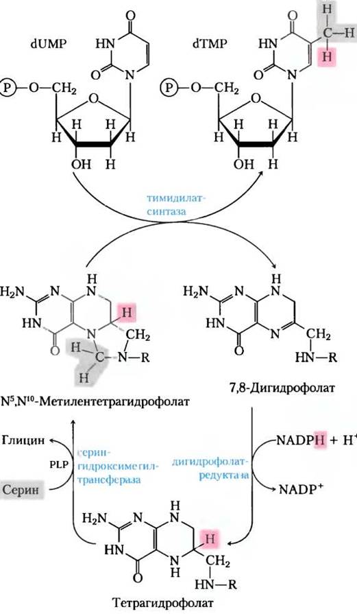
The conversion of dUMP to dTMP is catalyzed by thymidylate synthase. A one-carbon unit at the oxidation level of a hydroxymethyl group (-CH2OH) is transferred from N5, N10-methylenetetrahydrofolate to dUMP (see Fig. 18-17) and subsequently reduced to a methyl group (Fig. 22-44). This reduction is "funded" by The oxidation of tetrahydrofolate to dihydrofolate, which is quite unusual for tetrahydrofolate-dependent reactions (The Mechanism of this reaction is shown in Fig. 22-50). Dihydrofolate is reduced back to tetrahydrofolate by Dihydrofolate Reductase in a regeneration reaction that is almost universally present in processes involving tetrahydrofolate. In plants and at least one protozoan, thymidylate synthase and dihydrofolate reductase exist as a single bifunctional protein.
Fig. 22-44. Conversion of dUMP to dTMP by thymidylate synthase and dihydrofolate reductase. Serine hydroxymethyltransferase is required to generate the methylene form of N5, N10-methylenetetrahydrofolate. During dTMP synthesis, three hydrogen atoms of the methyl group originate from N5, N10-methylenetetrahydrofolate (pink and gray).
About 10% of the entire human population (including nearly 50% of impoverished populations) suffer from Folic acid deficiency. Significant folate deficiency can lead to cardiovascular disease, malignancies, and various neurological impairments. Several of these symptoms are linked to impaired thymidylate synthesis, which results in uracil being erroneously incorporated into DNA. Uracil in DNA is recognized and excised by the repair system (see Ch. 25, Vol. 3). High levels of uracil in DNA cause strand breaks, disrupting the normal function and regulation of nuclear DNA, which ultimately promotes Heart disease, neurological disorders, and Cancer. ■
The breakdown of purines and pyrimidines yields uric acid and urea
Purine nucleotides are degraded via a pathway in which they lose their phosphates through the action of 5'-nucleotidase (Fig. 22-45). Adenylate is converted to adenosine, which is deaminated to form inosine by adenosine deaminase, and inosine is hydrolyzed to hypoxanthine (its purine base) and D-ribose. Hypoxanthine is sequentially oxidized to xanthine and then to uric acid by xanthine oxidase, a flavoprotein containing a molybdenum atom and four iron-sulfur centers as prosthetic groups. Molecular oxygen serves as the electron acceptor in this complex reaction.
The Catabolism of GMP can also proceed with uric acid as the end product. GMP is first hydrolyzed to guanosine, which is subsequently cleaved to yield free guanine. The amino group is hydrolytically removed from guanine to form xanthine, which is then converted to uric acid by the action of xanthine oxidase (Fig. 22-45).
Figure 22-45. Catabolism of Purine nucleotides. Note that in primates, the majority of nitrogen is excreted via the urea cycle as urea (Ch. 18) rather than as uric acid, which is produced during nucleotide degradation. In fish, however, nitrogen is excreted primarily as NH+4 rather than as urea, the formation of which is shown here.

Uric acid is the final excreted product of purine catabolism in primates, birds, and certain other animals. In healthy adults, uric acid is excreted at a rate of about 0.6 g/day; a portion of this excreted product originates from dietary purines absorbed in the gut, while the rest derives from the purine bases of nucleic acids. In most mammals and other vertebrates, uric acid is further degraded to allantoin through the action of urate oxidase. In other organisms, this pathway proceeds further, as illustrated in Figure 22-45.
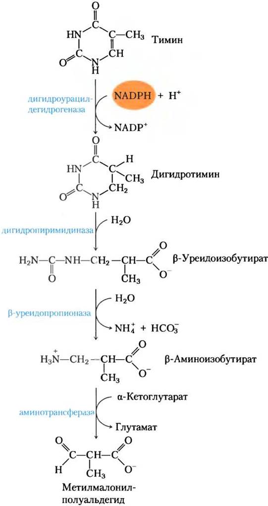
Pyrimidine breakdown pathways primarily lead to NH4+ and, consequently, to urea. Thymine, for example, is degraded to methylmalonyl semialdehyde (Fig. 22-46), an intermediate in valine catabolism. It is further metabolized via propionyl-CoA and methylmalonyl-CoA to succinyl-CoA (see Fig. 18-27).
Figure 22-46. Pyrimidine catabolism. Pathways of thymine degradation are shown. Methylmalonyl semialdehyde is subsequently broken down to succinyl-CoA.
Genetic Defects in purine metabolism have been identified in humans, some with severe clinical consequences. For instance, a deficiency in adenosine deaminase (ADA) leads to a severe form of immunodeficiency in which both T AND B lymphocytes fail to develop fully. The absence of ADA causes a 100-fold increase in dATP concentration, which strongly inhibits ribonucleotide reductase (Fig. 22-42). High levels of dATP result primarily in a shortage of other dNTPs in T lymphocytes. The causes of B-lymphocyte impairment are less understood. Due to ADA deficiency, the human immune system is severely compromised, and affected individuals can survive only in strictly sterile environments. ADA deficiency was one of the first Genetic Disorders targeted for Human Gene Therapy trials (see Box 9-2, Vol. 1). ■
Purine and pyrimidine bases are recycled via salvage pathways
Free purine and pyrimidine bases are constantly released within the cell during nucleotide catabolism. The bulk of these free purines is conserved and reused for nucleotide synthesis through a pathway much simpler than the de novo purine synthesis described earlier. One of the simplest nucleotide salvage pathways consists of a single reaction catalyzed by adenine phosphoribosyltransferase, in which free adenine reacts with PRPP to form the corresponding adenine nucleotide:
Adenine + PRPP —> AMP + PPi
Free guanine and hypoxanthine (the product of adenine deamination; Fig. 22-45) are salvaged via a similar pathway mediated by hypoxanthine-guanine phosphoribosyltransferase. Analogous salvage pathways exist for pyrimidine bases in microorganisms and, presumably, in mammals.
Genetic defects in hypoxanthine-guanine phosphoribosyltransferase, observed almost exclusively in males, give rise to a bizarre array of symptoms known as Lesch-Nyhan syndrome. Children with this genetic disorder, which becomes apparent around two years of age, often exhibit poor spatial orientation and mental retardation. Furthermore, they display extreme hostility and a manic drive toward self-mutilation: they bite and mangle their own fingers, toes, and Lips. Attempts to treat this syndrome were undertaken in the Cytology/cytology/16.html">Early stages of gene therapy development (see Box 9-2). ■
Excess uric acid causes Gout
The long-standing misconception that gout affects only aristocrats is false. Gout is a joint disorder caused by elevated levels of uric acid in the Blood and Tissues. Joint inflammation causes severe pain, and the deposition of sodium urate crystals can lead to symptoms characteristic of Arthritis.
The Kidneys are also affected, as excess uric acid accumulates within the renal tubules. Gout typically develops in men. Although the exact causes remain fully understood, the condition is frequently accompanied by inadequate urate excretion. In some cases, it may also stem from a genetic defect in one of the enzymes of purine metabolism.
Effective Treatment of gout requires a combination of dietary restrictions and pharmacotherapy. Foods particularly rich in nucleotides and nucleic acids, such as Liver or sweetbreads, must be excluded from the diet. Symptom relief is achieved through the administration of allopurinol (Fig. 22-47), a drug that inhibits xanthine oxidase, the enzyme catalyzing the conversion of purines to uric acid. Allopurinol acts as a substrate for xanthine oxidase, which converts it to oxypurinol (alloxanthine). Oxypurinol inactivates the reduced form of the enzyme by remaining tightly bound to its active site. Inhibition of xanthine oxidase leads to the accumulation of purine metabolites—xanthine and hypoxanthine—which are more soluble than uric acid and far less prone to crystalline deposition. Allopurinol was developed by Gertrude Elion and George Hitchings, who also created acyclovir (used in the treatment of AIDS patients) and other purine analogs applied in cancer Chemotherapy. ■
Gertrude Elion (1918–1999) and George Hitchings (1905–1998)

Figure 22-47. Allopurinol, an inhibitor of xanthine oxidase. Hypoxanthine is the natural substrate for xanthine oxidase. Merely minor modifications to hypoxanthine (highlighted in pink) yield allopurinol, a potent inhibitor of this enzyme. At the Active Site of xanthine oxidase, allopurinol is converted to oxypurinol, a potent competitive inhibitor that remains tightly bound to the reduced form of the enzyme.

Most enzymes targeted by chemotherapeutic agents belong to the nucleotide biosynthesis pathway
The growth of cancer cells is controlled differently from that of cells in most normal tissues. Because cancer cells require large amounts of nucleotides as DNA and RNA precursors, they are more sensitive to nucleotide biosynthesis inhibitors than normal cells. The range of important chemotherapeutic agents used in the treatment of cancer and other diseases that act as inhibitors of one or more enzymes in these pathways is continually expanding. Here we describe several well-studied Examples that illustrate effective treatment strategies and also help us understand how these enzymes function.
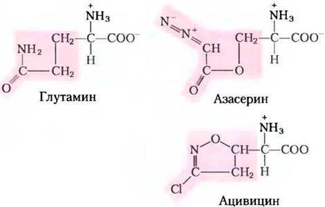
The first group of drugs includes compounds that inhibit glutamine amidotransferases. Recall that glutamine serves as a nitrogen donor in at least half a dozen different reactions in nucleotide biosynthesis. The glutamine-binding site and the mechanism by which NH4+ is released in these reactions are quite similar across many of these enzymes. Most of these enzymes are efficiently inhibited by glutamine analogs such as azaserine and acivicin (Fig. 22-48). Characterized by John Buchanan in the 1950s, azaserine was one of the earliest examples of mechanism-based enzyme inactivators (suicide inhibitors; see p. 298, Vol. 1, and Box 22–3). Acivicin also holds great promise as a chemotherapeutic agent.
Fig. 22-48. Azaserine and acivicin as inhibitors of glutamine amidotransferases. These glutamine analogs interfere with a variety of biosynthetic pathways involving Amino Acids and nucleotides.
Other valuable targets for pharmaceutical intervention are thymidylate synthase and dihydrofolate reductase—enzymes that operate exclusively in the cellular pathway for thymine synthesis (Fig. 22-49). Fluorouracil (5-fluorouracil) exerts an inhibitory effect specifically on thymidylate synthase and is a major chemotherapeutic drug. Fluorouracil itself does not inhibit the enzyme; rather, through cellular salvage pathways, it is converted to the deoxynucleoside monophosphate FdUMP, which binds to and inhibits the enzyme. Inhibition by FdUMP (Fig. 22-50) is a classic example of mechanism-based Enzyme inactivation. Another highly potent chemotherapeutic agent, methotrexate, inhibits dihydrofolate reductase. This folate analog acts as a competitive inhibitor, binding to dihydrofolate reductase with an affinity approximately 100 times greater than that for dihydrofolate. Aminopterin is a related compound that functions in a very similar manner.
Fig. 22-49. Thymidylate synthesis and folate metabolism as targets for chemotherapy. (a) During thymidylate synthesis, N5, N10-methylenetetrahydrofolate is converted to 7,8-dihydrofolate; N5, N10-methylenetetrahydrofolate is subsequently regenerated in a two-stage process (see Fig. 22-44). This cycle serves as a primary target for several pharmaceutical agents. (b) Fluorouracil and methotrexate are important chemotherapeutic drugs. Inside cells, fluorouracil (5-fluorouracil) is converted to FdUMP, which inhibits thymidylate synthase. Methotrexate, a structural analog of tetrahydrofolate, inhibits dihydrofolate reductase; the highlighted methyl and amino groups replace the carbonyl oxygen and hydrogen of folate, respectively (see Fig. 22-44). Another important folate analog, aminopterin, is identical to methotrexate except that it lacks the methyl group (highlighted). Trimethoprim is an inhibitor that binds tightly to bacterial dihydrofolate reductase and was developed as an antibiotic.

Fig. 22-50. Reaction mechanism: Conversion of dUMP to dTMP and its inhibition by FdUMP. Left: The conventional catalytic mechanism of thymidylate synthase. The nucleophilic sulfhydryl group of the enzyme participates in the reaction at step (1). The ring atoms of dUMP involved in the reaction are shown in red; B represents an amino acid side chain that acts as a base required to Abstract a proton in step (3). Hydrogen atoms derived from the methylene group of N5, N10-methylenetetrahydrofolate are shown in gray. An unusual feature of this reaction mechanism is a 1,3-hydride shift (step (3))—the transfer of a hydrogen ion (highlighted in pink) from the C-6 atom of H4-folate to the methyl group of thymidine, which oxidizes tetrahydrofolate to dihydrofolate. This very step (the hydride shift) is inhibited when FdUMP is used as the substrate (see below). Steps (1) and (2) proceed normally, but result in the formation of a stable covalent complex linking FdUMP, the enzyme, and tetrahydrofolate, thereby inactivating the enzyme. Mechanism of thymidylate synthase action.

The medical significance of nucleotide biosynthesis inhibitors extends well beyond cancer therapy. Potential targets include all rapidly proliferating cells (including bacteria and Protozoa). Trimethoprim, an antibiotic developed by Hitchings and Elion, binds to bacterial dihydrofolate reductase with an affinity roughly 100,000 times greater than to the mammalian enzyme. It is used to treat certain bacterial infections of the Urinary Tract and Middle ear. Parasitic protozoa, such as the trypanosomes responsible for African sleeping sickness (African trypanosomiasis), lack de novo nucleotide biosynthesis pathways, making these parasites exceptionally vulnerable to agents that block the salvage of environmental nucleotides. Allopurinol (Fig. 22-47) and several related purine analogs have shown promising results in treating African trypanosomiasis, providing further insight into parasite metabolism and enzyme function. ■
Summary of Section 22.4 BIOSYNTHESIS AND DEGRADATION of Nucleotides
■ The purine ring system is assembled stepwise, beginning with 5-phosphoribosylamine. All nitrogen atoms of the purine ring are contributed by the amino acids glutamine, glycine, and aspartate. The final two ring-closure steps yield the purine core.
■ Pyrimidines are synthesized from carbamoyl phosphate and aspartate, after which ribose 5-phosphate is attached to form pyrimidine ribonucleotides.
■ Nucleoside monophosphates are converted to their respective triphosphates via enzymatic phosphorylation reactions. Ribonucleotides are reduced to deoxyribonucleotides by ribonucleotide reductase, an enzyme exhibiting remarkably complex catalytic and regulatory properties. Thymine nucleotides are derived from dCDP and dUMP.
■ The End products of purine and pyrimidine catabolism are uric acid and urea.
■ Free purines can be reincorporated into nucleotides via salvage pathways. Genetic deficiencies in specific enzymes of these salvage pathways lead to severe disorders, such as Lesch-Nyhan syndrome and ADA deficiency.
■ The accumulation of uric acid crystals in the joints, potentially linked to an underlying metabolic defect, causes gout.
■ Enzymes of the nucleotide biosynthetic pathways serve as targets for numerous chemotherapeutic agents used in the treatment of cancer and other diseases.
Last update: 06/08/2026
Editorial and Educational Adaptation: This material has been compiled based on the primary/original source text. The project team performed an editorial review, corrected technical inaccuracies, structured sections, and adapted the content for an educational format.
What was processed:
- elimination of formatting defects (OCR errors, structural breaks, corrupted characters);
- editorial organization of content;
- standardization of terminology in accordance with academic sources;
- verification of factual statements against the original source text.
All mentions of the author, publication year, and origin of the primary text have been preserved in accordance with the source.