IMMUNOLOGY - Roitt I. - 2000
Chapter 16. Antiviral Immunity
T- AND B-CELL DEFENSIVE MECHANISMS
In the absence of T Cells, the host Organism is highly susceptible to viral attacks. For example, in athymic nude mice with a congenital lack of mature T cells, Herpes simplex virus (HSV) inoculated into the Skin causes a spreading lesion and ultimately penetrates the Central Nervous system, leading to the death of the animals. These mice can be protected by transferring HSV-specific T cells shortly after infection. The crucial importance of T and B cells in counteracting viral infections is discussed below.
Antibodies and Complement can limit viral spread and prevent reinfection
Antibodies can neutralize viral infectivity. If a virus succeeds in breaching innate immune barriers, it induces an adaptive (specific) Immune Response characterized by the appearance of cytotoxic T cells, helper T cells, and antiviral antibodies. Antibodies serve as the primary barrier preventing the spread of the virus to other Cells and Tissues, particularly its entry into the bloodstream. In mucosal lymphoid tissue, predominantly IgA Class antibodies are produced, which prevent reinfection.
Antibodies can be directed against any viral antigen synthesized within an infected Cell; however, containment of the infection is provided only by those antibodies specific to Glycoproteins expressed on the viral envelope or the membrane of infected cells. The mechanisms of humoral antiviral Immunity can vary. Thus, the mechanism by which viral particle infectivity is eliminated depends on their localization—extracellular or intracellular (Fig. 16.5). The in vivo action of the defensive mechanisms listed in Fig. 16.5 is evidenced by the fact that the injection of monoclonal virus-neutralizing antibodies very effectively suppresses viral Replication. The presence of virus-neutralizing antibodies in the bloodstream is also an important factor in preventing reinfection.
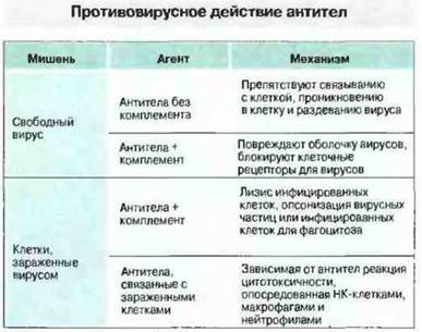
Fig. 16.5. Antibodies neutralize free Viral Particles or participate in the destruction of virus-infected cells.
Complement participates in the neutralization of certain extracellular Viruses. Complement is also capable of damaging the viral envelope, thereby effecting virolysis. Some viruses directly induce complement activation via the classical or alternative pathway. Nevertheless, complement is not considered a major protective factor against viruses, as deficiencies in complement system components are not associated with an increased predisposition to severe viral infections in humans.
Antibodies mobilize complement and/or effector cells to destroy virus-infected host cells. In addition to neutralizing extracellular viruses, the action of antibodies involves causing the destruction of virus-infected cells by activating The Complement System. As a result of this activation, the membrane-attack complex is assembled and infected cells undergo lysis (see Ch. 4). Complement-dependent cytolysis is possible only when There is a high density of viral antigen expression on The Cell membrane (approximately 5 x 10⁶ per cell). In contrast, lysis via ADCC requires the presence of only 103 IgG molecules on the target cell surface—a quantity sufficient for NK cell binding. These cells bind to the antibody-coated target via FcγRIII (CD16) and rapidly destroy it via perforins (see Ch. 10). The relative importance of each of these mechanisms in vivo remains difficult to determine. The best evidence for ADCC comes from murine studies investigating the protective effect of antiviral Monoclonal Antibodies; although lacking neutralizing activity in vitro, they proved capable of protecting C5-deficient animals when administered a high dose of virus. (This mouse strain was used to eliminate any antiviral effect of the terminal complement components.)
T cells participate in the generation and execution of antiviral immunity in several ways
T cells perform diverse Functions in immunity to viral infections. Antibody production in response to most Antigens is Thymus-dependent, as CD4+ T-cell participation is required for isotype switching and affinity maturation. Furthermore, these cells assist in the induction of CD8+ cytotoxic T cells, as well as in the recruitment and activation of macrophages at the site of viral infection.
CD8+ cytotoxic T cells. These represent the primary T-cell system for carrying out antiviral immunological surveillance in the body, and they operate very efficiently and selectively. CD8+ cytotoxic T cells, restricted by class I MHC antigens, accumulate at sites of viral replication and destroy the cells infected by them. This surveillance mechanism appears to be extremely important, given that virtually all somatic cells express class I MHC molecules.
Processing and presentation of viral Proteins. Presumably, any viral protein can be processed in the Cytoplasm of APCs to generate Peptides, which are then transported to the Endoplasmic reticulum and associate with class I MHC molecules. This provides a distinct advantage to the host organism, as viral proteins expressed by the cell early in its replication cycle become accessible to T-Cell Recognition long before the appearance of a new generation of viral particles. For example, T-cell immunity to murine cytomegalovirus infection is specific for the immediate-early viral protein pp89. Its protective epitope has been identified as a nine-amino-acid peptide Lb presented by a class I MHC molecule. Immunization of mice with a recombinant vaccinia virus carrying the pp89 Gene fully protects them against CMV infection. Deletion of the DNA segment encoding the Ld nonapeptide deprives the pp89 protein expressed by the vaccinia virus of its protective activity.
Structure/19.html">The Importance of T-cell mechanisms in antiviral immunity in vivo has been established in various ways:
✵ by adoptive transfer of antigen-specific T-cell subpopulations or clones into infected animals to test for their ability to clear the virus;
✵ in animals depleted of CD4+ or CD8+ T cells through the administration of specific monoclonal antibodies, and
✵ in mice selectively lacking embryonic CD4, CD8, and β2-microglobulin genes via gene knockout techniques.
It has been established that gene-knockout mice lacking specific lymphocyte subpopulations retain The ability to mount an Immune Response to viral infection. This can be viewed as a clear illustration of the extensive functional redundancy likely inherent in The Immune System. For instance, CD4+ T cells in the absence of CD8+ T cells can compensate for immunological deficiency and clear the infection.
CD4+ T cells are capable of performing vital effector functions in the immune response to viral infection. In the immune response to Epithelial Tissue Infections caused by herpes simplex virus type 1 (HSV-1), CD4+ T cells serve as the principal effector cell population. Much like in delayed-type hypersensitivity reactions (see Ch. 26), they mobilize and recruit macrophages, which accelerates viral clearance. Macrophages play a critical role in this process (Fig. 16.6). Key cytokines acting in the response to herpetic infection include IFNγ, which is required for monocyte activation, and tumor necrosis factor (TNF), which exerts a range of antiviral effects similar to those of IFNγ but through alternative pathways.

Fig. 16.6. During cutaneous herpes simplex virus (HSV) infection, CD4+ T cells, macrophages, and IFNγ act as protective factors. CD4+ T cells were harvested from mice infected with HSV 8 days prior to the experiment and transferred into syngeneic mice shortly after intradermal HSV challenge. One group of these mice was administered anti-CR3 antibodies (to block macrophage migration to the site of infection), another received anti-IFNγ antibodies (to block macrophage activation), and the remaining animals served as controls. An additional control group of mice was infected without receiving CD4+ T-cell transfer. Five days post-infection, The amount of residual infectious virus was quantified. The results demonstrate that the protective effect of CD4+ T cells is mediated via macrophages and IFNγ.
CD4+ cytotoxic T cells. Measles virus infection induces the generation of CD4+ cytotoxic T cells that recognize and lyse virus-infected target cells expressing class II MHC molecules. This indicates that the processing and presentation of measles virus antigens occur via the conventional pathway—phagocytosis and degradation (see Ch. 9). However, it is hypothesized that another, yet unidentified mechanism exists whereby measles virus proteins or peptides are translocated from the Cytosol into class II vesicles.
Protective antiviral mechanisms are summarized in Fig. 16.7, and The kinetics of their induction are illustrated in Fig. 16.8.

Fig. 16.7. IgA antibodies prevent viruses from penetrating the mucosal epithelial cells. Following initial infection, the virus may spread through the bloodstream to affect other tissues. Interferons, produced as factors of innate (IFN-$\alpha$ and IFN-$\beta$) and adaptive (IFN-$\gamma$) immunity, render neighboring cells resistant to infection by the spreading virus. Antibodies are required to eliminate free viral particles, whereas T cells and NK cells destroy virus-infected target cells. (ADCC — antibody-dependent cell-mediated cytotoxicity.)

Fig. 16.8. Dynamics of immune factors during a typical viral infection response. Following viral infection (e.g., Influenza or herpes), NK cells and interferons are the first to be detected in the Blood and infected tissues. Subsequently, activated cytotoxic T cells (Tc) appear in the regional Lymph Nodes or Spleen. This is followed by the detection of virus-neutralizing antibodies in the serum. Activated cytotoxic T cells disappear by the second or third week, but are succeeded by memory T cells, which can persist for many years.
Last update: 13/08/2026
Editorial and Educational Adaptation: This material has been compiled based on the primary/original source text. The project team performed an editorial review, corrected technical inaccuracies, structured sections, and adapted the content for an educational format.
What was processed:
- elimination of formatting defects (OCR errors, structural breaks, corrupted characters);
- editorial organization of content;
- standardization of terminology in accordance with academic sources;
- verification of factual statements against the original source text.
All mentions of the author, publication year, and origin of the primary text have been preserved in accordance with the source.