Genetics - A. V. Syvolob 2008
Human Genetics
Genetic Disorders
Investigating the types of Trait Inheritance and identifying the genes responsible for or modifying their expression is of great practical significance, particularly when addressing pathological phenotypic manifestations. Although pathological traits represent the extreme variants of normal trait expression, a distinct branch of human genetics—medical genetics—focuses specifically on The Role of heredity in The Development of pathological states and diseases. Depending on the relative contributions of genetic and environmental factors to the onset and progression of pathological conditions, all diseases can be broadly classified into three categories: Hereditary diseases (where the environmental contribution is minimal), diseases with a hereditary predisposition (where pathology develops through Gene-environment interactions), and non-hereditary diseases (pathologies triggered strictly by external factors).
Medical genetics aims to diagnose, treat, predict, and prevent hereditary diseases, including those with a hereditary predisposition, which encompass the vast majority of human disorders. Because pathology often arises from an interplay between genetic and environmental factors, these conditions are considered multifactorial traits. Effective Diagnosis of susceptibility to such disorders and the assessment of individual health risks are closely tied to the advancement of individual genome sequencing Methods.
Purely hereditary diseases are caused by Mutations—alterations in the genetic material. Conditions resulting from point mutations are classified as single-gene (mendelian) disorders, whereas those caused by changes in Chromosome Structure and number are grouped as Chromosomal Disorders. Notably, hereditary diseases are not necessarily transmitted to subsequent generations. For instance, the majority of chromosomal disorders are not inherited due to Reproductive System impairments in affected individuals. Similarly, most oncological diseases stem from somatic mutations and are likewise not passed on to offspring.
Most hereditary syndromes are caused by various pathological Gene Mutations. Such mutations are typically characterized by pleiotropic effects (where a single syndrome manifests as a complex of symptoms) and high penetrance, meaning their phenotypic expression is largely independent of environmental factors. Pathological symptoms may result from the absence of a mutant gene product, a quantitative increase or decrease in gene product expression, or the synthesis of an abnormal protein. Gene disorders are classified based on their phenotypic manifestations and inheritance patterns. This Classification relies either on systemic symptoms (e.g., hereditary Kidney or musculoskeletal disorders) or on biochemical disruptions (e.g., metabolic errors, structural protein abnormalities).
Among inborn errors of METABOLISM, disorders are categorized by impaired metabolism of Amino Acids (aminoacidopathies), CARBOHYDRATES, Lipids, Nucleic Acids, and minerals. Pathological manifestations can be triggered by mutations in genes encoding Enzymes (enzymopathies); Proteins that regulate The activity of enzyme-encoding genes or the enzymes themselves; transport proteins required for metabolic processes; or cellular receptors. Because Metabolic pathways are multi-step processes, mutations in different genes—ranging from those affecting initial cellular uptake to those influencing the direct action of metabolites on target Cells—can lead to similar pathological phenotypes. This phenomenon is known as the Selection/32.html">Genetic heterogeneity of hereditary disorders, and mutations in different genes that produce a clinically identical presentation are called genocopies.
A classic example of a hereditary Amino acid metabolism disorder is albinism, which is caused by a mutation in the tyrosinase gene (located on the long arm of chromosome 11). This enzyme converts Tyrosine into dihydroxyphenylalanine (DOPA), a key substrate for melanin synthesis. As a result, the condition is characterized by a complete lack of pigmentation, leaving affected individuals with milk-white Skin and white Hair. The disorder occurs with a frequency of 1 in 28,000–39,000 births and follows an Autosomal Recessive Inheritance pattern. Currently, about 40 mutations in the tyrosinase gene are known to cause albinism (some of which are shown in Fig. 7.6). Mutations in different Regions of the gene can determine various phenotypic Variants of the disease. For instance, a mutation at codon 81 (substituting Proline for leucine) leads to the classic type of albinism (designated as type IA), whereas a mutation at codon 406 (also a proline-to-leucine substitution) causes type IB albinism. Type IB, or the "yellow mutant," differs from the classic type by the presence of yellowish hair in affected individuals and a significantly lower skin sensitivity to sunlight.
Class="center">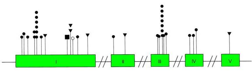
Fig. 7.6. Mutations in the tyrosinase gene. Roman numerals indicate exons, circles denote single-nucleotide substitutions, triangles represent microdeletions, and the square indicates an insertion
Another aminoacidopathy associated with impaired tyrosine metabolism is phenylketonuria (PKU). The disease is caused by mutations in the phenylalanine hydroxylase gene (with about 600 mutations known to date). Due to the lack of active enzyme, phenylalanine is not converted into tyrosine. The pathology is caused by phenylalanine itself, which is toxic at high concentrations, as well as by its toxic metabolite, phenylpyruvic acid. Affected newborns have a specific "mousy" odor, dysmorphic facial features, and hypopigmentation (fair skin and hair). Later, seizures appear and intellectual disability develops. The pathological manifestations of the mutations can be avoided if the child is placed on a special phenylalanine-free diet immediately after birth and maintained on it up to the age of 15. PKU occurs with a relatively high frequency—on average 1 in 10,000 (in some populations in Ukraine, the frequency reaches 1 in 4,500)—and is inherited as an autosomal recessive trait. Interestingly, the number of heterozygotes for the mutant phenylalanine hydroxylase gene in the population is slightly higher than expected from the Hardy-Weinberg equilibrium (see Chapter 8). This is explained by the fact that elevated phenylalanine levels in heterozygous women act as a factor that reduces the risk of Miscarriage.
An enzyme deficiency can lead to the gradual accumulation of certain toxic substrates within The Cell (specifically within the cell, unlike PKU). Such storage diseases include, for example, Tay-Sachs disease (amaurotic idiocy). Due to the absence of the enzyme hexosaminidase A in the Lysosomes of Nerve Cells, GM2 ganglioside—a component of The cell membrane—is not broken down. The ganglioside accumulates in the lysosomes, leading to the destruction of nerve cells. Symptoms typically begin to manifest a few months after birth, characterized by developmental delays and progressive neurological disorders. Death occurs on average by the age of three. There is also an adult-onset form of amaurotic idiocy, where the disease begins to develop between the ages of 20 and 40 and is accompanied by progressive mental deterioration. The infantile form is caused by frameshift mutations or splice site mutations in the hexosaminidase A gene, whereas the adult form is caused by missense mutations.
Cystic fibrosis is an example of a disease associated with impaired inorganic ion metabolism. Due to a mutation in the CFTR gene (mapped to the long arm of chromosome 7, which encodes the chloride ion channel protein), patients experience defects in sweat and other glands caused by high salt concentrations in secretions, as well as impaired nutrient absorption due to blockage of the pancreatic ducts. Consequently, a delay in the child's GROWTH AND DEVELOPMENT is observed. The most fatal consequence is the accumulation of mucus in the respiratory tract, which causes respiratory failure in patients and triggers the development of Pneumonia. The disease (autosomal recessive inheritance pattern) occurs with a frequency ranging from 1 in 2,000 to 1 in 90,000.
Порушення транспорту кисню гемоглобіном є причиною патологічних проявів гемоглобінопатій. Хвороби цього класу пов'язані з мутаціями в генах а- або ß-субодиниць гемоглобіну. Серпоподібноклітинна анемія зумовлена мутацією, яка викликає заміну 6-ї амінокислоти в ланцюзі ß-глобіну (глутамін замінюється на валін). Варіант гемоглобіну (гемоглобін S), до складу якого входить змінений ß-глобін, гірше виконує свою функцію. Крім того, він здатен полімеризуватися, утворюючи філаменти, які змінюють морфологію еритроцитів (вони набувають форму серпа - звідти назва хвороби). Гомозиготи за мутантним Геном мають широкий спектр симптомів: еритроцити серпоподібної форми, анемію, ниркову, серцеву та легеневу недостатність, розумову відсталість, паралічі, болі в суглобах і животі. Без спеціального лікарського нагляду хворі помирають у ранньому дитинстві. У гетерозигот еритроцити мають неправильну форму, оскільки містять два варіанти гемоглобіну - нормальний і мутантний. Але клінічні прояви захворювання в гетерозигот відсутні (починають виявлятися лише за умови зниження концентрації кисню в повітрі - наприклад, у високогірних умовах). Тобто серпоподібноклітинна анемія успадковується як ознака з неповним домінуванням. Хоча гемоглобін S є патологічним варіантом білка, його ген достатньо розповсюджений, особливо у країнах із великим рівнем захворюваності на малярію: кількість носіїв гена досягає 40 % у деяких країнах східної Африки. Це пояснюється високою резистентністю гетерозигот до малярії.
Another group of hemoglobinopathies comprises the thalassemias, disorders associated with aberrant expression of α- or β-Globins (distinguishing between α- and β-thalassemias, respectively). These pathologies arise either from deletions within globin genes or from mutations that reduce THE CELLULAR LEVEL of active mRNAs. The diseases are widespread in certain Mediterranean regions, where the incidence can reach up to 30% (the name originates from the Greek word for sea). Thalassemias are characterized by hemolytic anemia, jaundice, Bone Marrow hyperplasia, and skeletal abnormalities. Depending on the patient's genotype, symptoms range from subclinical (mild forms) to severe and fatal. Notably, Sickle-Cell Anemia and β-thalassemia serve as a classic example of how different mutations in the same gene can yield entirely distinct phenotypic manifestations.
Mutations in genes encoding structural tissue proteins that lead to the absence, deficiency, or production of an abnormal protein also account for a vast number of hereditary syndromes. For instance, Marfan Syndrome is caused by mutations in the fibrillin gene (located on the long arm of chromosome 15), which encodes an Extracellular matrix protein of Connective Tissue. Affected individuals present with skeletal abnormalities, tall stature, arachnodactyly (spider fingers), and cardiovascular defects (with aortic wall weakness being the most dangerous symptom). Marfan syndrome is an autosomal dominant disorder with an estimated prevalence of about 1 in 10,000. Interestingly, approximately 25% of affected children are born to unaffected parents, pointing to a high de novo mutation rate in the fibrillin gene.
A distinct category of inherited genetic disorders is known as Trinucleotide Repeat Expansion diseases. The underlying cause of these disorders (Table 7.4), which do not share a common Clinical presentation, is the massive expansion (see Chapter 4) in the copy number of a trinucleotide repeat within a specific gene. In most cases, these conditions are inherited as dominant traits manifesting in adulthood. If a clinically healthy parent gives birth to an affected child, it typically means that the parent carried a repeat number exceeding the normal range yet below the threshold for clinical symptoms (the so-called premutation state); once a sufficient repeat threshold is reached, the probability of further expansion during subsequent rounds of DNA Replication increases dramatically. Consequently, all disorders of this type exhibit The phenomenon of anticipation—with each successive generation, the symptoms become more severe and manifest at an earlier age, driven by the increasing copy number of the trinucleotide repeat.
Table 7.4. Selected trinucleotide repeat expansion disorders
|
Disorder |
Symptoms |
Inheritance |
Repeat unit |
Copy number |
|
|
Normal |
Pathological |
||||
|
Myotonic dystrophy |
Progressive Muscle weakness, myotonia, cataracts, Cardiac Arrhythmias |
Autosomal dominant |
CTG |
5-37 |
80-3000 |
|
Huntington's disease |
Progressive degeneration of the Basal Ganglia accompanied by movement disorders, psychiatric changes, and epileptic seizures |
Autosomal dominant |
CAG |
6-34 |
40-121 |
|
Spinocerebellar ataxia type 1 |
Progressive degeneration of cerebellar and Spinal Cord pathways with gradual loss of motor coordination |
Autosomal dominant |
CAG |
6-30 |
41-81 |
|
Friedreich's ataxia |
Progressive degeneration of the posterior and lateral columns of the spinal cord with progressive loss of motor coordination |
Autosomal recessive |
GAA |
10-21 |
200-900 |
|
Fragile X syndrome |
Fragile X chromosome, intellectual disability |
X-linked, incomplete dominance (intellectual disability traits are observed in 60% of heterozygous females) |
CGG |
6-52 |
230-2000 |
A major category of hereditary pathologies comprises chromosomal disorders. Various karyotypic abnormalities occur in approximately 0.6% of newborns on average. Only 10% of these anomalies are unaccompanied by overt pathological conditions. However, the actual rate of chromosomal aberration formation is significantly higher: estimates suggest that roughly 25% of zygotes harbor an abnormal karyotype, with the vast majority of such embryos perishing during the preimplantation period. Among spontaneous abortions, 50% display chromosomal anomalies.
Numerical or structural chromosome abnormalities can arise during gametogenesis in one of the parents. In such cases, the anomaly will be present in all Cells of the embryo, an Organism referred to as a complete mutant. Occasionally, Chromosomal aberrations occur during embryonic development, resulting in only a fraction of the embryonic cells possessing an abnormal karyotype—a phenomenon known as genetic mosaicism.
Quantitative chromosomal anomalies result from errors in chromosome segregation (see Chapter 4). The only human monosomy compatible with life is monosomy of the X chromosome. Monosomies for any autosome are lethal: the embryo perishes at very early Stages of development, as these karyotypic abnormalities are completely absent even among spontaneously aborted fetuses. Trisomies for most autosomes likewise cause developmental defects incompatible with life. Among live births, trisomies are observed exclusively for Chromosomes 21, 18, and 13 (Fig. 7.7).
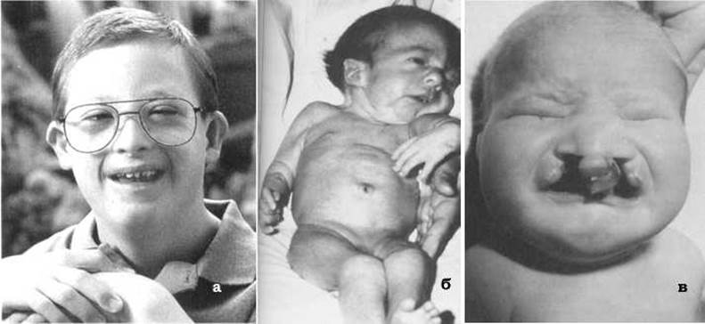
Fig. 7.7. Characteristic phenotypic features of patients with trisomies for chromosomes 21 (a), 18 (b), and 13 (c).
Trisomy 21—Down syndrome—is the most prevalent chromosomal disorder, occurring with an average frequency of 1 in 500–700 newborns. The sex ratio among affected individuals is 1:1. Patients exhibit a characteristic phenotype: an upward slant of the eyes, epicanthic folds, a flat and broad nasal bridge, short stature, and a small HEAD with a flattened occiput. Many patients suffer from cardiovascular anomalies and immunodeficiencies, which are the leading causes of mortality. Males are sterile, whereas females are occasionally fertile. A hallmark symptom of Down syndrome is intellectual disability (ranging from mild to moderate debility, with a maximum IQ of 75). Mosaic forms of Down syndrome (accounting for about 3% of all cases) present with significantly milder symptoms. In some instances, one of the parents of a child with Down syndrome (more frequently the mother) is a carrier of a balanced Robertsonian translocation involving chromosome 21 and chromosomes 13–15, or less commonly chromosome 22. Such families can produce children with Down syndrome, chromosomally normal children, and healthy carrier children possessing the balanced Robertsonian translocation.
Trisomy 18—Edwards syndrome—occurs with a frequency of 1 in 4,500–7,000 newborns. Affected infants are small at birth, frail, and experience delayed physical and mental development. Typical craniofacial anomalies include a receding chin, microstomia, hypoplastic jaws, and a prominent occiput, accompanied by malformed low-set ears, limb deformities, a short Sternum, and muscular developmental defects. Patients present with severe cardiac and renal malformations. Only 1–2% of children survive to the age of 1 year. A distinctive feature of Edwards syndrome is a higher live-birth rate of affected females compared to males (3:1), reflecting a higher intrauterine mortality rate among male fetuses.
Trisomy 13—Patau syndrome. The clinical presentation is characterized by multiple Congenital Malformations: cleft palate and cleft lip, microphthalmia (underdeveloped small eyes) or sometimes anophthalmia, ear anomalies, microcephaly, growth and mental retardation, as well as cardiac, renal, and gastrointestinal defects. The syndrome occurs with a frequency of 1 in 14,500, with the majority of fetuses perishing during intrauterine development. Infants are frequently born prematurely, have low birth weight, and rarely survive past the first year of life; those who survive suffer from profound idiocy. Cases have been documented where Patau syndrome resulted from Robertsonian translocations involving chromosome 13. Mosaicism for an extra chromosome 13 is often associated with intellectual disability in the absence of major external physical anomalies.
Quantitative sex chromosome anomalies are represented by X chromosome monosomy and X and Y chromosome polysomies. The main syndromes caused by Changes in the normal number of sex chromosomes are listed in Table 7.5.
Table 7.5. Main syndromes associated with changes in the number of sex chromosomes
|
Syndrome |
Karyotype* |
Incidence |
Symptoms |
|
Turner syndrome (X monosomy) |
45, X0 |
1/3000 - 1/5000 |
Female phenotype. Underdeveloped Ovaries and external genitalia, absent secondary sexual characteristics, Infertility, short stature, and a "masculine" body build. Cardiovascular defects are common. Intelligence is normal, and psychological infantilism is often observed. |
|
Trisomy X |
47, XXX |
1/1000 |
Female phenotype. Mild and diverse physical abnormalities. Fertility is preserved. Mild mental retardation is observed in 75% of cases. Increased risk of developing psychoses. |
|
47, XXY |
1/1000 |
Male phenotype. Underdeveloped Testes, lack of Spermatogenesis, and infertility. "Eunuchoid" body type: narrow shoulders, wide pelvis, female-pattern fat distribution and hair growth. Varying degrees of mental retardation. |
|
|
Y polysomy |
47, XYY |
1/250 - 1/1000 |
Male phenotype. Normal physical, mental, and sexual development. Elevated androgen levels; some men exhibit psychopathic personality traits, aggressiveness, and antisocial behavior. |
* Total number of chromosomes, sex chromosome Complement.
For most trisomies (both viable and lethal), a maternal Water/144.html">Origin of the extra chromosome has been proven, resulting from chromosome nondisjunction most frequently During the first meiotic division. For example, 88% of trisomy 21 cases are caused by impaired chromosome segregation in the mother, with 65% of nondisjunction events occurring in Meiosis I. Chromosome nondisjunction during spermatogenesis accounts for only 8% of chromosome 21 trisomies (3% in meiosis I and 5% in meiosis II). However, paternal sex chromosome nondisjunction is the primary cause of Turner syndrome and 46% of Klinefelter syndrome cases. All maternal-effect trisomies show a clear positive correlation between maternal age and the risk of developing a fetus with the corresponding abnormal karyotype. External factors such as infectious diseases, mutagenic agents, and harmful habits (smoking, alcoholism, etc.) may also increase the risk of maternal chromosome nondisjunction.
The described quantitative chromosome anomalies are forms of aneuploidy. Polyploidy accounts for 23% of spontaneous abortions. Tetraploids are not observed among live births, and there are only isolated cases of triploid infants born with multiple severe developmental anomalies that are incompatible with life.
In addition to quantitative chromosome anomalies, congenital pathological conditions can be caused by structural chromosomal aberrations. Such chromosomal disorders are subdivided into partial aneuploidy syndromes (excluding unbalanced Robertssonian translocations) and microcytogenetic syndromes.
Partial aneuploidy syndromes (around 100 are currently described) are so named because the pathological symptoms of these disorders are caused either by an excess (partial trisomy) or a deficiency (partial monosomy) of a fairly large segment of a specific chromosome. The main clinical picture, similar to complete aneuploidy, is characterized by a combination of syndrome-specific features along with a broad set of nonspecific symptoms, such as congenital malformations, mental retardation, dysplasias, and cardiovascular defects. This symptom nonspecificity indicates that the overall pathology is driven primarily by the fact of chromosomal imbalance itself rather than by specific lost or duplicated gene loci.
Microcytogenetic syndromes include disorders caused by submicroscopic deletions or duplications of specific chromosomal regions. These syndromes occur with a relatively low frequency (1/50,000 to 1/100,000) and are characterized by a distinct clinical presentation. Examples include hereditary retinoblastoma (a microdeletion in chromosome 13 causing congenital malignant tumors of the retina), Beckwith-Wiedemann syndrome (a microduplication in the short arm of chromosome 11 leading to characteristic malformations such as omphalocele, microcephaly, hypoglycemia, and multiple internal organ defects), as well as Angelman and Prader-Willi syndromes (microdeletions in the long arm of chromosome 15).
Among the mentioned disorders, Angelman and Prader-Willi syndromes attract special attention. Patients with Prader-Willi syndrome suffer from uncontrolled appetite resulting in obesity, facial dimorphism, small hands and feet, hypotonia, and mental retardation. Angelman syndrome is characterized by severe neurological, psychiatric, and intellectual disorders, including impaired motor coordination (puppet-like movements), epileptic seizures, unprovoked bouts of laughter, and lack of speech development. Despite different phenotypic manifestations, the microdeletions in chromosome 15 found in these syndromes are identical. The pathological manifestations depend on which parent transmitted the altered chromosome: if the microdeleted chromosome is inherited from the father, the symptoms correspond to Prader-Willi syndrome; if from the mother, to Angelman syndrome. Thus, not only the inheritance of an abnormal chromosome matters, but also its parental origin. This phenomenon is termed Genomic Imprinting, and disorders inherited in the manner of Angelman and Prader-Willi syndromes are known as imprinting disorders. Imprinting is explained by the differential activity of certain alleles on maternal and paternal chromosomes due to Epigenetic Inheritance effects (see Chapter 6). Consequently, imprinted genes exhibit functional monosomy, and organisms are functionally hemizygous for these genes.
A distinct group of genetically determined pathological conditions involves maternal-fetal incompatibilities. The most well-known example of such pathology is Rh incompatibility. When the mother is Rh-negative (lacking the Rh antigen) and the fetus is Rh-positive, the mother's body produces Antibodies against the Rh antigen. The first Pregnancy may proceed without any complications. During a second pregnancy involving another Rh-positive fetus, an incompatibility conflict arises between the maternal and fetal organisms because the mother has already developed Immunity against Rh-positive cells. This conflict can lead to miscarriage or the birth of a child with pathologies.
Last update: 11/08/2026
Editorial and Educational Adaptation: This material has been compiled based on the primary/original source text. The project team performed an editorial review, corrected technical inaccuracies, structured sections, and adapted the content for an educational format.
What was processed:
- elimination of formatting defects (OCR errors, structural breaks, corrupted characters);
- editorial organization of content;
- standardization of terminology in accordance with academic sources;
- verification of factual statements against the original source text.
All mentions of the author, publication year, and origin of the primary text have been preserved in accordance with the source.