Human Anatomy - Kotsan I. Ya. 2009
Urogenital Apparatus
Genitals - Male Genitals
Spermatogenesis
Spermatogenesis, The process of male germ Cell development, is described in detail in the course of developmental biology. Here, we provide only A brief Overview.
Spermatogenesis takes place within the convoluted seminiferous tubules of the Testis, which constitute the bulk of its mass. The inner wall of a seminiferous tubule is lined with spermatogenic epithelium, interspersed with Sertoli Cells, which serve as supporting cells.
The Cells of the spermatogenic epithelium rest on a basement membrane. In adult males, the basement membrane of the convoluted seminiferous tubule is externally surrounded by loose Connective Tissue containing Actin-bearing contractile cells. The spermatogenic epithelial cells, representing various stages of spermatogenesis, are arranged in several layers. Spermatogenesis begins with the primary germ cell, the spermatogonium (Fig. 176). Spermatogonia located in the basal layer undergo several successive rounds of mitotic division. Some daughter cells remain as stem cells—retaining the capacity for division and differentiation—while the majority migrate toward the lumen and differentiate into primary spermatocytes. Each primary spermatocyte divides twice (the First and Second meiotic divisions), resulting in the Formation of secondary spermatocytes and spermatids. The latter ultimately transform into spermatozoa (sperm cells). From a single spermatogonium with a diploid (double) set of Chromosomes, Meiosis yields four small spermatids with a haploid (single) set of chromosomes. During Fertilization, the nuclei
of the male and Female Germ Cells fuse, restoring the diploid chromosome set in the zygote Nucleus. According to current data, human spermatogenesis lasts between 64 and 72 days. The number of spermatozoa produced in the Testes is extremely large; for instance, the semen of a healthy adult male contains up to 100 million spermatozoa per 1 mm3.
The supporting cells (Sertoli cells) provide trophic support for the spermatogenic epithelium and phagocytose and degrade the breakdown products of spermatids.
Like spermatogonia, the supporting cells rest on the basement membrane; however, they are larger than spermatogonia, project slightly above them, and partially envelop them. The Plasma Membranes of adjacent supporting cells are tightly connected, thereby isolating the layer of spermatogonia from the layer of dividing cells.
The interconnected layer of supporting cells forms the Blood-testis barrier.
A human spermatozoon consists of a HEAD, neck, and tail (Fig. 177). The head forms the anterior part of the spermatozoon, is consistently widened, and contains a nucleus with a haploid set of chromosomes. At the anterior pole of the head, beneath The Plasma Membrane, lies the acrosome, which is derived from the Golgi apparatus. The Enzymes contained within the acrosome facilitate the penetration of the spermatozoon through the thick zona pellucida of the ovum. The neck of the spermatozoon contains two centrioles (upper, or proximal, and lower, or distal). The axial filament (axoneme) originates from the lower centriole and extends through the entire length of the tail. The Base of the axial filament is surrounded by numerous Mitochondria arranged in a spiral pattern around it. Currently, some authors suggest that this is actually a single large mitochondrion helically wrapped around the axial filament. The mitochondria provide the energy required for sperm motility. A small amount of Cytoplasm surrounds the axial filament. The axial filament, running through all PARTS OF THE sperm tail, serves as its contractile element. Typically, the sperm tail is greatly elongated, exceeding the length of the head many times over.
As they pass through the sperm ducts, spermatozoa enter a fluid secreted by the accessory glands. Together with the spermatozoa, this fluid is referred to as semen, or seminal fluid. In addition to spermatozoa, its cellular elements include leukocytes and a small number of epithelial cells shed from the walls of the sperm ducts.
Class="center">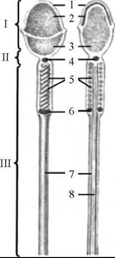
Fig. 177. Diagram of The Structure of a spermatozoon
I — head; II — neck; III — tail; 1 — membrane; 2 — acrosome; 3 — nucleus; 4 — proximal (upper) centriole; 5 — mitochondria; 6 — terminal ring; 7 — tail sheath; 8 — axial filament

Fig. 176. Diagram of spermatogenesis
1 — wall of the convoluted seminiferous tubule; 2 — supporting cell (Sertoli cell); 3 — spermatogonium; 4 — spermatocytes; 5 — spermatids; 6 — spermatozoon
Diagram of the sperm ducts:
straight seminiferous tubules of the testis → rete testis → efferent ductules of the testis → epididymal duct → vas deferens → ejaculatory duct → prostatic and other parts of the Urethra.
Last update: 08/08/2026
Editorial and Educational Adaptation: This material has been compiled based on the primary/original source text. The project team performed an editorial review, corrected technical inaccuracies, structured sections, and adapted the content for an educational format.
What was processed:
- elimination of formatting defects (OCR errors, structural breaks, corrupted characters);
- editorial organization of content;
- standardization of terminology in accordance with academic sources;
- verification of factual statements against the original source text.
All mentions of the author, publication year, and origin of the primary text have been preserved in accordance with the source.