Review of Medical Physiology - William F. Ganong 2002
Cardiovascular System
The Nature of the Heartbeat and Electrical Activity of the Heart
Cardiac Arrhythmias
Normal Heart Rate
In a healthy individual, The Heart rate is set by the SA node (normal sinus rhythm [NSR]). At rest, the heart beats at approximately 70 beats per minute. Heart rate decreases during Sleep (bradycardia) and increases during physical exertion, fever, emotional stress, and many other conditions (tachycardia). The mechanisms controlling heart rate are discussed in Chapter 31. In young, healthy individuals with a normal respiratory rate, the heart rate varies with the respiratory cycle: it increases during inspiration and decreases during expiration, especially as breathing depth increases. Sinus arrhythmia (Fig. 28-10) is a normal physiological variant caused by fluctuations in parasympathetic tone on the heart. During inspiration, impulses from pulmonary stretch receptors travel via the vagus nerves to the Medulla Oblongata, where they inhibit the cardioinhibitory area. The tonic activity of the Vagus nerve, which normally slows the heart rate, is thus reduced, leading to an increased heart rate.
Pathological conditions affecting the sinoatrial node lead to marked bradycardia accompanied by dizziness and fainting (sick sinus syndrome).
Ectopic Pacemakers
Under pathological conditions, the AV node and other PARTS OF THE conduction system can assume The Role of cardiac pacemakers. Furthermore, in damaged Muscle fibers of the atria and ventricles, Action Potential generation may be suppressed, triggering re-entrant excitation.
Class="center">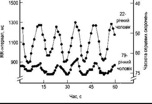
Fig. 28-10. Sinus arrhythmia in a young and an elderly man. The breathing rate was maintained at five respiratory cycles per minute. With each inspiration, the R-R interval shortens, indicating an increased heart rate. Note the less pronounced arrhythmia in the elderly man. This tracing was recorded following the administration of ß-blockers, though similar results can be obtained without them (reproduced with permission from Pfiefer MA et al: Differential changes of Autonomic Nervous system function with age in man. Am J Med 1983;75;49).
As noted above, the SA node exhibits a higher rate of impulse generation than other Regions of the conduction system. Therefore, it normally acts as the primary pacemaker of the heart. When impulse propagation from the atria to the ventricles is completely interrupted, third-degree (complete) heart block occurs, and the ventricles beat at a slow, independent rhythm (idioventricular rhythm, Fig. 28-11). Complete block may result from damage to the AV node (AV nodal block) or from lesions in the conduction pathways distal to the node (infranodal block). In patients with AV nodal block, surviving tissue within the conduction system takes over as the pacemaker, maintaining a ventricular rate of about 45 beats per minute. In patients with infranodal block due to bundle of His lesions, the ventricular pacemaker is located in the peripheral Branches of the conduction system. Consequently, the ventricular rate is slower, averaging 35 beats/min, but may drop as low as 15 beats/min in some patients. Such individuals may experience periods of asystole lasting a minute or longer, leading to cerebral ischemia manifested by dizziness and fainting (Stokes-Adams syndrome). Third-degree heart block can be caused by myocardial infarction affecting the septal region or by surgical trauma to the bundle of His during correction of a congenital ventricular septal defect.

Fig. 28-11. Heart blocks
If impulse conduction from the atria to the ventricles is merely delayed rather than completely interrupted, incomplete heart block develops. In first-degree heart block, all atrial impulses reach the ventricles, but the PR interval is significantly prolonged. In second-degree block, not all impulses pass through to the ventricles; every second or third impulse may be blocked (2:1 block, 3:1 block, etc.). Another type of second-degree block is characterized by progressive prolongation of the PR interval culminating in a dropped ventricular beat (Wenckebach phenomenon). Following the dropped beat, the PR interval usually returns to normal or is only slightly prolonged (see Fig. 28-11).
Occasionally, impulse propagation through one of the bundle branches may be impaired, resulting in right or left bundle branch block. In bundle branch block, excitation initially travels through the intact branch to activate the corresponding ventricle before spreading to the blocked side. While the overall ventricular rate remains normal, the QRS complexes become widened and deformed (see Fig. 28-11). Blocks can also occur at the level of the anterior and posterior fascicles of the left bundle branch, causing so-called fascicular block, or hemiblock. A block of the left anterior fascicle results in left axis deviation, whereas a block of the posterior fascicle causes right axis deviation. Combinations of fascicular blocks and bundle branch blocks (bifascicular or trifascicular blocks) are frequently observed. The precise anatomical site of a conduction defect can be identified using His bundle electrography.
Implanted Pacemakers
In cases of severe bradycardia associated with sick sinus syndrome or third-degree heart block, electronic pacemakers are frequently implanted. These devices have become highly sophisticated and reliable, proving exceptionally effective for patients suffering from sinus node dysfunction, AV block, as well as bifascicular and trifascicular blocks. They are also used in patients with neurogenic syncope who exhibit pauses of more than 3 seconds upon carotid sinus stimulation, and in certain cardiomyopathy patients primarily to improve hemodynamics rather than merely to restore normal heart rhythm.
Ectopic Foci of Excitation
Under normal conditions, myocardial Cells possess the capacity for spontaneous impulse generation. However, the probability of spontaneous discharge in the bundle of His and Purkinje fibers is low because the SA node, acting as the primary pacemaker, fires at a higher frequency. Nevertheless, under pathological circumstances, the bundle of His, Purkinje fibers, and ordinary cardiomyocytes can spontaneously generate impulses, a condition referred to as enhanced automaticity. When an ectopic focus fires spontaneously, it triggers a premature contraction ahead of the next scheduled normal beat, temporarily disrupting the cardiac rhythm (atrial, junctional, or ventricular extrasystoles, or premature beats). If the ectopic focus fires repeatedly at a rate exceeding that of the SA node, it causes sustained regular tachycardia (paroxysmal atrial, ventricular, or junctional tachycardia, or atrial flutter).
Re-entry
The primary mechanism underlying paroxysmal arrhythmias is impaired impulse propagation, which causes an excitation wave to circulate continuously in a closed loop (circus movement). For example, following a transient unilateral block in a segment of the conduction system, an impulse may travel around via the unblocked pathway. If the block subsequently resolves, the impulse can travel in a retrograde direction through the previously blocked area back to its origin and continue circulating in a loop. A ring of tissue, as illustrated in Fig. 28-12, provides a classic example of this phenomenon. When re-entry occurs within the AV node, the atria are re-excited, producing what is known as an "echo" beat. Simultaneously, impulses propagate back down to the ventricles, resulting in paroxysmal nodal tachycardia. Circus movements can also occur within the atrial or ventricular myocardium. In individuals with an accessory pathway connecting the atria and ventricles (Bundle of Kent; see below), a circus movement can travel in one direction through the AV node and return via the accessory pathway, thus involving both the atria and ventricles in the loop.
Atrial Arrhythmias
Excitation originating in ectopic atrial foci prematurely stimulates the AV node and propagates down to the ventricles. Atrial extrasystoles are characterized by an altered P wave Morphology, while the subsequent QRST complex is usually normal (Fig. 28-13). Such ectopic impulses may also depolarize the SA node. Following this, the SA node must undergo repolarization and subsequent diastolic depolarization to threshold before it can initiate the next normal beat. Consequently, the pause between the extrasystole and the following normal contraction equals the normal cycle length (compensatory pause), after which regular rhythm resumes (see below). If the premature impulse fails to reach the SA node before its next scheduled normal discharge, a non-compensatory pause occurs, which equals the interval between two normal beats.
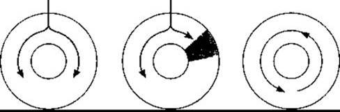
Fig. 28-12. Depolarization of a cardiac tissue ring. Normally, an impulse propagates around the ring in both directions (left), rendering the tissue immediately refractory. In the case of a transient unilateral block (middle), the impulse from the opposite side travels around the entire ring. When the temporary block resolves (right), the impulse can pass through the previously affected area and circulate indefinitely (circus movement).

Fig. 28-13. Atrial arrhythmias. Shown are an atrial premature contraction with the P wave superimposed on the T wave of the preceding beat (arrow); atrial tachycardia; atrial flutter with AV block (4:1); and atrial fibrillation with an irregular ventricular response (reproduced with permission from Goldschlager N, Goldman MJ: Principles of Clinical Electrocardiography. 13th ed. McGraw-Hill, 1989).
Atrial tachycardia can be triggered by The Emergence of a regularly firing ectopic focus or by a re-entry mechanism. In such cases, the heart rate may reach 220 beats per minute. Occasionally, particularly in patients receiving digitalis therapy, tachycardia may be accompanied by atrioventricular block (paroxysmal atrial tachycardia with block).
In atrial flutter, the heart rate typically ranges from 200 to 350 beats per minute (see Fig. 28-13). The most common form of this arrhythmia involves a large counterclockwise circus movement of impulses within the right atrium, producing characteristic atrial waves (sawtooth-like notches). Atrial flutter is usually accompanied by AV block (2:1 or greater) because the adult AV node cannot conduct more than approximately 230 impulses per minute.
During atrial fibrillation, the atrial rate is extremely rapid (300-500 beats per minute), and contractions are irregular and chaotic. Because the AV node is activated irregularly, the ventricles also contract irregularly at a rate of 80-160 beats per minute (see Fig. 28-13). This condition may present as paroxysmal episodes or become chronic, and in some cases, There is a genetic predisposition. The exact cause of atrial fibrillation remains a subject of debate; however, it is most commonly driven by multiple simultaneous micro-reentrant wavelet circuits in both atria. In some cases, paroxysmal atrial fibrillation is triggered by impulse generation in one or more ectopic foci. Many of these foci are located within the Pulmonary Veins, about 4 cm from the heart. Atrial muscle fibers extend along the pulmonary veins, and these fibers serve as the source of these impulses.
Consequences of Atrial Arrhythmias
Sporadic atrial premature beats may occur occasionally in most healthy individuals and have no clinical consequences. In paroxysmal atrial tachycardia and atrial flutter, however, the ventricular rate can become so rapid that the ventricles do not have sufficient time to fill with Blood during diastole. Consequently, Cardiac Output drops, and symptoms of Heart Failure appear. The relationship between heart rate and cardiac output is discussed in detail in Chapter 29. Heart failure can also result from atrial fibrillation when the ventricles beat at an excessively high rate. Acetylcholine released from vagal nerve endings inhibits impulse conduction through the atrial muscle and the AV node. Therefore, reflex stimulation of the vagus nerve—achieved by gentle pressure on the eyeballs (oculocardiac reflex) or massage of the carotid sinus—can often restore a normal sinus rhythm in cases of tachycardia and occasionally in atrial flutter. At the same time, vagal stimulation enhances AV block and slows the ventricular rate. Digitalis also suppresses AV conduction and is used clinically to reduce the ventricular rate in atrial fibrillation.
Ventricular Arrhythmias
Premature beats originating from ectopic foci in the ventricles are typically characterized by an altered configuration and prolonged duration of the QRS complexes (Fig. 28-14). This is due to the slow propagation of impulses from the ectopic focus through the ventricular muscle to other areas of the ventricle. In most cases, the bundle of His is not invaded, and retrograde conduction to the atria does not occur. Eventually, the next normal impulse from the SA node depolarizes the atria. Most often, the P wave becomes buried within the premature QRS complex. When this normal impulse reaches the ventricles, they are still in a refractory state resulting from the ectopic depolarization; however, the subsequent SA nodal impulse elicits a normal contraction. Consequently, a premature ventricular contraction is followed by a compensatory pause, which is often longer than the pause following a premature atrial contraction. Furthermore, ventricular extrasystoles do not disturb the regular rhythm of the SA node, whereas premature atrial contractions do.

Fig. 28-14. Top: Premature ventricular contraction (PVC). The lines beneath the tracing indicate the compensatory pause, demonstrating that the duration of the premature beat plus the preceding normal beat equals the duration of two normal cardiac cycles. Bottom: Ventricular tachycardia.
If atrial and ventricular premature beats occur early in diastole—when the ventricles have had little time to fill with blood and the ventricular muscle is still in a state of relative refractoriness—they may be too weak to produce a palpable pulse at the wrist. They may fail to even open the aortic and pulmonary Valves, resulting in the absence of a second heart sound (see Chapter 29).
Paroxysmal ventricular tachycardia (see Fig. 28-14) is characterized by rapid, regular ventricular depolarization, usually driven by a circus movement of impulses within the ventricles. Tachycardias in which the ectopic foci are located above the ventricles (supraventricular tachycardias, such as nodal paroxysmal tachycardia) can be differentiated from paroxysmal ventricular tachycardia using an esophageal lead (EPG). Supraventricular tachycardias reveal a deflection H that is absent in ventricular tachycardias. Ventricular premature beats are quite common and, in the absence of CORONARY HEART DISEASE, are typically benign. Paroxysmal ventricular tachycardia is much more dangerous because it significantly reduces cardiac output and can readily degenerate into ventricular fibrillation.
In ventricular fibrillation (Fig. 28-15), the contractions of the ventricular muscle fibers are completely uncoordinated and ineffective, driven by extremely rapid impulse generation from numerous ectopic foci or by re-entrant circus movements. During fibrillation, the ventricles (like the atria) resemble a "bag of worms." Ventricular fibrillation can be triggered by an electric Shock or by an extrasystole occurring during a critical period known as the vulnerable period. The vulnerable period coincides chronologically with the middle of the T wave—a time when some ventricular fibers are depolarized, others are incompletely repolarized, and still others are fully repolarized. These are optimal conditions for re-entry and circus movement of impulses. Because fibrillating ventricles cannot effectively pump blood, Circulation ceases entirely. Without prompt intervention, ventricular fibrillation lasting more than a few minutes is fatal. Ventricular fibrillation is the primary cause of sudden cardiac death in patients with myocardial infarction.
Further evidence of cardiac vulnerability during repolarization is the observation that patients with a prolonged QT interval have a significantly higher incidence of ventricular arrhythmias and sudden death. Genetic defects have been identified for three inherited forms of long QT syndrome. The first defect involves the blockade of a specific type of K+ channel (the HERG channel), which slows repolarization (Fig. 28-16). The second defect is associated with the blockade of another type of K+ channel and is linked to deafness (see Chapter 9). The third defect enhances The activity of cardiac Na+ channels, which likewise delays repolarization.
Although ventricular fibrillation is induced during execution by electric chair, it can frequently be terminated and normal sinus rhythm restored through the application of an electric shock. Automated external defibrillators are now standard equipment not only in hospitals but also in ambulances and should be applied as rapidly as possible. Additionally, implantable cardioverter-defibrillators can be surgically placed in patients at high risk for ventricular fibrillation.
Resuscitation of Cardiac and Respiratory Function
In patients experiencing ventricular fibrillation or cardiac arrest, cardiac output and coronary blood flow can be partially maintained through external chest compressions (cardiopulmonary resuscitation). Effective external massage can be applied across the chest wall. The rescuer should place the heel of one hand over the lower half of the Sternum (near the Base of the xiphoid process) and the other hand on top of the first (Fig. 28-17). Compressions should be directed straight down, depressing the sternum by 4 to 5 cm toward the spine at a rate of 80 to 100 compressions per minute. Direct manual compression of the ventricles is also effective when the chest is already open, though performing an emergency thoracotomy is otherwise unjustified. If breathing has also ceased, both Cardiac Activity and ventilation must be supported. Chest compressions should be alternated with Mouth-to-mouth artificial Respiration (see Chapter 37) at a ratio of one rescue breath to every five chest compressions.

Fig. 28-15. Ventricular fibrillation triggered by a premature ventricular contraction (PVC) during the vulnerable period in a patient with myocardial infarction. The patient was successfully defibrillated immediately, terminating the fibrillation.

Fig. 28-16. Long QT syndrome caused by a genetic defect leading to blockade of HERG K+ channels. This creates a predisposition to ventricular arrhythmias driven by slowed K+ efflux from The Cell, prolonged action potential duration, and a consequent prolongation of the QT interval (modified from Keating M, Sanguinetti MC: Molecular genetic insights into cardiovascular disease. Science 1996;272:681).
Accelerated AV Conduction
A fascinating phenomenon observed in some healthy individuals who are prone to attacks of paroxysmal atrial arrhythmia is accelerated AV conduction (Wolff-Parkinson-White syndrome). Normally, the AV node serves as the sole electrical pathway between the atria and ventricles. Individuals with Wolff-Parkinson-White syndrome possess an additional muscular bridge or atypical cardiomyocyte connection between the atria and ventricles (the bundle of Kent) that conducts impulses faster than the AV node, causing one ventricle to be activated prematurely. The manifestations of this early activation merge with the normal QRS complex, resulting in a shortened PR interval and a widened QRS complex with an initial slurring or delta wave (Fig. 28-18), although the total duration from the onset of the P wave to the end of the QRS complex (the "PJ interval") remains normal. In this syndrome, paroxysmal atrial tachycardia often develops following a premature atrial contraction. Premature depolarization spreads from the atria through the AV node while the accessory pathway is still refractory, as the accessory bundle typically has a longer refractory period than the AV node. However, once the excitation reaches the ventricular end of the accessory pathway, that pathway is no longer refractory, and the impulse propagates retrogradely back to the atrium, thereby establishing a re-entrant circuit. Less commonly, premature depolarization finds the AV node in a refractory state; it then reaches the ventricles via the bundle of Kent, Setting up a circus movement in which impulses travel from the ventricles back to the atria through the AV node.
Attacks of paroxysmal supraventricular tachycardia, usually nodal tachycardia, occur in individuals with a short PR interval and a normal QRS complex (Lown-Ganong-Levine syndrome). In this condition, depolarization spreads from the atria to the ventricles via an accessory pathway that bypasses the AV node but connects directly with the intraventricular conduction system distal to the AV node.
Antiarrhythmic Drugs
A wide variety of pharmacological agents are currently available that successfully treat arrhythmias by slowing impulse propagation within the cardiac conduction system and myocardium. These drugs suppress the activity of ectopic foci and reduce the differences between normal and re-entrant pathways, thereby abolishing circus movement circuits. Some of these agents block Na+ channels (Class I antiarrhythmics), whereas others block β-adrenergic receptors or otherwise inhibit cardiac sympathetic innervation (Class II drugs). Other agents prolong the refractory period of The cardiac muscle (Class III drugs), and still others block Ca2+ channels (Class IV drugs). However, it is now evident that all of these agents can exert proarrhythmic effects in certain patients—meaning they can actually provoke various arrhythmias. Consequently, radiofrequency catheter ablation of arrhythmogenic pathways (see below) is increasingly favored over pharmacological therapy for managing cardiac arrhythmias.

Fig. 28-17. Technique for performing external cardiac massage. The black circle on the heart diagram indicates the target area for applying force. The circles on the person lying down—located over the cardiac apex and to the right of the upper sternum—indicate the electrode placement sites for defibrillation.

Fig. 28-18. Accelerated AV conduction. Top: Normal sinus rhythm. Middle: Short PR interval; wide, fused QRS complex; normal PJ interval (Wolff-Parkinson-White syndrome). Bottom: Short PR interval, normal QRS complex (Lown-Ganong-Levine syndrome).
Many class IV drugs are also used to treat angina pectoris and myocardial infarction because they induce relaxation of vascular smooth muscle, cause vasodilation, and reduce cardiac afterload by lowering blood pressure (see Chapter 29).
Endogenous adenosine, which binds to specific receptors (see Chapter 4), slows AV nodal conduction and is therefore administered intravenously for supraventricular arrhythmias. This effect is mediated by the efflux of potassium ions from atrial cardiomyocytes and conduction system cells, but not from ventricular cardiomyocytes. The action is linked to the Activation of a1 adenosine receptors.
Radiofrequency catheter ablation of re-entrant pathways
Today, electrode-tipped catheters can be advanced into the heart chambers to precisely locate the ectopic focus or accessory pathway involved in impulse re-entry and The Development of supraventricular tachycardia. The accessory pathway can be destroyed by passing radiofrequency current through it. In experienced hands, this Treatment method is highly effective and carries a low rate of complications. It is particularly indicated for supraventricular arrhythmias, including Wolff-Parkinson-White syndrome and atrial flutter. It is also successfully used for the ablation of ectopic foci in the pulmonary veins that trigger paroxysmal atrial fibrillation (see above).
Last update: 10/08/2026
Editorial and Educational Adaptation: This material has been compiled based on the primary/original source text. The project team performed an editorial review, corrected technical inaccuracies, structured sections, and adapted the content for an educational format.
What was processed:
- elimination of formatting defects (OCR errors, structural breaks, corrupted characters);
- editorial organization of content;
- standardization of terminology in accordance with academic sources;
- verification of factual statements against the original source text.
All mentions of the author, publication year, and origin of the primary text have been preserved in accordance with the source.