Medical Genetics - V. M. Zaporozhan 2005
Etiology of Hereditary Diseases
Types of mutations caused by changes in chromosome number and structure
Chromosomal aberrations
All Mutations associated with changes in chromosome number and Structure can be divided into three groups:
— chromosomal aberrations caused by changes in Chromosome structure;
— genomic mutations caused by changes in chromosome number;
— mixoploidy — mutations resulting from the presence of Cell clones with different chromosomal sets.
Chromosomal aberrations (chromosomal mutations) are structural changes in Chromosomes (Scheme 2.3), which generally result from unequal Crossing-over during Meiosis. They can also be triggered by chromosome breaks caused by ionizing radiation, certain Chemical Mutagens, Viruses, and other Mutagenic Factors. Chromosomal aberrations may be either unbalanced or balanced.
Unbalanced mutations involve the loss or gain of genetic material, altering Gene dosage or activity, which leads to phenotypic changes.
Chromosomal rearrangements that do not affect gene structure or activity and leave the phenotype unchanged are termed balanced. However, chromosomal aberrations disrupt homologous chromosome pairing (synapsis) and crossing-over during meiosis, leading to The formation of Gametes with unbalanced chromosomal mutations. Carriers of balanced chromosomal aberrations may experience Infertility, a high rate of spontaneous abortions, and an increased risk of having offspring with Chromosomal Disorders.
Class="center">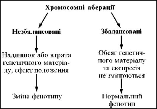
Scheme 2.3. Phenotypic effects of chromosomal aberrations
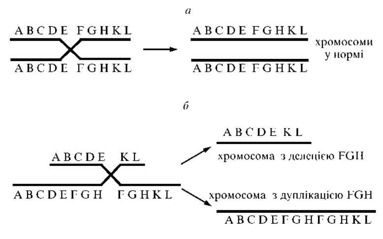
Fig. 2.2. Mechanism of formation of chromosomes with deletion and duplication resulting from unequal crossing-over:
a — normal crossing-over scheme; b — unequal crossing-over resulting in the formation of chromosomes with deletion and duplication
The following types of chromosomal mutations are distinguished:
1. Deletion — the loss of a chromosomal segment.

2. Duplication — the doubling of a chromosomal segment.

Deletions and duplications are frequently the result of abnormal crossing-over (Fig. 2.2).
3. Inversion — the rotation of a chromosome segment by 180° (wherein genes within a specific region are arranged in reverse order compared to normal). If the inverted segment includes the centromere, the inversion is called pericentric; if it does not involve the centromere, it is paracentric (Fig. 2.3).
If an inversion does not alter the total amount of chromosomal material or cause a position effect, individuals remain phenotypically normal. Pericentric inversion of chromosome 9 is a common variant that does not lead to phenotypic changes. Other inversions may impair pairing and crossing-over, resulting in chromosome breakage and the formation of unbalanced gametes.
4. Ring chromosome (Fig. 2.4) arises from the loss of two telomeric fragments. The "sticky" ends of the chromosome then fuse to form a ring (Fig. 2.5).
This mutation can be either balanced or unbalanced, depending on The amount of genetic material lost.
Ring chromosomes are unstable because reduplication generates dicentric rings that subsequently break. Various chromosomal rearrangements then occur within the gametes (Fig. 2.6).
5. Isochromosomes (Fig. 2.7) — the loss of one chromosome arm and the duplication of the other. They most likely arise from horizontal rather than longitudinal division of the centromere.
This results in a metacentric chromosome with two identical arms. An isochromosome of the long arm of the X chromosome is the most common variant, with the karyotype designated as 46,X,i(Xq). Isochromosome X is observed in 15% of all cases of Turner syndrome.
6. Translocation refers to The transfer of a chromosome segment to a non-homologous chromosome, into a different linkage group. Several types of translocations are distinguished:
a) reciprocal translocations — a mutual exchange of segments between two non-homologous chromosomes (Fig. 2.8, a).
The frequency of reciprocal translocations in populations is 1:500. For unknown reasons, the reciprocal translocation involving the long arms of chromosomes 11 and 22 is the most common. Carriers of balanced reciprocal translocations frequently experience spontaneous abortions or give birth to children with multiple Congenital Malformations due to the formation of gametes with unbalanced mutations. The genetic risk for carriers of such translocations ranges from 1 to 10 %;
b) non-reciprocal translocations (Transpositions) — the movement of a chromosome segment either within the same chromosome (Fig. 2.8, b) or to another chromosome without mutual exchange;
c) a special type of translocation is the Robertsonian translocation (or centric fusion).
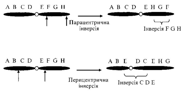
Fig. 2.3. Paracentric and pericentric inversions
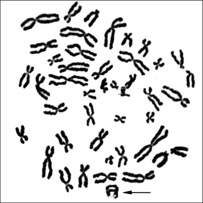
Fig. 2.4. Ring chromosome in a metaphase plate (indicated by an arrow)
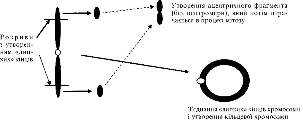
Fig. 2.5. Mechanism of ring chromosome formation
These occur between any two acrocentric chromosomes from group D (pairs 13, 14, and 15) and group G (pairs 21 and 22). During centric fusion, two homologous or non-homologous chromosomes lose their short arms and one centromere, while their long arms fuse together (Fig. 2.9). Instead of two chromosomes, a single one is formed containing the genetic material of the long arms of both chromosomes. The total chromosome count in carriers of a balanced Robertsonian translocation is 45. The short arms of all ten chromosomes in groups D and G contain identical genes encoding rRNA. Each cell possesses A large number (up to 105) of copies of these genes. The loss of the short arms of two chromosomes does not lead to significant phenotypic changes, as the loss of these genes is compensated by The activity of identical genes on the remaining eight acrocentric chromosomes.
Thus, carriers of Robertsonian translocations are healthy, but they have an increased frequency of spontaneous abortions and a high risk of having children with chromosomal disorders. The frequency of Robertsonian translocations in the population is 1:1000.
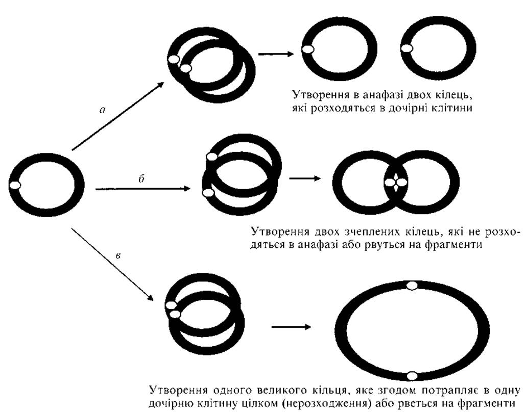
Fig. 2.6. Variants of ring chromosome segregation in mitosis:
a — normal Replication; b — replication and two sister chromatid exchanges during crossing-over; c — reduplication and one sister chromatid exchange during crossing-over
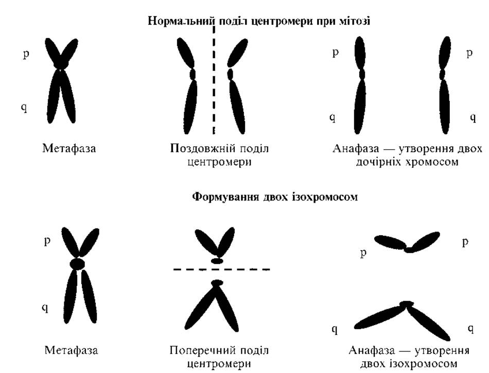
Fig. 2.7. Mechanism of isochromosome formation
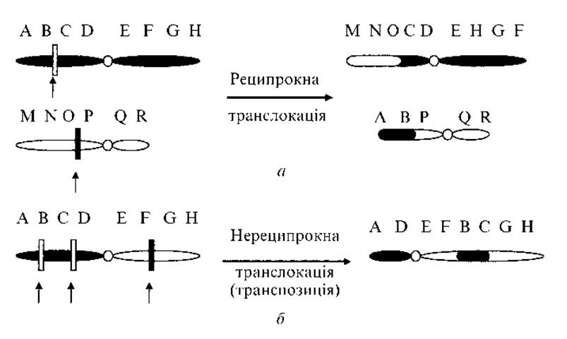
Fig. 2.8. Mechanism of translocation formation:
a — reciprocal translocation; b — non-reciprocal translocation
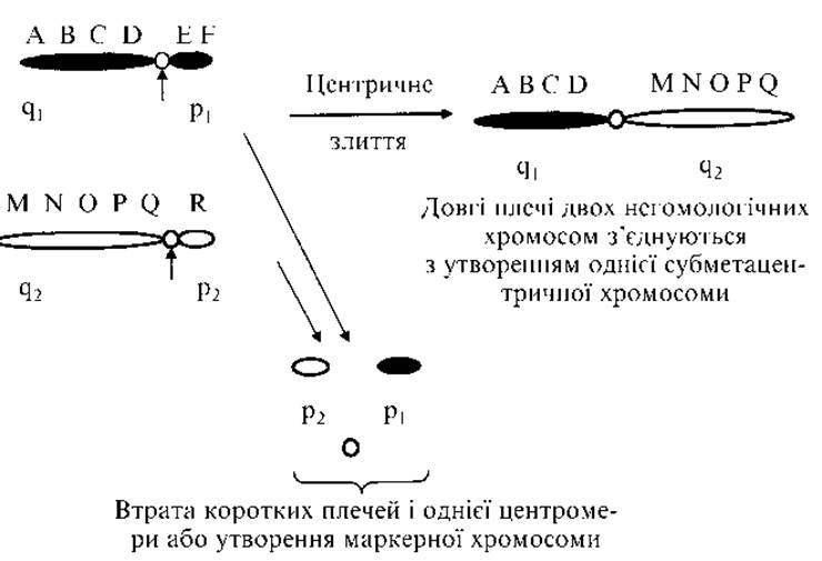
Fig. 2.9. Centric chromosome fusion (Robertsonian translocation)
An example is the fusion of the long arms of chromosomes 14 and 21 (14q21q). A carrier of such a balanced translocation has only 45 chromosomes and a normal phenotype (Fig. 2.10). An individual with a normal karyotype has two chromosomes from the 14th pair and two from the 21st pair (14,14,21,21). Normal gametes contain one chromosome 14 and one chromosome 21 (14,21). In a balanced translocation carrier, instead of four chromosomes, there will be three (14,14q21q,21).
Carriers theoretically produce 6 types of gametes (Table 2.3) with varying probabilities of formation.
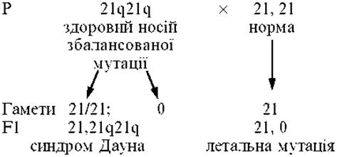
Sometimes one of the parents is a carrier of a balanced translocation characterized by centric fusion of two homologous chromosomes of group D or G. Such individuals produce Two Types of gametes. For instance, a 21q21q translocation yields the following gametes:
1) 21q21q;
2) 0 — i.e., a gamete lacking chromosome 21.
Following Fertilization by a normal gamete, two types of zygotes are formed: 1) 21,21 q21 q — the translocation form of Down syndrome; 2) 21,0 — monosomy of chromosome 21, a lethal mutation. The probability of having an affected child is 100 %.
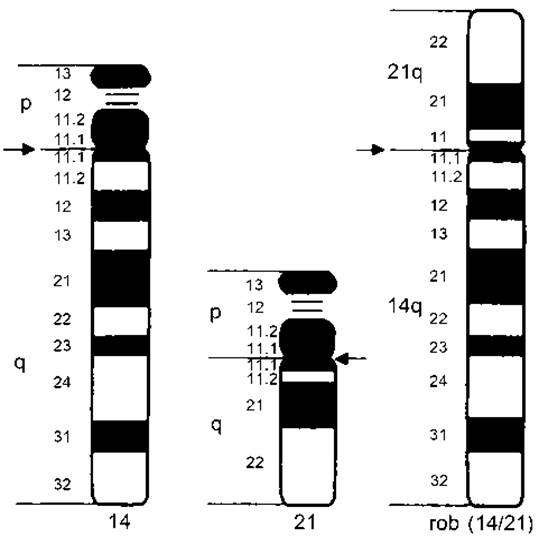
Fig. 2.10. Balanced Robertsonian translocation of chromosomes 14 and 21 (the carrier has 3 chromosomes: a normal 14, a normal 21, and a chromosome combining the long arms of chromosomes 14 and 21)
Table 2.3. Possible types of gametes and zygotes in a carrier of the balanced 14q21q translocation
|
Possible types of gametes in a carrier of a balanced translocation with karyotype 14,14q21q,21 |
Karyotype of zygotes formed after the fusion of possible gamete types with a cell containing a normal chromosome Complement (14,21) |
|
14,21 — normal chromosome complement |
14,14,21,21 — normal karyotype |
|
14q21q — balanced chromosomal mutation |
14,14q21q,21 — balanced chromosomal mutation, normal phenotype |
|
14,14q21q — gamete with an unbalanced mutation (extra long arm of chromosome 14) |
14,14,14q21q,21 — unbalanced chromosomal mutation with an extra long arm of chromosome 14, a lethal mutation leading to embryo demise at early developmental stages |
|
14q21q,21 — gamete with an unbalanced mutation (extra long arm of chromosome 21) |
14,14q21q,21,21 — translocation form of Down syndrome |
|
14 — missing chromosome of the 21st pair |
14,14,21 — monosomy for chromosome 21, a lethal mutation for Germ Cells or early embryos |
|
21 — missing chromosome of the 14th pair |
14,21,21 — monosomy for chromosome 14, a lethal mutation for germ cells or early embryos |
Deletions and duplications alter the gene dosage in an Organism (partial monosomy or trisomy). Inversions, translocations, and transpositions change the arrangement of genes within chromosomes.
7. Centric fission is the reverse phenomenon of centric fusion. A single chromosome splits into two, requiring the formation of a new centromere; otherwise, the acentric chromosome fragment is lost during Cell Division.
8. Dicentric chromosomes (Fig. 2.11) contain two centromeres. Two sister chromatids lose their telomeres and fuse into a single chromosome (Fig. 2.12). During Mitosis and Meiosis, the segregation of such chromosomes is impaired, leading to breaks.
9. A marker chromosome is an additional chromosome (specifically, a fragment of a chromosome containing a centromere). It typically resembles a very short acrocentric chromosome, and less frequently, a ring or other structure. If the marker chromosome contains exclusively heterochromatin, the phenotype remains unchanged. However, if it contains euchromatin (expressed genes), this is associated with The Development of a chromosomal disorder (analogous to the duplication of a chromosomal region).
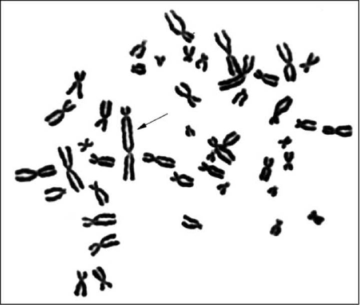
Fig. 2.11. Dicentric chromosome in a metaphase plate (indicated by the arrow)
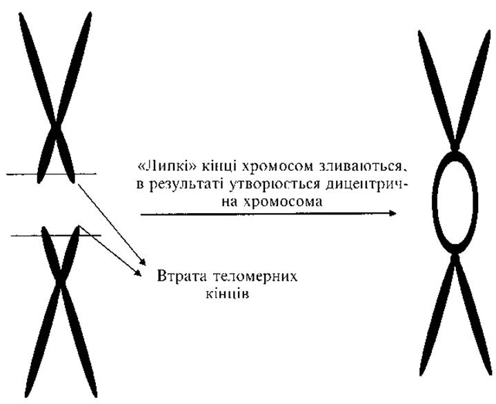
Fig. 2.12. Mechanism of dicentric chromosome formation
Significance of chromosomal mutations in evolution
Chromosomal mutations play a vital role in evolution. Throughout evolutionary history, karyotypes undergo active restructuring via inversions, Robertsonian translocations, and other mutational events. The more evolutionarily distant organisms are from one another, the greater the divergence in their chromosome sets.
For instance, at least one Robertsonian rearrangement occurred during the evolution of humans from apes. Humans have 23 pairs of chromosomes, whereas great apes have 24. The two arms of the large human chromosome 2 correspond to two separate chromosomes in apes (namely, chromosomes 13 and 14 of the gorilla and orangutan). Human and chimpanzee chromosomes 4, 5, 12, and 17 differ from one another by pericentric inversions.
Last update: 11/08/2026
Editorial and Educational Adaptation: This material has been compiled based on the primary/original source text. The project team performed an editorial review, corrected technical inaccuracies, structured sections, and adapted the content for an educational format.
What was processed:
- elimination of formatting defects (OCR errors, structural breaks, corrupted characters);
- editorial organization of content;
- standardization of terminology in accordance with academic sources;
- verification of factual statements against the original source text.
All mentions of the author, publication year, and origin of the primary text have been preserved in accordance with the source.