Harper's Illustrated Biochemistry, Volume 1 - Murray R. 1993
Structure and Function of Proteins and Enzymes
Enzymes: Regulation of Activity
Allosteric Regulation
Principles
The catalytic activity of certain regulatory Enzymes can be modulated by low-molecular-weight allosteric effectors, which typically exhibit either minimal structural similarity to the substrates or Coenzymes of the enzyme they regulate, or none at all. The inhibition of an enzyme catalyzing one of the reactions in a pathway by the end product of that pathway is termed feedback inhibition. In a biosynthetic pathway from A to D catalyzed by the enzymes Enz1, ... Enz3:
Class="center">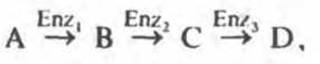
at high concentrations of D, The conversion of A to B is typically inhibited. This is not a simple "reversal" of the reaction caused by the accumulation of intermediates, but rather a consequence of the ability of product D to bind to the enzyme Enz1, acting as its inhibitor. Thus, D Functions as a negative allosteric effector of the enzyme, or a feedback inhibitor. Consequently, the inhibition of Enz1 by D regulates the synthesis of D. Typically, D binds to the inhibited enzyme at an allosteric site distinct from the catalytic site.
Kinetically, feedback inhibition may be competitive, noncompetitive, partially competitive, or mixed. Feedback inhibition is a hallmark of biosynthetic pathways. Very frequently, the feedback inhibitor is the small molecule immediately preceding the synthesis of a macromolecule (for example, an amino acid in Protein Synthesis, or a nucleotide in nucleic acid synthesis). Feedback regulation typically occurs at the first functionally irreversible1 step unique to that particular biosynthetic pathway.
Examples of feedback inhibition in microorganisms include the inhibition of phosphoribosyl ATP pyrophosphorylase by Histidine, anthranilate synthase by Tryptophan, and aspartate transcarbamoylase by CTP. In each case, the regulatory enzyme participates in a biosynthetic pathway leading to a single end product—His, Trp, or CTP.
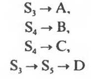
Biosynthetic pathways are frequently branched, with their initial reactions giving rise to the synthesis of two or more metabolites. Figure 10.4 indicates the likely sites in a branched biosynthetic pathway where simple feedback inhibition operates (Amino Acids, Purines, or Pyrimidines may serve as inhibitors). S1, S2, and S3 are precursors to all four end products (A, B, C, and D), S4 is a precursor to B and C, and S5 is a precursor exclusively to D. These sequences are linear and subject to end-product feedback inhibition.
Finer regulation is achieved through multiple feedback loops (Fig. 10.5). For instance, if B is present in excess, the demand for S2 decreases. Consequently, the ability of B to inhibit the process that generates this very product appears biologically advantageous. However, if an excess of B inhibits not only the reactions exclusively dedicated to the synthesis of B, but also those that concurrently lead to the synthesis of A, C, and D, it would impair the synthesis of all four products. This, clearly, would be disadvantageous. Nevertheless, mechanisms have evolved to overcome this difficulty.
Several variations of feedback inhibition exist. In cumulative inhibition, the inhibitory effect of two or more end products on the same regulated enzyme is strictly additive.
1 This refers to a reaction whose equilibrium (in the thermodynamic sense) lies far to one side, i.e., a reaction characterized by a large negative value of ∆G.
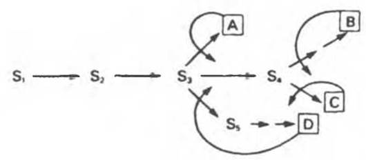
Fig. 10.4. Feedback inhibition at various points in a branched biosynthetic pathway. S1–S5 are intermediates generated during The Biosynthesis of end products A–D. Solid arrows denote enzymes catalyzing the indicated conversions. Curved arrows indicate feedback loops and potential sites of feedback inhibition by specific end products.
In concerted or multivalent inhibition, complete inhibition is observed only when two or more end products are simultaneously present in excess.
In cooperative inhibition, an excess of each individual end product exerts an inhibitory effect on the regulatory enzyme, but the combined inhibitory action of two or more end products vastly exceeds the additive effect characteristic of cumulative inhibition.
Another regulatory mechanism is observed in Enzymes of the aspartate family, which comprises multiple enzyme forms (isozymes), each possessing distinct regulatory characteristics. Three aspartokinases are synthesized in E. coli. One of these (AKL) is specifically and completely inhibited by Lysine, another (AKT) by Threonine, and the third (AKH) by homoserine, a precursor of Met, Thr, and Ile (Fig. 10.6). An excess of Lys inhibits AKL, leading to a decrease in the synthesis of ß-aspartyl phosphate. However, this alone is not yet sufficient to channel metabolites toward the synthesis of homoserine and subsequent products. Switching to this biosynthetic branch is achieved via feedback inhibition of enzymes participating in the downstream steps of the pathway. Lysine also inhibits the first enzyme in the reaction sequence leading from ß-aspartyl phosphate to lysine. This facilitates the unhindered synthesis of homoserine and, consequently, of threonine and isoleucine. The next regulatory locus is located at the branch point where one branch leads to Methionine and the other to threonine and isoleucine.
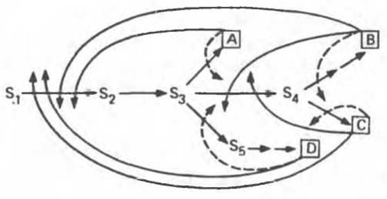
Fig. 10.5. Multiple feedback inhibition at various points in a branched biosynthetic pathway. In addition to simple feedback loops (dashed curved arrows), loops (solid curved arrows) regulating The activity of enzymes common to the biosynthesis of multiple end products are indicated.
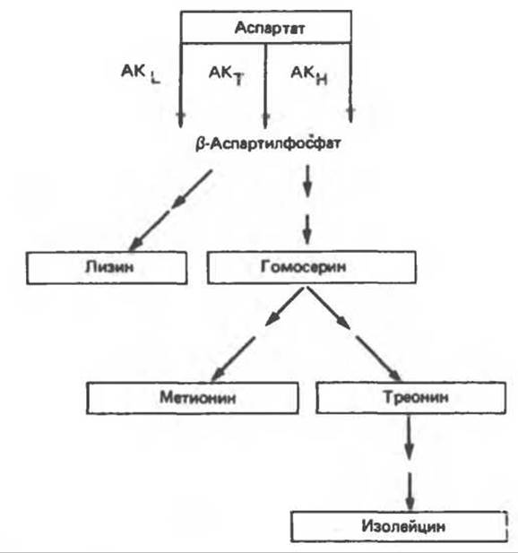
Fig. 10.6. Regulation of aspartokinase (AK) activity in E. coli Cells. The enzyme isoforms undergo selective inhibition by individual end products: lysine (AKL), threonine (AKT), and homoserine (AKH).
The actual operation of these Metabolic Regulation pathways is supported by data on the patterns of end-product inhibition in the aspartate-initiated biosynthetic pathway across various Bacteria (Table 10.2).
The best-characterized allosteric enzyme, aspartate transcarbamoylase, catalyzes the first unique reaction in pyrimidine biosynthesis (Fig. 10.7). Aspartate transcarbamoylase (ATCase) is feedback-inhibited by cytidine triphosphate (CTP). Upon Treatment with mercurial Reagents, ATCase loses its sensitivity to CTP while retaining full activity in the synthesis of carbamoylaspartate. This indicates that CTP binds not to the substrate-binding sites, but to a distinct allosteric site. ATCase consists of two catalytic and three or four regulatory protomers. Each catalytic protomer contains four aspartate-binding sites, and each regulatory protomer contains at least two CTP-binding (regulatory) sites. Each type of protomer is under independent Genetic control. This was demonstrated through the Selection of mutants lacking normal CTP regulation; revertants possessing nearly normal regulatory properties were subsequently isolated from these mutants.
Table 10.2. Variants of allosteric regulation of aspartokinase
|
Feedback inhibitor |
Repressor |
|
|
E. coli (kinase I) |
Homoserine |
|
|
E. coli (kinase II) |
Lys |
Lys |
|
E. coli (kinase III) |
Thr |
|
|
R. rubrum |
Thr |
|
|
B. subtilis |
Thr + Lys |
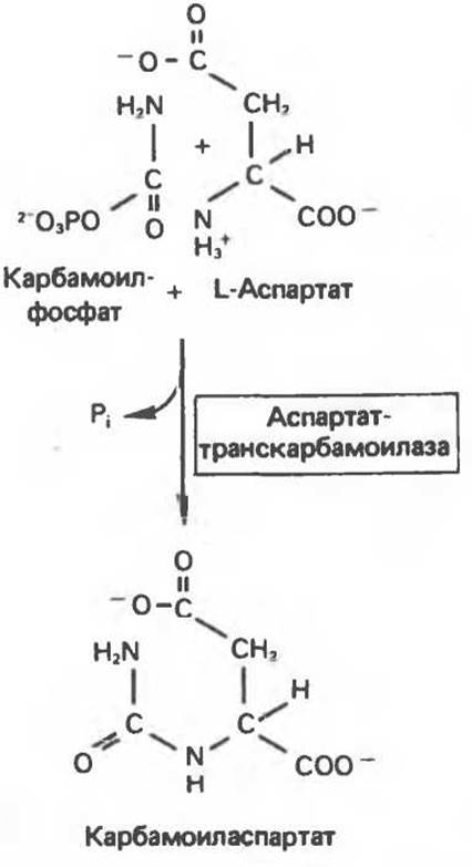
Fig. 10.7. Reaction catalyzed by aspartate transcarbamoylase (ATCase).
Evidence for the Existence of Allosteric Sites in Regulatory Enzymes
Around 1963, Monod drew attention to the lack of structural similarity between feedback inhibitors and their target enzyme substrates. The lack of isostericity with the substrate implies that the corresponding effectors are allosteric. Based on this, Monod suggested that enzymes regulated by such allosteric effectors (particularly feedback inhibitors) bind the effector at an allosteric site that is physically distinct from the catalytic site. Thus, Allosteric enzymes are those whose catalytic site activity is modulated by allosteric effectors binding at an allosteric site. Evidence supporting the existence of physically separate allosteric sites in regulatory enzymes includes the following.
1. When modified by chemical or physical Methods, regulatory enzymes often lose their sensitivity to allosteric effectors while retaining full catalytic activity. Selective Denaturation of allosteric sites has been demonstrated upon treatment with mercury-containing reagents, urea, X-rays, Proteolytic Enzymes, solutions of extreme Ionic strength or pH, as well as prolonged storage at 0–5 °C, freezing, or heating.
2. Allosteric effectors frequently protect the catalytic site from denaturation under conditions where the substrate offers no such protection. It is difficult to conceive of a situation where an effector binding at the catalytic site protects the enzyme while the substrate lacks this capability. Therefore, it is more logical to assume that the effector binds to a separate, allosteric site on the enzyme.
3. Mutant strains of bacteria and mammals have been discovered in which regulatory enzymes exhibit significantly different regulatory properties compared to wild-type enzymes, yet retain identical catalytic properties. This indicates that the structures of the allosteric and catalytic sites are determined by different Regions of the Gene.
4. It has been shown that the binding of substrates and allosteric effectors to regulatory enzymes occurs independently.
5. In certain enzymes (such as ATCase), the allosteric and catalytic sites are localized on different protomers.
Kinetics of Allosteric Inhibition
Fig. 10.8 illustrates the dependence of the reaction rate, catalyzed by a typical allosteric enzyme, on Substrate Concentration in the presence and absence of an allosteric inhibitor. In the absence of the inhibitor, a hyperbolic saturation curve is observed. In its presence, the curve becomes sigmoidal; at high substrate concentrations, the reaction rate may approach values close to those observed without the inhibitor. Note the analogy here with the oxygen saturation curves of Myoglobin and Hemoglobin (Chapter 6).
Kinetic analysis reveals that feedback inhibition can be competitive, noncompetitive, partially competitive, or of another nature. If, at high concentrations of S, the enzyme activity is approximately the same in the presence and absence of the allosteric inhibitor, the kinetics superficially resemble competitive inhibition. However, because the substrate saturation curve remains sigmoidal rather than hyperbolic, the double-reciprocal (Lineweaver-Burk) plotting method is inapplicable to allosteric inhibitors; it was originally formulated for competitive inhibition at the catalytic site. Since allosteric effectors bind to a separate (allosteric) site on the enzyme, the original kinetic model no longer holds.
The sigmoidal dependence of V on [S] in the presence of an allosteric inhibitor is due to cooperativity. At low concentrations of S, activity in the presence of the inhibitor is significantly lower than in its absence. However, as [S] increases, the inhibitory effect becomes less pronounced. Similar kinetics are observed when There are two or more interacting substrate-binding sites: the binding of a substrate molecule to one catalytic site facilitates the binding of a second substrate molecule to another site. Substrate-binding cooperativity was described in Chapter 6 using Hemoglobin as an example. The sigmoidal Nature of the oxygen saturation curve of hemoglobin is due to cooperative interactions among four O2-binding sites located on different protomers.
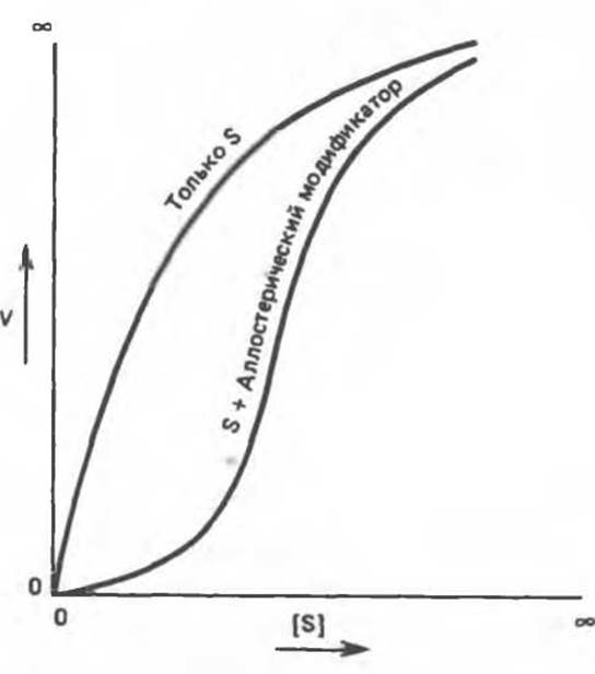
Fig. 10.8. Sigmoidal substrate saturation curve in the presence of an allosteric inhibitor.
Models of Allosteric Regulation
Attempts to describe The kinetics of allosteric inhibition as "competitive" or "noncompetitive" with respect to the substrate rely on a purely mechanical analogy that can be misleading. Therefore, we will classify regulatory enzymes into two categories: K-series and V-series enzymes. Substrate saturation of K-series allosteric enzymes appears as a competitive process in the sense that Km increases (substrate affinity decreases), while Vmax remains entirely unchanged. In the case of V-series allosteric enzymes, Vmax decreases (catalytic efficiency drops), whereas the apparent Km value remains constant. Changes in Km or Vmax are presumably driven by Conformational Changes in the catalytic site induced by the binding of the allosteric inhibitor at the allosteric site. In K-series allosteric enzymes, these conformational changes weaken the bond between the substrate and substrate-binding residues. In V-series allosteric enzymes, the primary effect involves an alteration in the orientation of catalytic residues, leading to a lowered Vmax. Nevertheless, intermediate cases can occur where conformational changes affect both Km and Vmax.
Various models have been proposed to describe The regulation of allosteric enzymes, yet it is unlikely that any single model can account for The behavior of all regulatory enzymes. Because the sigmoidal nature of the substrate saturation curve confers distinct regulatory advantages, any mutation leading to a sigmoidal saturation curve tends to be conserved in a population. One can hardly expect all such Mutations to dictate the exact same inhibition mechanism. Consequently, the sigmoidity of kinetics alone provides no direct insight into the underlying mechanism of inhibition.
Physiological Consequences of Cooperativity
Substrate-binding cooperativity produces consequences analogous to those seen in the cooperative O2 binding of hemoglobin. At low substrate concentrations, the allosteric effector acts as a potent inhibitor. Thus, it exerts its regulatory effect most efficiently precisely when it is most needed: at low intracellular substrate concentrations. As the substrate concentration rises, The Need for stringent regulation diminishes, leading to decreased inhibition and consequently greater product formation. By analogy with hemoglobin, a sigmoidal substrate saturation curve in the presence of an inhibitor implies that relatively small shifts in substrate concentration result in large changes in activity. This allows for fine-tuning of catalytic activity in response to minor fluctuations in substrate level. Finally, much like the oxygenation curves of Hemoglobins from different animal species, the sigmoidal saturation curves of regulatory enzymes from diverse sources can be shifted to the left or right, optimizing their fit to the prevailing in vivo substrate concentrations.
Feedback Regulation in Mammalian Cells
In mammalian cells, much like in bacterial cells, end products regulate their own synthesis via feedback mechanisms. In some instances (notably ATCase), feedback inhibition targets the first enzyme of the biosynthetic pathway. However, we must distinguish between feedback regulation—a broad term implying no specific mechanism—and feedback inhibition, which denotes a specific regulatory mechanism operating in many bacterial and mammalian enzymes through direct inhibition. For instance, dietary Cholesterol suppresses its own synthesis from acetate in mammalian Tissues. This type of regulation, however, does not directly inhibit the first enzyme of the cholesterol biosynthetic pathway. Instead, inhibition affects a rate-limiting enzyme (HMG-CoA reductase); operating early in biosynthesis, this mechanism involves the repression—by cholesterol or its metabolites—of Gene Expression encoding HMG-CoA reductase. Cholesterol added directly in vitro to an isolated HMG-CoA reductase system exerts no effect on its catalytic activity.
Last update: 06/08/2026
Editorial and Educational Adaptation: This material has been compiled based on the primary/original source text. The project team performed an editorial review, corrected technical inaccuracies, structured sections, and adapted the content for an educational format.
What was processed:
- elimination of formatting defects (OCR errors, structural breaks, corrupted characters);
- editorial organization of content;
- standardization of terminology in accordance with academic sources;
- verification of factual statements against the original source text.
All mentions of the author, publication year, and origin of the primary text have been preserved in accordance with the source.