Principles of Biochemistry, Volume 3 - A. Lehninger 1985
Molecular Mechanisms of Genetic Information Transfer
Protein Synthesis and Its Regulation
Newly synthesized proteins are directed to their cellular destinations
Some newly synthesized Proteins are released directly into The Cell Cytosol, others are directed to various cell Organelles, still others are secreted from the cell, and some are integrated into various cell membranes, where they function as transport proteins or membrane Enzymes. Therefore, it is essential that a newly synthesized protein finds its way to its designated cellular destination. How does it accomplish this?
Many proteins contain specific polypeptide "leaders" at their N-terminus that act as signals directing these proteins to their destination. These signal sequences can aptly be compared to a postal zip code on a letter. Proteins synthesized by Ribosomes of the rough Endoplasmic reticulum in pancreatic Cells and exported from these cells—for example, trypsinogen and procarboxypeptidase (sec. 24.1,6)—possess polypeptide leader sequences at their N-termini. These signal sequences consist of 15–30 amino acid residues, many of which contain hydrophobic R-groups (Fig. 29-20). During the synthesis of any protein, including those destined for export, the signal leaders, being located at the N-terminus, are formed first. These leaders are recognized by specific receptor sites on the outer surface of The endoplasmic reticulum, and this process occurs even before the ribosome fully completes Protein Synthesis. The hydrophobic, lipid-soluble portion of the leader sequence penetrates through the membrane into the cisternae of the endoplasmic reticulum, dragging the growing polypeptide chain along with it. Inside the cisternae, the signal leader is cleaved by a specific peptidase. Afterward, the mature protein is directed to the Golgi apparatus, encapsulated, and finally leaves the cell in the form of a secretory vesicle. Many other exported proteins that function outside the cell—such as Blood Plasma Proteins, polypeptide Hormones, Antibodies, and mucoproteins—may reach their destination via a similar pathway.
Class="center">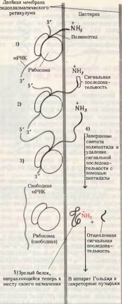
Fig. 29-20. Stages of synthesis of a protein destined for export from the cell (1–5). The formation and subsequent transformations of the signal sequence—a polypeptide leader located at the N-terminus of many proteins synthesized by ribosomes of the rough endoplasmic reticulum—are shown. Signal sequences help the newly formed polypeptide chain penetrate through the membrane into the cisternae. During or after The entry of the polypeptide into the cisterna, the signal sequence is cleaved from it by a peptidase.
Very similar processes occur in Bacteria. The outer membrane of E. coli cells consists of Lipids and proteins. The latter are synthesized on ribosomes bound to the inner surface of the inner membrane. Signal sequences at the N-termini of these proteins ensure their passage through the inner membrane and across The Cell wall to the appropriate sites on the outer membrane, where polypeptide insertion takes place. Genetic studies of signal sequences and the peptidases that ultimately remove them have been made possible by the availability of corresponding bacterial mutants.
Last update: 06/08/2026
Editorial and Educational Adaptation: This material has been compiled based on the primary/original source text. The project team performed an editorial review, corrected technical inaccuracies, structured sections, and adapted the content for an educational format.
What was processed:
- elimination of formatting defects (OCR errors, structural breaks, corrupted characters);
- editorial organization of content;
- standardization of terminology in accordance with academic sources;
- verification of factual statements against the original source text.
All mentions of the author, publication year, and origin of the primary text have been preserved in accordance with the source.