Principles of Biochemistry, Volume 1 - A. Lehninger 1985
Biomolecules
Cells
The endoplasmic reticulum forms channels in the cytoplasm
The Cytoplasm of virtually all Eukaryotic Cells contains a highly complex, three-dimensional labyrinth of membrane-bounded channels known as the Endoplasmic reticulum, whose numerous folds and branches permeate the entire cytoplasm (Fig. 2-12). The spaces inside the endoplasmic reticulum, referred to as cisternae, act as channels for The transport of various substances, typically from The Cell to the external environment. In some cells, however, the cisternae serve as storage sites for nutrient reserves. There are two types of endoplasmic reticulum: rough and smooth. The outer surface of the rough endoplasmic reticulum membrane is studded with Ribosomes, whereas the smooth endoplasmic reticulum lacks ribosomes entirely. The ribosomes associated with the rough endoplasmic reticulum participate in the Biosynthesis of Proteins that either remain temporarily within the cell or are immediately secreted from it. Proteins synthesized on membrane-bound ribosomes are translocated across the membrane into the lumen of the cisternae, from which they are ultimately dispatched into the extracellular space. The endoplasmic reticulum also plays a role in Lipid Biosynthesis. In various types of eukaryotic cells, it varies in Morphology and performs diverse Functions. For instance, in Skeletal Muscle cells, where contraction is triggered by Ca+ ions, the endoplasmic reticulum facilitates muscle relaxation by mediating the reabsorption of Ca2+ ions.
Class="center">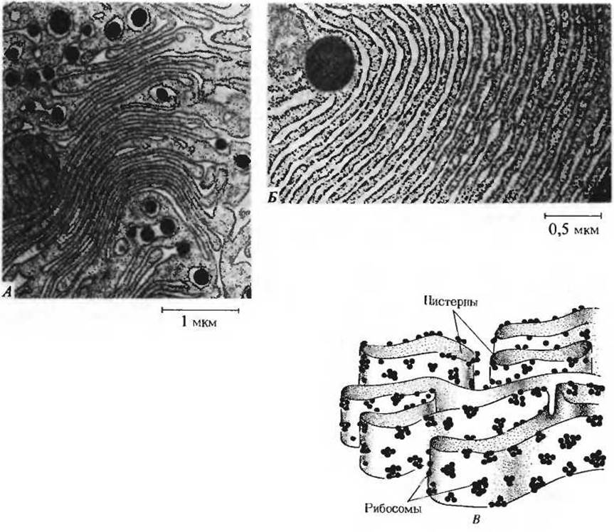
Fig. 2-12. A. Endoplasmic reticulum (rough) of a pancreatic cell. Cells of this type are highly active in synthesizing proteins on ribosomes attached to the outer surface of the membrane and subsequently secreting them into the cisternae. A portion of a mitochondrion is visible on the left. B. Individual ribosomes and cisternae at a higher magnification. C. Schematic diagram illustrating the three-dimensional Structure OF THE endoplasmic reticulum, showing how continuous, labyrinthine channels pervading a substantial part of the cellular cytoplasm are formed from narrow cisternae.
Last update: 06/08/2026
Editorial and Educational Adaptation: This material has been compiled based on the primary/original source text. The project team performed an editorial review, corrected technical inaccuracies, structured sections, and adapted the content for an educational format.
What was processed:
- elimination of formatting defects (OCR errors, structural breaks, corrupted characters);
- editorial organization of content;
- standardization of terminology in accordance with academic sources;
- verification of factual statements against the original source text.
All mentions of the author, publication year, and origin of the primary text have been preserved in accordance with the source.