Fundamentals of Biochemistry - Filippovich, Y. B. 1999
Hormones and Their Role in Metabolism
Peptide Hormones
Structure AND Functions of peptide Hormones. Several dozen natural peptide hormones are currently known, and this list is gradually expanding.
Thanks to the widespread application of Methods from rapidly developing Protein Chemistry in recent years, A number of peptide hormones have been obtained in a homogeneous state, their Amino Acid Composition has been studied, their Primary Structure (as well as secondary, tertiary, and quaternary structure in the case of protein hormones) has been elucidated, and some of them have been synthesized. Moreover, major breakthroughs in chemical Peptide Synthesis have made it possible to artificially produce numerous Peptides that are isomers or analogues of Natural peptides. Studying the hormonal activity of the latter has provided exceptionally important insights into the relationship between The structure of peptide hormones and their function. Examples of this will be discussed below.
The most important peptide hormones include oxytocin, vasopressin, gastrin, Glucagon, Insulin, adrenocorticotropic hormone, melanocyte-stimulating hormone, parathyroid hormone, thyrotropin, and Growth Hormone. In recent years, a large family of neurohormones, opioid peptides, and releasing factors has been discovered and investigated. Let us examine the structure and functions of these hormones.
Oxytocin is a 9-membered peptide isolated by V. du Vigneaud and co-workers from the posterior Pituitary Gland. In 1953 (simultaneously with H. Tuppy), the same scientist proposed the complete structural formula of oxytocin:
Class="center">
As can be seen from the formula above, oxytocin contains a ring closed by a disulfide bond between the 1st and 6th Cysteine residues in its molecule.
Within just 20–30 seconds after intravenous administration of a mere 1 mcg, oxytocin induces contraction of the Muscle fibers surrounding the alveoli of the Mammary Glands in laboring women, leading to milk ejection. In addition, as childbirth approaches, the sensitivity of the uterine Muscles to oxytocin increases, causing them to contract under its influence. Therefore, this hormone promotes normal progression of labor.
Oxytocin has been synthesized, and numerous isomers and analogues of it have also been created. This has made it possible to thoroughly elucidate The Significance of each amino acid residue and their combinations for the functional activity of the hormone. It turned out that the opening of the disulfide bridge in the oxytocin molecule is accompanied by its complete inactivation. The amidated aspartic acid residue (position 5) is of paramount importance for the biological action of oxytocin. Acylation of the amide groups (at positions 4, 5, and 9) leads to a decrease in oxytocin activity. The presence of a free OH group in the Tyrosine residue promotes the expression of full physiological activity. Removal of the tyrosine side chain from the oxytocin molecule leads to its acquisition of The ability to inhibit the action of other Pituitary Hormones. Replacing isoleucine (position 3) with leucine, norleucine, valine, or alloisoleucine confirms that the isoleucine side chain serves for the specific binding of the hormone to the receptor. The attachment of Other Amino Acids or short peptides to the free NH2 group of the cysteine residue (position 1) leads to an extended duration of action of the hormone in the Organism compared to the control.
Vasopressin is a 9-membered peptide, also isolated from the posterior pituitary gland. The credit for elucidating its structure and developing its synthesis method belongs to V. du Vigneaud (1953–1956). The structure of vasopressin closely resembles that of oxytocin (see Fig. 22 on p. 50).
As can be easily noticed, with the exception of positions 3 and 8, the structure of vasopressin entirely replicates that of oxytocin. This is the structure of vasopressin isolated from the pituitary glands of humans, monkeys, cattle, horses, camels, sheep, and dogs. In the vasopressin molecule obtained from the porcine pituitary gland, an Arginine residue (position 8) is replaced by a Lysine residue. This indicates the presence of species Specificity in the structure of vasopressin.
Being structurally similar to oxytocin, vasopressin is also somewhat similar in its functional activity: it stimulates the contraction of vascular smooth muscles. However, this function is not the primary one for vasopressin. Its main action is directed toward the Regulation of Water METABOLISM. Vasopressin ensures the proper level of water reabsorption in the distal renal tubules of higher vertebrates, regulating the body's Water Balance and Blood Plasma osmotic pressure. Naturally, it is capable of increasing blood pressure, which is reflected in its name.
Gastrin is a 17-membered peptide secreted by the mucosa of the gastric antrum. Its primary structure has been deciphered, and it has been obtained synthetically:

Gastrin stimulates the secretion of gastric juice. Its activity in this regard is 500 times that of histamine, a previously known stimulant of gastric secretion. Gastrin exerts an excitatory effect almost exclusively on Hydrochloric acid production and only slightly increases Pepsin output. It promotes pancreatic secretion and enhances the tone and contraction of the Muscles of the Stomach and Small Intestine. This specific hormonal activity of gastrin is almost entirely attributed to the presence of the C-terminal tetrapeptide (residues 14–17) in its molecule.
Since a similar situation is observed in many peptide hormones as well as Proteins with other biological functions, it is of great interest to determine the purpose of the remaining part of the polypeptide that does not directly participate in carrying out the hormonal, biocatalytic, or other function.
Glucagon is a 29-residue peptide synthesized in the α-Cells of the pancreatic islets. The first mention of this hormone dates back to 1923, when J. Murlin and co-workers detected its presence in insulin preparations. In 1953, F. Staub obtained glucagon as a homogeneous crystalline preparation, and its primary structure was elucidated somewhat later. It is as follows:

Excessive secretion of glucagon by the Pancreas, or its artificial administration to animals and humans, leads to a transient increase in blood glucose levels—hyperglycemia. This effect of glucagon is due to the fact that it promotes The conversion of the less active form of Liver phosphorylase into the more active one (see p. 334). As a result, the action of phosphorylase a enhances Glycogen breakdown in The Liver and increases the glucose content (in the form of glucose-1-phosphate) in the blood. Naturally, liver glycogen stores decrease in the process, and Glycogenolysis is intensified throughout the body. Thus, glucagon promotes carbohydrate degradation.
Insulin is a protein produced in the ß-cells of the pancreas. Its structure has been studied in detail. Insulin was the first protein whose primary structure was elucidated by F. Sanger (see p. 61). It was also the first protein obtained by chemical synthesis.
The presence of a Carbohydrate Metabolism-influencing hormone in the pancreas was first noted by von Mering and O. Minkowski (1889). Later, L. V. Sobolev (1901) established that the islet tissue serves as the source of insulin in the pancreas, which is why in 1909 this hormone, not yet isolated as an individual entity, was named insulin (from Lat. insula — island). In 1922, F. Banting and C. Best first prepared an active insulin formulation, and by 1926 methods for its isolation in a highly purified state had been developed, including in the form of crystalline preparations containing 0.36% Zn.
The Molecular Weight of crystalline insulin is 36,000. Its molecule is a multimer composed of six protomers and two Zn atoms. The protomers form dimers that interact with the imidazole rings of the His10 residues of the B chain, facilitating their aggregation into a hexamer (Fig. 136). Upon dissociation, the multimer yields three subparticles, each with a molecular weight of 12,000. In turn, each subparticle splits into two equal parts with MW = 6,000. All of these modifications of insulin—protomer, dimer, and hexamer—possess full hormonal activity. Therefore, the insulin molecule is often equated with the protomer, which exhibits full biological activity (MW = 6,000), especially since insulin exists in a monomeric form under physiological conditions. Further fragmentation of the insulin molecule (with MW = 6,000) into chain A (consisting of 21 amino acid residues) and chain B (consisting of 30 amino acid residues) leads to the loss of hormonal properties.

Fig. 136. Structure of insulin and its receptor:
I — Tertiary Structure of the protomer; A1–A21 — chain A; B1–B30 — chain B; the dashed line indicates the receptor-binding region; II — Quaternary Structure of the hexameric molecule; each block corresponds to a protomer; one block is omitted to show the interaction of zinc ions (dashed lines) with three Histidine residues and of a calcium ion with three glutamic acid residues (both residues are in chain B, at positions 10 and 13, respectively); both ions are also bound to three water molecules each (not shown in the figure); III — transport systems sensitive to the regulatory Action of Insulin on target cells: 1 — stimulation of glucose transport; 2 — stimulation of the Na+, K+-pump; 3 — increase in The rate of Na+/H+ exchange; 4 — inhibition of the Ca2+-pump; 5 — stimulation of Amino Acid Transport; 6 — increase in The activity of the Na+/Ca2+ exchange system; P — receptor; H — hormone; IV — hypothetical STRUCTURE OF THE Insulin Receptor (α2β2 chains linked by Disulfide Bonds; α — 93,000, ß — 135,000 Da). It is a glycoprotein (MW = 460 kDa) embedded in Cell/30.html">The Plasma Membrane of The Cell; the α-subunits are exposed externally, while the ß-subunits span the membrane; the polypeptide chain fragment of the ß-subunit localized in the Cytosol exhibits tyrosine kinase activity, specifically toward phosphatase; the latter, upon activation, dephosphorylates a number of carbohydrate metabolism Enzymes and alters their activity
Insulins isolated from the pancreas of various animals are nearly identical in primary structure (see Table 8). When insulin Biosynthesis in the human pancreas is insufficient (normally, 2 mg of insulin is synthesized daily), a characteristic disease develops, known as Diabetes Mellitus. This condition is accompanied by elevated blood glucose levels (hyperglycemia) and increased urinary glucose excretion (glucosuria). Concurrently, various secondary phenomena develop—muscle glycogen content drops, The biosynthesis of peptides, proteins, and Lipids slows down, and Mineral Metabolism is disrupted, among other disturbances.
Administering insulin via injection or per os as a liposome-encapsulated preparation produces the opposite effect: a decrease in blood glucose levels, an increase in muscle glycogen stores, enhanced anabolic processes, and the normalization of mineral metabolism, among others. All of the phenomena listed above result from insulin-induced Changes in the glucose permeability of cell membranes, which feature high- and low-affinity Ca2+-dependent insulin receptors on their surface. By increasing glucose penetration into cells and subcellular particles, insulin enhances its utilization across various Tissues, whether through glycogen biosynthesis or its dichotomous or apotomous breakdown (Fig. 136).
The interaction of insulin with The cell membrane receptor stimulates the activity of the protein kinase domain of the insulin receptor (Fig. 136, IV), which impacts the intracellular metabolism of CARBOHYDRATES, lipids, and proteins. The mechanism behind this becomes clear upon examining Fig. 136, III. The adenylate cyclase MECHANISM OF ACTION is not typical for insulin.
Adrenocorticotropic hormone (ACTH) is a 39-residue peptide produced by the anterior pituitary gland. Although discovered in 1928, it was not until a little less than three decades later that its primary structure was successfully elucidated—first for ovine ACTH (C. Li et al., 1955) and subsequently for porcine ACTH (P. Bell et al., 1956). The primary structures of bovine, human, and shark ACTH are now also known. As an example, let us examine the structure of human ACTH:
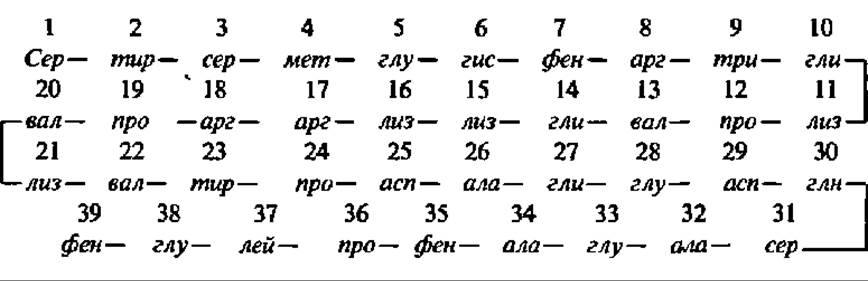
As C. Hofmann demonstrated, the species specificity of ACTH is determined by The sequence of amino acid residues at positions 25–33. The segment of the ACTH molecule spanning amino acid residues 1 through 13 is absolutely essential for the hormone's activity. Positioned between amino acid residues 14 and 20 is the hormone's binding site (residues 15–18, i.e., —lys—lys—arg—arg—). The peptide sequence consisting of 19 amino acid residues (positions 21–39) can be removed without any loss of hormonal activity, yet it dictates its immunological specificity.
ACTH exerts multifaceted effects: it increases the activity of phosphorylase, lipase, and glucose-6-phosphate dehydrogenase, and enhances the synthesis of proteins and Ribonucleic Acids, among other functions. However, its primary physiological role is to regulate the rate and scope of corticosteroid biosynthesis by the Adrenal Glands. Conversely, a drop in blood corticosteroid concentrations below a certain threshold stimulates ACTH production in the anterior pituitary. Furthermore, ACTH is known to primarily stimulate the biosynthesis of glucocorticosteroids, thereby altering the ratio among the various corticosteroids produced by the adrenal glands.
There is reason to believe that, on the one hand, ACTH promotes corticosteroid biosynthesis by ensuring a higher intracellular level of NADPH, which is required for their de novo synthesis from Cholesterol. On the other hand, ACTH activates membrane-bound adenylate cyclase, thereby stimulating cAMP biosynthesis (see below), which influences the intensity of protein kinase reactions during steroidogenesis.
Melanocyte-stimulating hormone (MSH) is an 18-residue peptide produced by the intermediate lobe (or, in certain species such as the pig, the posterior lobe) of the pituitary gland in animals. Human MSH is a 22-residue peptide closely resembling animal MSH.
Although evidence regarding the existence of MSH in the intermediate pituitary lobe dates back to 1932, it was first isolated in 1955 from the posterior pituitary of a pig by J. Porath, who developed a detailed method for purifying pituitary peptides. It was designated as a-MSH. That same year, a similar MSH was also isolated from the posterior pituitary and designated as ß-MSH.
The molecular structure of porcine a-MSH, elucidated by J. Harris and P. Roos (1956), is as follows:

The primary structures of a- and ß-MSH from humans, monkeys, cattle, horses, camels, and sharks are now also known, and both porcine a- and ß-MSH have been synthesized. Additionally, approximately two dozen MSH analogs have been obtained.
The Mechanism of action of ß-MSH on melanocytes in lower vertebrates is currently viewed through the lens of ß-MSH inducing the transition of melanin-containing granules from a gel to a sol state. This process leads to the dispersion of pigment throughout the cell and subsequent darkening. For lower vertebrates, the ability to alter integumentary coloration serves as an ADAPTATION TO ENVIRONMENTAL conditions. The Physiological Role of MSH in higher vertebrates remains unclear.
Parathyroid hormone (PTH) is a protein synthesized by the Parathyroid glands. J. Collip (1925) was the first to point out the hormonal properties of an acidic extract derived from the parathyroid glands. However, parathyroid hormone was isolated in a pure state much later, thanks to the efforts of G. Aurbach and H. Rasmussen (1959–1964). It was found to be a protein with a molecular weight (M) of 9,500, consisting of 84 amino acid residues. The Introduction/19.html">Primary structure of bovine parathyroid hormone was finally elucidated in 1970 by J. Potts et al.:

Human and porcine parathyroid hormones share a similar structure, with only minor differences (just a few Amino Acid Substitutions).
Parathyroid hormone regulates the blood levels of calcium cations and phosphoric and citric acid anions. Prolonged dietary calcium deficiency or impaired intestinal calcium absorption leads to a drop in blood calcium levels. This triggers an increased Synthesis and Secretion of the hormone by the parathyroid glands, which mobilizes calcium salts (in the form of citrates and phosphates) from Bone tissue. Maintaining a normal blood Ca2+ level is achieved by parathyroid hormone-stimulated renal phosphate excretion, which in turn slows down the deposition of calcium phosphate in bones.
The biological activity of parathyroid hormone is ensured by an N-terminal fragment of at least 28 amino acid residues in length, with the first two N-terminal amino acids being particularly crucial.
The targets of parathyroid hormone action are receptor proteins on the plasma membrane of target cells and the subsequent activation of adenylate cyclase, resulting in elevated intracellular cAMP levels (see below). This is accompanied by enhanced protein kinase reactions and the activation of membrane-bound Ca2+-dependent ATPase, leading to the redistribution of calcium among cellular compartments and, ultimately, across tissues and Organs.
Thyrotropin (thyroid-stimulating hormone) is a protein secreted by the anterior pituitary gland. It is a glycoprotein with M = 28,300, composed of two unequal subunits (M = 13,600 and 14,700) that are exceptionally rich in disulfide bridges (5 and 6, respectively). The primary structures of bovine and porcine thyrotropin have been elucidated. Thyrotropin deficiency (pituitary hypofunction) leads to diminished thyroid activity, a decrease in its size, and a 50% reduction in the blood levels of its secreted hormone, thyroxine (see below).
Thus, thyrotropin stimulates Thyroid Gland activity. In turn, the secretion of thyrotropin is regulated through a negative feedback loop by THYROID HORMONES. Consequently, the functions of these two Endocrine glands are finely coordinated.
The administration of thyrotropin triggers multiple metabolic shifts: within 15–20 minutes, the secretion of thyroid hormones increases, along with the uptake of iodine required for their synthesis (see below); thyroid oxygen consumption rises, glucose oxidation increases, and Phospholipid Metabolism and de novo RNA Synthesis are activated. It has now been established that the mechanism of action of thyrotropin, much like that of many other peptide hormones, relies on the activation of adenylate cyclase located in close proximity to the receptor protein to which thyrotropin binds. As a result, a series of processes is accelerated within The Thyroid Gland, including the Biosynthesis of Thyroid hormones.
Growth hormone (somatotropic hormone, STH) is a protein secreted by the anterior pituitary gland of vertebrates. Its presence in pituitary extracts was noted as early as 1921 by H. Evans and J. Long; however, it was not until two decades later (1944) that it was obtained as a purified preparation, and several years after that (1948) in crystalline form.
Depending on the animal species (cattle, sheep, pigs, rats, etc.), the molecular weight of the crystalline growth hormone preparation ranges from 20,000 to 22,000.
The human pituitary gland contains between 3.7 and 6.0 mg of STH. Possessing an M of 21,000, it consists of a single polypeptide chain of 191 amino acid residues. Its primary structure was elucidated by C. Li et al. (1969) and refined by G. Niall et al. (1973). The primary structures of bovine and ovine STH (191 amino acid residues each) are also known. Furthermore, the Amino acid sequences of growth hormone in numerous fish species (Lake Baikal omul, chum salmon, trout, eel, cod, salmon, pike, etc.) have been studied, revealing the multiplicity of genes encoding slightly variant polypeptide chains of STH even within a single fish species. These findings open up a new avenue in the chemistry of peptide hormones, shedding light on the underlying causes of sequence multiplicity not only in STH but in many other instances as well.
The tertiary structure of human growth hormone (HGH) is characterized by the close proximity of its C- and N-terminal amino acids, the presence of four robust helices spanning half of its polypeptide chain, and an unstructured sequence comprising the other half of the molecule. The recognition of the HGH receptor involves its 1st and 2nd α-helices along with a portion of the disordered loop connecting them; subsequently, a second receptor molecule binds via the 1st and 3rd helices, driving receptor dimerization and hormonal signal Transduction. Normal human blood levels of HGH vary widely, ranging from 0–3 µg/ml after overnight fasting up to 100 µg/ml following glucose intake.
Growth hormone exerts a pronounced anabolic effect, influencing virtually all cells in the body by upregulating biosynthetic processes. It enhances the Biosynthesis of Proteins, DNA, RNA, and glycogen, while promoting lipid mobilization from fat depots and accelerating the Breakdown of Higher Fatty acids and glucose. Furthermore, HGH improves renal Tubular Function and normalizes the body's mineral and water balance. While all these factors contribute to somatic growth, the ultimate scope of HGH action extends far beyond mere growth regulation.
Molecular-level studies have demonstrated that HGH stimulates the activity of RNA Polymerases and the cellular polyribosome apparatus. As evidenced by experiments using radioactive phosphorus tracers, the earliest metabolic response to growth hormone is the synthesis of mRNA and rRNA precursors within the Cell Nucleus. In addition, HGH significantly affects cell membrane permeability, substantially increasing the intracellular pool of free amino acids, which in turn facilitates de novo Protein Synthesis. It is possible, however, that the primary trigger for all these events is the activation of membrane-bound adenylate cyclase. Moreover, HGH has been shown to elevate blood levels of specific growth-promoting factors known as somatomedins—proteins with an M ≈ 7,000. The mechanism of their action is currently an active area of research.
The obesity hormone, leptin (from the Greek leptos, meaning thin), was discovered by a research team led by J. Friedman in 1992, although initial insights into the "ob" (obese) Gene Mutations causing obesity in mice emerged in the early 1950s. Synthesized in adipocytes as a 167-residue precursor, this protein hormone is secreted into the bloodstream as an active peptide hormone consisting of 145 amino acid residues, whose sequence has been fully elucidated. Parenteral or intravenous administration of just a few micrograms per animal daily leads to increased Energy Expenditure, suppressed food intake, elevated locomotor activity, and weight loss in mice due to a reduction in body fat stores. Upon cessation of leptin administration, all measured parameters fairly rapidly return to baseline levels. It is hypothesized that the obesity hormone is secreted into the blood by adipocytes in fluctuating amounts According to the body's physiological demands. Which cells or tissues act as the "lipostat," as well as the exact Mechanisms of Hormone-receptor interaction and the subsequent metabolic responses, remain unknown. It is worth noting that the potential use of leptin in treating human obesity requires dedicated clinical investigation.
Mechanism of action of peptide hormones. Peptide hormones do not penetrate target cells; instead, they interact with specific protein receptors located on the cell surface within the plasma membrane. Consequently, their mechanism of action differs fundamentally from that of Steroid Hormones.
The vast majority of peptide hormones trigger the activity of adenylate cyclase embedded within the plasma membrane upon binding to their receptor complex:
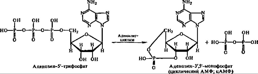
The resulting compound, cyclic adenosine monophosphate (cAMP)—discovered in 1957 independently by E. Sutherland and co-workers and D. Markham and co-workers—acts as the intracellular messenger transmitting the hormonal signal to the cell's metabolic machinery; in essence, it serves as a secondary messenger in signal transduction (with the primary messenger being the hormone-binding receptor protein). Specifically, cAMP functions as an allosteric regulator of protein Kinases, which in turn phosphorylate chromatic Histones and non-histone proteins (affecting the cell's genomic metabolic activity and, notably, mRNA biosynthesis rates), ribosomal proteins and Translation factors (influencing the intensity of Protein synthesis in the ribosomal machinery), numerous enzymes (determining their activation state), and so forth. Because these processes affect fundamental aspects of metabolism, the biochemical and physiological phenomena observed during peptide hormone deficiencies or excesses are readily explained. Several specific examples of this mechanism were examined earlier in relation to glycogen phosphorolysis, lipolysis, and other pathways.
Hormonal signal transmission to adenylate cyclase is mediated by a hormone-receptor complex, the structure of which is illustrated in Fig. 137. G-proteins play a key role in coupling hormonal and other extracellular signals to the generation of secondary messengers. They can either stimulate (Gs proteins) or inhibit (Gi proteins) signal transduction. Many of these proteins have been isolated in a homogeneous state, and their subunit composition has been deciphered: they consistently contain α (M = 39–52 kDa), β (35 kDa), and γ (8–10 kDa) subunits. The primary Structure and Functional activity of several subunits have been established (Fig. 138). Regarding the receptors that couple these signals to heterotrimeric G-proteins, over a hundred are currently known. They not only Relay signals to G-proteins but are also capable of amplifying the signal as long as the Ligand remains bound, since a single receptor can activate multiple G-proteins while the signaling molecule is attached. Thus, G-protein-coupled interactions form a multivector and multilevel network for signal transmission and Processing. Despite their functional differences, these receptors exhibit a high degree of structural Homology, with each featuring seven transmembrane domains arranged in α-helical polypeptide Conformations.

Fig. 137. Mechanism of action of peptide hormones:
P – hormone receptor; G – regulatory guanine nucleotide-binding protein (hence the term G-protein), which couples the membrane receptor to The secondary messenger system; AC – adenylate cyclase; H – hormone. Upon hormone-receptor binding, the signal is transmitted to the regulatory G-protein, where bound GDP is replaced by GTP. This activates adenylate cyclase and stimulates cAMP synthesis.
The concentration of cAMP, as well as another secondary messenger (cGMP), is regulated by cyclic nucleotide phosphodiesterases, which accelerate the Hydrolysis of cAMP and cGMP, thereby terminating their regulatory effects. Conversely, cAMP phosphodiesterase activity is inhibited by 2',5'-oligoadenylate. This establishes a multi-tiered regulatory system for protein kinase reactions within the cell:
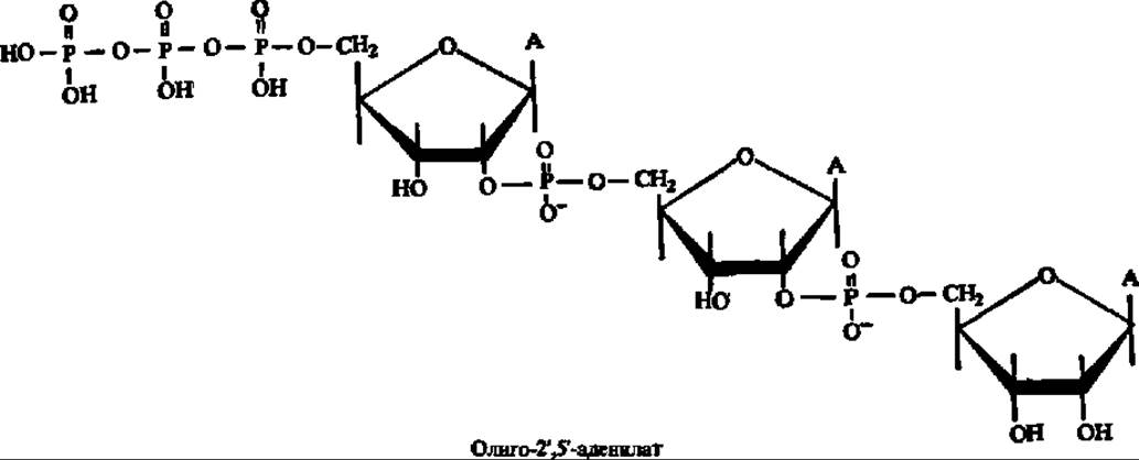

Fig. 138. Structure and mechanism of action of the G-protein:
I – triggered by the hormone receptor, GDP is released from the G-protein and replaced by GTP, causing the G-protein to dissociate into an α-subunit and a β/γ-subunit complex; II – the α-subunit associates with adenylate cyclase and activates it, driving cAMP production; III – the slow-acting GTPase center hydrolyzes GTP, causing the α-subunit to lose its stimulatory effect on adenylate cyclase, which is thereby inactivated, halting cAMP synthesis; IV – the α-subunit dissociates from adenylate cyclase and reassociates with the β/γ-subunit complex, returning the system to its initial state, primed to receive and process another hormonal signal.
However, certain peptide hormones operate via non-adenylate cyclase pathways. For instance, insulin binds to a glycoprotein receptor in the target cell plasma membrane (M = 460,000, composed of 4 subunits), thereby altering its permeability (see Fig. 136). This enhances the cellular uptake of substrates such as glucose and amino acids, fully activating the corresponding intracellular enzymes. Oxytocin acts in a similar manner: The formation of the hormone-receptor complex triggers an influx of Ca2+ ions, initiating the contraction of myoepithelial muscle fibers in the mammary gland alveoli.
Biosynthesis of peptide hormones. The biosynthesis of all peptide hormones, with the exception of releasing factors (discussed later in this section), proceeds according to the established pathways for protein synthesis. Nevertheless, three key distinct features of peptide hormone biosynthesis deserve mention.
The first feature is that peptide hormones are typically generated via the proteolytic Cleavage of high-molecular-weight precursors known as preprohormones. In this process, signal peptides (see p. 300) that facilitate the export of hormone precursors from glandular cells are first cleaved off, followed by the removal of prohormone peptide fragments.
The second feature concerns the biosynthesis of peptide hormones whose molecules consist of multiple polypeptide chains, such as insulin. Research has shown that the joining of these polypeptide chains is mediated by insulin-Glutathione transhydrogenase:

However, it has more recently been established that the formation of disulfide (SS) bonds in biologically active proteins generally occurs via enzymatic pathways. Therefore, this specific reaction may not be uniquely exclusive to peptide hormones.
The third feature is that the Biosynthesis of certain peptide hormones is regulated by neurohormones produced in a specialized Brain region, the Hypothalamus. These are oligopeptides numbering around ten, of which the structures of four have been elucidated. Because they either stimulate or inhibit the synthesis of Anterior Pituitary Hormones upon reaching the pituitary via the capillary portal system directly from the hypothalamus, they are termed releasing hormones (liberins) and inhibiting hormones (statins), respectively (though the original designation, releasing factors, which denotes substances facilitating the release of tropic peptide hormones, is still retained). Thus, thanks to the monumental efforts of two research groups (R. Guillemin and co-workers and A. Schally and co-workers; shared half of the 1977 Nobel Prize in Physiology or Medicine), the structure of thyrotropin-releasing hormone (TRH)—a tripeptide that drastically enhances pituitary thyrotropin secretion—was determined in the late 1970s:

It should also be borne in mind that multifunctional peptide hormones are subject to feedback regulation by the specific hormones whose biosynthesis they accelerate.
The discovery of releasing factors triggered a massive surge in research on the Regulatory Functions of peptides. Their most prominent breakthrough was the identification of the family of endogenous opioid peptides (endorphins and enkephalins), which induce feelings of pleasure, positive mood, and euphoria through their direct morphine-like action on opiate receptors in the Central Nervous system. They also exert analgesic effects, influence blood pressure, motor and respiratory functions, body Temperature, etc. Ranging from 5 to 31 amino acid residues in length, opioid peptides are generated from proopiomelanocortin ($M = 29 ext{ kDa}$, 265 amino acid residues, synthesized in the brain) via its selective hydrolysis. Since their characteristic N-terminal sequence—Tyr-Gly-Gly-Phe—is quite common in dietary proteins, they can also be produced during The breakdown of the latter.
The discovery of opioid peptides and a number of other Neuropeptides raised the issue of potential societal implications stemming from advancements in this field of endocrinology, which is now widely regarded as the "neuropeptide revolution".
Last update: 06/08/2026
Editorial and Educational Adaptation: This material has been compiled based on the primary/original source text. The project team performed an editorial review, corrected technical inaccuracies, structured sections, and adapted the content for an educational format.
What was processed:
- elimination of formatting defects (OCR errors, structural breaks, corrupted characters);
- editorial organization of content;
- standardization of terminology in accordance with academic sources;
- verification of factual statements against the original source text.
All mentions of the author, publication year, and origin of the primary text have been preserved in accordance with the source.