Human Anatomy - Kotsan I. Y. 2009
Skin
Mammary glands
The mammary gland (glandula mammaria; Greek mastos, hence mastitis — inflammation of the mammary gland) is a specialized feature for nourishing newborns in mammals. The number of Mammary Glands depends primarily on the number of offspring born. Monkeys and humans have a single pair of glands located on the chest, which is why they are also referred to as the pectoral (or breasts) glands. In males, the mammary gland remains rudimentary throughout life, whereas in females, it enlarges with the onset of Puberty. The mammary gland reaches its maximum development by the end of Pregnancy, although milk secretion (Lactation) occurs in the postnatal period.
Class="center">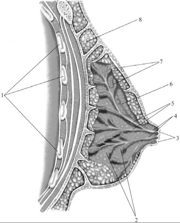
Fig. 284. Sagittal section of the mammary gland
1 — Ribs; 2, 7 — milk-producing glands; 3 — nipple; 4 — lactiferous sinus; 5 — lactiferous ducts; 6 — body of the mammary gland; 8 — adipose tissue
The mammary gland is a modified sweat gland situated on the Fascia of the pectoralis major Muscle, to which it is connected by loose Connective Tissue, allowing for its mobility (Fig. 284). Its base extends from the III to the VI rib, reaching the medial edge of the Sternum. Slightly below the center of the gland on its anterior surface lies the nipple (papilla mammaria), the apex of which is pierced by lactiferous ducts opening onto it, surrounded by a pigmented area of Skin known as the areola (areola mammae). The skin of the areola appears bumpy due to the areolar glands (glandulae areolares) embedded within it. Large Sebaceous Glands lie between the areolar glands. The skin of the areola and nipple contains numerous smooth muscle Cells arranged partly circularly and partly longitudinally along the nipple; upon their contraction, the nipple becomes erect, facilitating suckling.
The glandular body of the mammary gland consists of 15–20 cone-shaped lobes, interspersed with adipose and loose connective tissue. Each lobe Functions as a complex alveolar-tubular gland, with its excretory duct directed radially toward the nipple. Just before reaching the nipple, the duct dilates to form the lactiferous sinus, which then opens via its excretory duct onto The surface of the nipple.
The Blood supply to the mammary gland is provided by the lateral mammary branches from the III–VII posterior intercostal Arteries and the medial mammary branches from the internal thoracic artery. Deep Veins OF THE mammary gland accompany the corresponding arteries, while superficial veins form a wide-meshed plexus beneath the skin. Lymphatic vessels from the mammary gland drain into the parasternal, axillary, internal thoracic (parasternal), and lower cervical Lymph Nodes.
Sensory Innervation of the gland (skin) is supplied by the intercostal nerves (II–VI) and supraclavicular nerves (from the Cervical plexus). Trophic (sympathetic) and secretory (parasympathetic) fibers enter the gland alongside sensory nerves and Blood Vessels.
Last update: 08/08/2026
Editorial and Educational Adaptation: This material has been compiled based on the primary/original source text. The project team performed an editorial review, corrected technical inaccuracies, structured sections, and adapted the content for an educational format.
What was processed:
- elimination of formatting defects (OCR errors, structural breaks, corrupted characters);
- editorial organization of content;
- standardization of terminology in accordance with academic sources;
- verification of factual statements against the original source text.
All mentions of the author, publication year, and origin of the primary text have been preserved in accordance with the source.