Anatomy of the Vessels and Nerves of the Head and Neck (Angioneurology) - Chornokulskyi S.T. 2009
Spinal nerves
Cervical plexus
Formed by the anterior rami of the four upper cervical Spinal Nerves (C1–C4). It is located on the anterolateral surface of the deep Neck Muscles beneath the sternocleidomastoid Muscle, at the level of the four upper cervical vertebrae. Here, the anterior rami of the plexus form three arched loops.
The muscular, cutaneous, and mixed branches (nerves) are distinguished.
1. The muscular Branches of the plexus (rr. musculares) innervate the longus capitis and longus colli muscles, the anterior and lateral rectus capitis muscles, the anterior intertransversarii Muscles of the neck, the scalene muscles (anterior, middle, posterior), and the levator scapulae muscle.
Another muscular branch is the inferior ROOT (radix inferior), which, upon joining the superior root (radix superior) from the XII cranial nerve, forms the ansa cervicalis (see "Hypoglossal nerve").
Class="center">
Fig. 50. Cutaneous branches of the cervical plexus and the External Jugular Vein (7):
1 - clavicle (clavicula);
2 - lesser occipital nerve (n. occipitalis minor);
3 - greater auricular nerve (n. auricularis magnus);
4,5 - anterior and posterior branches of the greater auricular nerve (rr. anterior et posterior n. auricularis magnus);
6 - transverse cervical nerve (n. transversus colli);
7 - external jugular vein (v. jugularis externa);
8. Cervical branch of the Facial Nerve (r. cervicalis n. facialis);
9 Superficial cervical loop (ansa cervicalis superficialis);
10,11,12 - Medial, intermediate, and lateral supraclavicular nerves (nn. supraclaviculares medialis, intermedius et lateralis)
2. The cutaneous branches of the cervical plexus wrap around the posterior border of the sternocleidomastoid muscle and are represented by the following nerves:
a) Lesser occipital nerve (rr. occipitalis minor) innervates the Skin of the inferolateral region of the occiput adjacent to the greater occipital nerve, as well as the skin of the posterior surface of the auricle.
b) Greater auricular nerve (n. auricularis magnus), the largest among the cutaneous branches, runs upward over The surface of the sternocleidomastoid muscle, reaching the skin of the auricle and the external acoustic meatus.
c) Transverse cervical nerve (n. transversus colli) runs forward, giving off superior branches (rr. superiores) to the skin of the neck above the Hyoid bone and inferior branches (rr. inferiores) to the skin of the anterolateral surface of the neck below the hyoid bone, where they connect with the cervical branch of the facial nerve, forming the superficial cervical loop (ansa cervicalis superficialis).
d) Supraclavicular nerves (nn. supraclaviculares), branching into medial, intermediate, and lateral rami (mediales, intermedii et laterales), innervate the skin within the supraclavicular fossae, the lateral triangle of the neck, and the skin over the deltoid and pectoralis major muscles down to the level of the IIIrd rib.
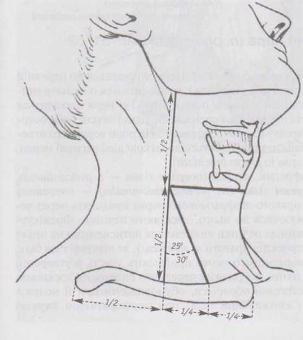
Fig. 51. PROJECTION OF THE right n. phrenicus and the Location of its trigger/pain point in neuralgia and Liver diseases:
✵ The upper projection point of the nerve is located at the midpoint of the line: angle of the Mandible to the midpoint of the right clavicle.
✵ The lower point is located at the midpoint of the medial half of the clavicle, or in the fossa supraclavicularis minor
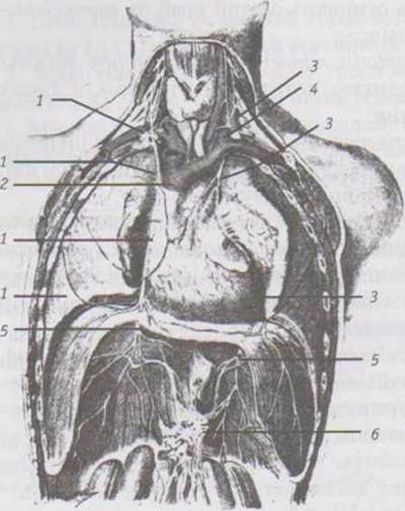
Fig. 52. Phrenic nerves (right and left), topography:
1 - right phrenic nerve (n. phrenicus dexter);
2 - SUPERIOR VENA CAVA (v. cava superior);
3 - left phrenic nerve (n. phrenicus sinister);
4 - left Internal jugular vein (v. jugularis interna sinistra);
5 - phrenicoabdominal branches (rr. phrenicoabdominales);
6 - celiac plexus (plexus coeliacus)
Phrenic nerve (n. phrenicus) figs. 51, 52
This is the only mixed branch of the cervical plexus. The nerve descends along the anterior surface of the anterior scalene muscle, passes between the Subclavian Artery and vein, and enters the thoracic cavity. Here, the nerve lies in the superior Mediastinum anterior to the root of the corresponding lung, between the Pericardium and mediastinal Pleura, reaching the Diaphragm. At the level of the superior thoracic aperture, accessory phrenic nerves join the n. phrenicus as branches of the subclavian nerve (from the plexus brachialis).
The nerve innervates the muscle of the diaphragm, the pericardium (pericardial branch — r. pericardiacus), the pleura, and its phrenicoabdominal branches (rr. phrenicoabdominales) supply the diaphragmatic Peritoneum. Sensory branches of the right phrenic nerve pass through the celiac plexus, branching off to it and reaching the liver. The phrenic sign (+) in liver diseases is elicited by pressure on the right lesser supraclavicular fossa (the projection of the right n. phrenicus), where pain is felt. Sensory branches of the right phrenic nerve participate in The formation of the deep cardiac plexus, and via branches of the celiac plexus, they reach the walls of The Stomach, particularly the serous membrane, causing intense pain upon its irritation ("dagger-like pain" in perforated gastric ulcer).
Topography of the Vessels and nerves of the neck (superficial layer)
For independent study


Blood supply (vascularization) and Innervation of the HEAD and Neck
(Synthetic-analytical session)
The Organs of the head and neck are supplied with blood by three Branches of the aortic arch: (truncus brachiocephalicus, a. carotis communis sinistra, a. subclavia sinistra).
Arteries (A.):
✵ Of the facial skin, the skin of the frontal and parietal scalp regions, and the facial muscles (figs. 12, 14, 20):
— A. facialis → aa. labiales inferior et superior, a. lateralis nasi, a. angularis;
— A. temporalis superficialis → rr. frontalis et parietalis, a. transversa faciei, a. zygomaticoorbitalis;
— A. maxillaris → r. mentalis (from a. alveolaris inferior), a. infraorbitalis, a. buccalis;
— A. ophthalmica → a. supraorbitalis, a. supratrochlearis, a. dorsalis nasi, aa. palpebrales med. et lat.
✵ Of the skin of the occipital region (A. occipitalis, A. auricularis posterior)
✵ Of the auricle and external acoustic meatus:
— A. temporalis superficialis → rr. auriculares anteriores;
— A. auricularis posterior → r. auricularis;
— A. maxillaris → a. auricularis profunda;
— A. occipitalis → r. auricularis.
✵ Skin and SUPERFICIAL MUSCLES OF the neck (Fig. 12, 16, 19a):
— A. lingualis → r. suprahyoideus; a. facialis → a. submentalis;
— A. thyroidea superior → r. infrahyoideus;
— A. sternocleidomastoidea; A. occipitalis; A. auricularis posterior; A. subclavia a. transversa colli.
Veins (V.) (see pp. 54–56) Nerves (N.):
✵ Innervation of the facial skin (see pp. 85–86; Fig. 36, 37 and N. vagus — p. 101)
✵ Innervation of the skin of the occipital region and neck:
— Nn. occipitales major, minor et tertius, N. auricularis magnus, N. transversus colli, Nn. supraclaviculares, as well as the posterior rami of the C3 and C4 cervical nerves.
✵ Innervation of the facial muscles and m. platysma — N. facialis (VII)
→ A. maxillaris → a. masseterica, aa. temporales profundae, rr. pterygoidei. Corresponding veins drain into the plexus pterygoideus. Nerves: N. mandibularis, N. massetericus, Nn. temporales profundi, Nn. pterygoidei lateralis et medialis.
✵ Dura mater
The meningeal Arteries of the dura mater also supply the adjacent cranial bones and lie within their sulci arteriosi. A. maxillaris → a. meningea media; A. ophthalmica → a. ethmoidalis anterior → r. meningeus anterior; A. pharyngea ascendens → a. meningea posterior; A. occipitalis → r. meningeus; A. vertebralis → rr. meningei. The dural veins are generally paired with the corresponding arteries, draining blood into the Dural Venous Sinuses and partially into the plexus pterygoideus (see also pp. 46–53). Nerves: N. trigeminus → N. ophthalmicus r. tentorius, N. ethmoidalis anterior → r. meningeus anterior; N. maxillaris → r. meningeus; N. mandibularis → r. meningeus (N. spinosus). Within the posterior cranial fossa — N. vagus → r. meningeus (dyspeptic and cardiac responses to changes in intracranial pressure during meningitis, concussions, etc.). All sensory fibers of the posterior ramus of the 1st cervical spinal nerve also innervate the dura mater of the posterior cranial fossa, making the suboccipital nerve exclusively motor (rigidity of the suboccipital muscles in meningitis).
✵ Brain
Supplied by arteries originating from two sources: a. carotis interna and a. vertebralis. For CEREBRAL ARTERIES AND veins, see pp. 34–36, 37–39, 46–53.
✵ Eye and associated structures
Eyeball: A. ophthalmica → aa. ciliares breves et longae, aa. ciliares anteriores, a. centralis retinae. For veins, see p. 48. Nerves: N. ophthalmicus → Nn. ciliares longi, and to the intrinsic eye muscles — Nn. ciliares breves (see ganglion ciliare, pp. 66–67). The visual pathway originates from retinal receptors (pp. 63–64).
✵ Lacrimal gland: A. ophthalmica → a. lacrimalis. Nerves: N. ophthalmicus → N. lacrimalis, with secretory innervation via rr. orbitales (from ganglion pterygopalatinum), p. 76.
✵ Extraocular muscles: A. ophthalmica → rr. musculares; A. maxillaris → a. infraorbitalis. Nerves: Nn. oculomotorius, trochlearis, abducens, and their connections with the N. ophthalmicus provide proprioceptive sensitivity.
✵ Eyelids, conjunctiva: A. ophthalmica → aa. palpebrales lat. et med., arcus palpebrales superior et inferior; Aa. conjunctivales anteriores et posteriores. Nerves: N. trigeminus → N. ophthalmicus → Nn. supraorbitalis, supratrochlearis et infratrochlearis.
✵ Middle ear, walls of the tympanic cavity: paries jugularis — a. tympanica inferior passes through the canaliculus tympanicus (from a. pharyngea ascendens); paries tegmentalis — a. tympanica superior passes through the hiatus canalis N. petrosi minoris (from a. meningea media); paries caroticus — a. tympanica anterior passes through the fissura petrotympanica (from a. maxillaris), rr. caroticotympanici pass through the corresponding canaliculi (from a. carotis interna); paries mastoideus — a. tympanica posterior passes through the canaliculus chordae tympani (from a. stylomastoidea); paries membranaceus — a. auricularis profunda (from a. maxillaris); paries labyrinthicus — a. labyrinthi passes through the meatus acusticus internus (from a. basilaris). Veins accompany the arteries. Nerves: plexus tympanicus (from N. tympanicus, Glossopharyngeal nerve, ganglion tympanicum, Nn. caroticotympanici). The respective pathways originate from the receptors of the Auditory and Vestibular systems in the labyrinth (pp. 92–95).
✵ Mucous membrane of the Nasal cavity: Aa. ethmoidales ant. et post., rr. septales anteriores (from a. ophthalmica); A. sphenopalatina → rr. septales posteriores (from a. maxillaris). Veins correspond to the arteries, draining blood into the pterygoid venous plexus. Nerves: Nn. ethmoidales ant. et post. (from N. nasociliaris); secretory — Nn. nasales posteriores, N. nasopalatinus (from ganglion pterygopalatinum). The Pathways of the olfactory analyzer originate within the mucosa of the regio olfactoria, while the terminal nerve (N. terminalis) originates within the organum vomeronasale of the cartilaginous nasal septum.
✵ Hard and Soft palate: A. palatina ascendens (from a. facialis); A. pharyngea ascendens, aa. palatinae major et minores (from a. palatina descendens ← a. maxillaris); A. sphenopalatina → a. nasopalatina. Veins — v. facialis → v. jugularis interna. Nerves: plexus pharyngeus; Nn. palatini major et minores, N. nasopalatinus (from ganglion pterygopalatinum). Innervation of the soft palate muscles: N.m. tensoris veli palatini (N. mandibularis) supplies the muscle of the same name, while the remaining muscles are supplied by N. vagus (rr. pharyngei).
✵ Upper Teeth: Aa. alveolares superiores, posteriores et anteriores (from a. maxillaris). Nerves: see pp. 77, 80.
✵ Lower teeth: A. alveolaris inferior (from a. maxillaris). All specified alveolar arteries give off rr. dentales to the teeth and rr. peridentales to the periosteum of the tooth sockets and Gums. Veins from the teeth of the jaws (corresponding to the arteries) drain blood into the pterygoid venous plexus. Nerves: see pp. 82-84.
✵ Tongue: A. lingualis → rr. dorsales linguae, a. profunda linguae, a. sublingualis. Veins: v. lingualis → v. jugularis interna. Nerves: the tongue is innervated by 5 nerves. Mucosa, anterior 2/3 — N. lingualis ← N. mandibularis (general sensitivity), chorda tympani ← N. intermedius (taste, sensory), posterior 1/3 — rr. linguales ← N. glossopharyngeus (general and taste sensitivity), N. laryngeus superior ← N. vagus; tongue muscles — N. hypoglossus (XII) → rr. linguales.
✵ Oral mucosa: (see arteries and VEINS OF THE facial skin and palate).
Nerves: upper lip — rr. labiales superiores ← N. infraorbitalis; Cheeks and angle of the Mouth — N. buccalis ← N. mandibularis; lower lip — rr. labiales inferiores ← N. mentalis ← N. mandibularis; floor of the Oral Cavity — N. sublingualis → N. lingualis.
✵ Glands of the head and neck: the lacrimal gland, glands of the nasal cavity and palate are innervated from ganglion pterygopalatinum (see p. 76). The submandibular and sublingual glands are innervated from ganglia submandibulare et sublinguale (see p. 81). Arteries: a. lacrimalis, (for the mucosa of the nasal cavity and palate see above).
✵ Parotid gland: A. temporalis superficialis → r. parotideus; A. auricularis posterior → r. parotideus. Veins: vv. parotidei → v. retromandibularis → v. jugularis interna.
Nerves: rr. parotidei (from ganglion oticum, see pp. 83—84).
✵ Thyroid and Parathyroid glands: Aa. thyroideae superiores et inferiores dextrae et sinistrae, a. thyroidea ima ← truncus brachiocephalicus. Veins: vv. thyroideae superiores et mediae dextrae et sinistrae → v. jugularis interna, v. thyroidea inferior → v. brachiocephalica sinistra. Nerves: plexus caroticus externus et subclavius, Nn. laryngei superior et inferior.
✵ Pharynx: A. pharyngea ascendens; Aa. palatinae ascendens et descendens. Veins: vv. pharyngeae → v. jugularis interna. Nerves: plexus pharyngeus (Nn. laryngopharyngei), N. glossopharyngeus, N. vagus. All pharyngeal muscles except m. stylopharyngeus (← N. glossopharyngeus) are innervated by N. vagus.
✵ Larynx: Aa. laryngeae superiores et inferiores dextrae et sinistrae. Veins: corresponding veins → v. jugularis interna. Nerves: Nn. laryngopharyngei. All laryngeal muscles except m. cricothyroideus (supplied by N. laryngeus superior [r. externus]) are innervated by N. laryngeus inferior.
✵ Muscles of the neck (superficial and middle groups): A. sternocleidomastoidea; A. lingualis; A. thyroidea superior; A. submentalis; A. transversa colli. Deep neck muscles: A. vertebralis; A. cervicalis profunda, A. cervicalis ascendens. Veins: corresponding veins drain into: vv. jugularis interna, subclavia, brachiocephalica. Nerves: superficial muscles: m. platysma ← r. colli, VII, m. sternocleidomastoideus ← N. accessorius, plexus cervicalis. Suprahyoid muscles: mm. mylohyoideus, venter anterior m. digastrici ← N. mylohyoideus; m. stylohyoideus, venter posterior m. digastrici ← N. facialis; m. geniohyoideus + all Infrahyoid muscles ← ansa cervicalis. All deep neck muscles are innervated by plexus cervicalis (rr. musculares). Voluntary contraction of striated muscles of the head and neck innervated by Cranial Nerves is mediated by impulses from the corticonuclear tract, whereas muscles innervated by branches of the cervical plexus are mediated by the corticospinal tract. Proprioceptive sensitivity of the muscles of the head and partially the neck is provided by N. trigeminus.
Last update: 11/08/2026
Editorial and Educational Adaptation: This material has been compiled based on the primary/original source text. The project team performed an editorial review, corrected technical inaccuracies, structured sections, and adapted the content for an educational format.
What was processed:
- elimination of formatting defects (OCR errors, structural breaks, corrupted characters);
- editorial organization of content;
- standardization of terminology in accordance with academic sources;
- verification of factual statements against the original source text.
All mentions of the author, publication year, and origin of the primary text have been preserved in accordance with the source.