Anatomy of the Vessels and Nerves of the Head and Neck (Angioneurology) - Chornokulskyi S.T. 2009
Arteries of the Head and Neck. Vessels of the Systemic Circulation
Subclavian Artery
(a. subclavia) (Fig. 15, 16)
On the right, it is a branch of the brachiocephalic trunk, and on the left, of the aortic arch; consequently, the left subclavian artery is 4–5 cm longer than the right and features a thoracic part. Superior to the apertura thoracis superior, the subclavian artery is symmetrical and lies in the corresponding groove of the first rib beneath the clavicle. At the lateral border of the 1st rib, the subclavian artery continues as the axillary artery. According to its topography, the a. subclavia is conventionally divided into three parts:
1) up to the interscalene space (from the Water/144.html">Origin of the artery to the anterior scalene Muscle; branches: vertebral artery, internal thoracic artery, and thyrocervical trunk);
2) within the interscalene space, giving off the costocervical trunk;
3) beyond the interscalene space, giving off the transverse cervical artery.
3) beyond the interscalene space, the branch is the transverse cervical artery.
Branches of the 1st part
I. Vertebral artery (a. vertebralis) (Fig. 12, 17, 18).
It has 4 parts:
— prevertebral part (pars prevertebralis) extending to the transverse process of the VI cervical vertebra;
— transverse process (cervical) part, pars transversalis (cervicalis), running through the transverse foramina of the VI–II cervical vertebrae;
— atlanto-occipital part (pars atlantica), situated in the transverse foramen and the corresponding groove of the atlas arch;
— intracranial part (pars intracranialis).
Class="center">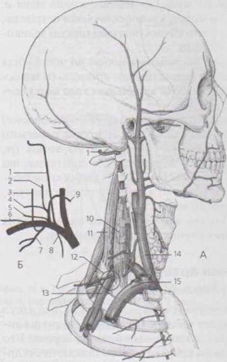
Fig. 15. Subclavian artery (A), branching diagram of the subclavian artery (B):
1 - vertebral artery (a. vertebralis);
2 - ascending cervical artery (a. cervicalis ascendens);
3 - deep cervical artery (a. cervicalis profunda);
4 - superficial cervical artery (a. cervicalis superficialis);
5 - suprascapular artery (a. suprascapularis);
6 - transverse cervical artery (a. transversa colli);
7 - costocervical trunk and supreme intercostal artery (truncus costocervicalis et a. intercostalis suprema);
8 - internal thoracic artery (a. thoracica interna);
9 - inferior thyroid artery (a. thyroidea inferior);
10 - anterior scalene muscle (m. scalenus anterior);
11 - middle scalene muscle (m. scalenus medius);
12 - site of compression of the subclavian artery against the 1st rib;
13 - subclavian artery (a. subclavia);
14 - common carotid artery (a. carotis communis);
15 - subclavian vein (v. subclavia)
After passing through the posterior atlanto-occipital membrane, the dura mater of the Spinal Cord, and the foramen magnum, the a. vertebralis enters the cranial cavity. The right and left Arteries converge anterior to the Medulla Oblongata and fuse to form the unpaired Basilar artery (a. basilaris), which lies in the basilar sulcus of the Pons.
Of the four PARTS OF THE a. vertebralis, only the transverse process part and the intracranial part give off branches.
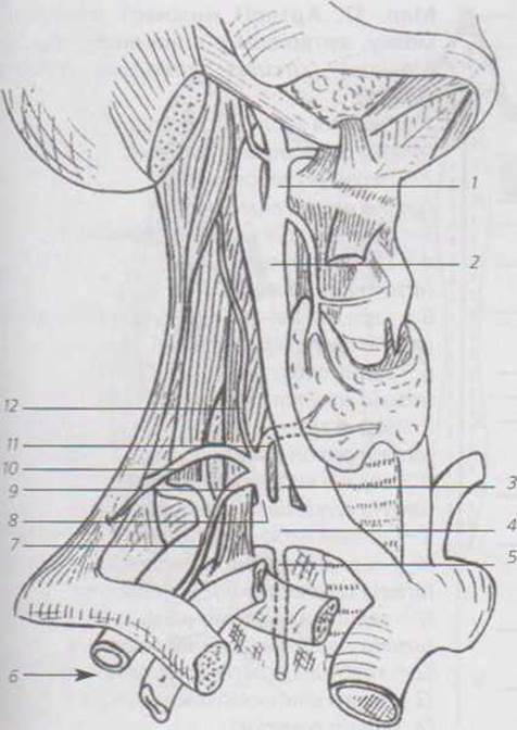
Fig. 16. Topography of the right subclavian and common carotid arteries:
1 - External Carotid Artery (a. carotis externa);
2 - common carotid artery (a. carotis communis);
3 - vertebral artery (a. vertebralis);
4 - subclavian artery (a. subclavia);
5 - internal thoracic artery (a. thoracica interna);
6 - axillary artery (a. axillaris);
7 - transverse cervical artery
(a. transversa colli);
8 - thyrocervical trunk (truncus thyrocervicalis);
9 - suprascapular artery (a. suprascapularis);
10 - superficial cervical artery (a. cervicalis superficialis);
11 - inferior thyroid artery (a. thyroidea inferior);
12 - ascending cervical artery (a. cervicalis ascendens)
(Independent work: sketch all arteries in red)
✵ In the transverse process part, the following branches are given off:
— spinal (radicular) branches (rr. spinales, radiculares), which reach the spinal cord and the roots of the Spinal Nerves after passing through the intervertebral foramina;
— muscular branches (rr. musculares), which supply Blood to the Deep Muscles of the Neck.
Branches of the intracranial part
1. Meningeal branches (rr. meningei) supplying the dura mater and cranial bones.
2. Posterior spinal artery (a. spinalis posterior) descends along the posterolateral sulcus of the spinal cord down to the conus medullaris, vascularizing the spinal cord and its Meninges (Fig. 18, 19).
3. Anterior spinal artery (a. spinalis anterior) runs inferiorly, converging and fusing with the corresponding artery of the opposite side to form a diamond-shaped anastomosis known as Zakharchenko's ring (Fig. 18 b). Below this anastomosis, the a. spinalis anterior continues as a single vessel, descending within the anterior median fissure of the spinal cord to the filum terminale, supplying the spinal cord and its meninges.
4. Posterior inferior cerebellar artery (a. cerebellaris inferior posterior) supplies the medulla oblongata and the posteroinferior region of the Cerebellum.
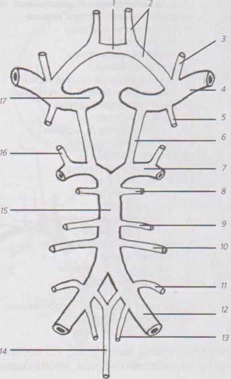
Fig. 17. Arteries of the Inferior surface of the Brain, arterial circle of the brain (circle of Willis) (circulus arteriosus cerebri Willisii):
1 - anterior communicating artery (arteria communicans anterior);
2 - anterior cerebral artery (arteria cerebri anterior);
3 - ophthalmic artery (arteria ophthalmiea);
4 - middle cerebral artery (arteria cerebri media);
5 - anterior choroidal artery (arteria choroidea anterior);
6 - posterior communicating artery (arteria communicans posterior);
7 - posterior cerebral artery (arteria cerebri posterior);
8 - superior cerebellar artery (arteria cerebellaris superior);
9 - labyrinthine artery (arteria labyrinthi);
10 - anterior inferior cerebellar artery (arteria cerebellaris inferior anterior);
11 - posterior inferior cerebellar artery (arteria cerebellaris inferior posterior);
12 - vertebral artery (arteria vertebra lis);
13 - posterior spinal artery (a. spinalis posterior);
14 - anterior spinal artery (a. spinalis anterior);
15 - basilar artery (arteria basilaris);
16 - posterior choroidal branch (ramus choroideus posterior);
17 - Internal Carotid Artery (arteria carotis interna)
Last update: 11/08/2026
Editorial and Educational Adaptation: This material has been compiled based on the primary/original source text. The project team performed an editorial review, corrected technical inaccuracies, structured sections, and adapted the content for an educational format.
What was processed:
- elimination of formatting defects (OCR errors, structural breaks, corrupted characters);
- editorial organization of content;
- standardization of terminology in accordance with academic sources;
- verification of factual statements against the original source text.
All mentions of the author, publication year, and origin of the primary text have been preserved in accordance with the source.