IMMUNOLOGY TEXTBOOK - Mercury Podillia 2013
STRUCTURE AND PRINCIPLES OF FUNCTIONING OF THE IMMUNE SYSTEM
In modern medicine, immunology has assumed a prominent role as a rapidly developing field, inspiring high expectations among physicians across various specialties. Clinical immunology is both a clinical and laboratory discipline dedicated to the examination, Diagnosis, and Treatment of patients with pathological processes arising from immune system dysfunctions, as well as cases where immunological interventions form a crucial part of therapy and/or Prevention.
Impairments in the development, differentiation, and functioning of immunocompetent Cells, the synthesis of their products, or The regulation of these processes lead to immune dysfunction. These disorders may remain asymptomatic or manifest clinically, with severity ranging from mild to fatal. Such abnormalities can affect the primary Cells of the immune system—T AND B lymphocytes, phagocytes, natural killer cells—and their products, including Complement Proteins, IMMUNOGLOBULINS, and cytokines.
A significant proportion of these disorders is associated with congenital or acquired defects in the production or function of immunocompetent cells. Other cases of immunodeficiency stem from the malignant transformation and uncontrolled proliferation of immunocompetent cells, accompanied by the excessive accumulation of their products. Clinical manifestations of immune regulation disorders are diverse and include unregulated complement activation, as well as the unregulated production and reception of cytokines.
The immune system comprises the following Organs: the Bone Marrow, thymus, spleen, Lymph Nodes, and clusters of lymphoid tissue. They are classified into primary (central) organs—the bone marrow and thymus—and secondary (peripheral) organs—the spleen, lymph nodes, and lymphoid tissue aggregates. All of these are interconnected by the circulatory and lymphatic systems, forming a unified immunoregulation network.
Immunity is an evolutionarily shaped set of interaction reactions between the immune system and biologically active agents (Antigens) aimed at preserving the phenotypic constancy of the body's internal environment (Homeostasis).
The Main Functions of the immune system include monitoring the antigenic constancy of the body's internal environment, protecting the Organism against pathogenic microorganisms, and providing antitumor surveillance. Both nonspecific defense mechanisms and specific immune responses tailored to particular infectious or tumor antigens are involved in carrying out these functions. The specific Immune Response enhances and focuses nonspecific defense mechanisms.
Organs of the Immune System
Central Organs of the Immune System—the bone marrow and thymus—perform critical functions by ensuring the self-renewal of the immune system. These organs are sites where progenitor cells proliferate, differentiate, and mature before entering Circulation and populating the peripheral immune organs as mature, immunocompetent cells.
Bone marrow. All Blood Cells, including immunocompetent cells, originate from pluripotent stem cells that give rise to various hematopoietic lineages, notably the myeloid-monocytic and lymphocytic lineages. The direction of differentiation of early progenitors is governed by influences from their microenvironment and bone marrow stromal cells.
The Effect of specific cytokines on progenitor cells in vitro manifests as The stimulation of growth for distinct colonies comprising specific types of leukocytes. Hence their name—colony-stimulating factors (CSFs): GM-CSF, G-CSF, and M-CSF. Granulocyte-macrophage colony-stimulating factor stimulates the proliferation of early common progenitors of myelomonocytopoiesis as well as progenitor cells of individual lineages. Even more versatile is the so-called multi-CSF (interleukin-3), which stimulates all hematopoietic lineages. These growth factors and other cytokines are produced by bone marrow stromal cells, macrophages, and activated lymphocytes. Interleukin-1 and interleukin-6 act synergistically with colony-stimulating factors to stimulate progenitor Cell proliferation and induce The production of growth factors.
The thymus is the only immune organ that undergoes rapid age-related involution. During the first 50 years of life, approximately 3% of true thymic tissue is lost annually, gradually being replaced by adipose and Connective Tissue, with a corresponding decline in T lymphocyte production. Peak T-cell production persists up to two years of age and then drops sharply. However, the number of circulating T lymphocytes remains stable. This is because a significant portion of the T-cell population consists of long-lived cells that do not require constant renewal. Consequently, T-cell numbers can be sustained in adults even in the absence of the thymus. Furthermore, mature T lymphocytes undergo what is known as "clonal expansion"—selective proliferation in response to encountering their specific antigen—which increases their population. Once the peripheral T-cell pool is established, the loss of the thymus no longer leads to a catastrophic drop in immunity. This is supported by immunological evaluations of adults who have undergone thymectomy.
Peripheral Organs of the Immune System. The peripheral immune organs—lymph nodes, spleen, and mucosa-associated lymphoid tissue—serve as sites where antigens encounter immunocompetent cells, where antigens are recognized, specific immune responses develop, immunocompetent cells interact and proliferate (clonal expansion), antigen-dependent differentiation occurs, and immune response products accumulate.
Lymph nodes function as unique lymph filters, trapping microorganisms and other particulate matter present in the lymph. At the same time, lymph nodes serve as sites for the interaction of immunocompetent cells during a specific immune response, the synthesis of antibody immunoglobulins, and the orchestration of cell-mediated immunity events.
A single lymph node weighs about 1 gram and contains approximately 2,000 million lymphocytes, which corresponds to 25% of all circulating blood lymphocytes. Every hour, an amount of lymphocytes equivalent to three times the node's weight exits into the lymph. The majority (90%) of the cells in this efferent lymph are lymphocytes that left the bloodstream within that specific lymph node. Labeled lymphocytes injected into the blood reappear in the lymph within a few hours, peaking at 20 hours. Among the cells within a lymph node, roughly 10% are macrophages and about 1% are dendritic cells.
Lymph node tissue consists of an outer cortical zone, where cell clusters form follicles (partially featuring germinal centers), and an inner medullary zone with a lower lymphocyte density combined with macrophages clustered along lymphatic and vascular sinuses. This structural arrangement allows for the free circulation and recirculation of lymphocytes between the lymph, blood, and Tissues. Specific Zones of the lymph node are populated by strictly designated cell types.
Spleen. Much like lymph nodes, the spleen contains T-dependent and B-dependent zones. Periarteriolar lymphoid sheaths constitute the T-dependent areas. The spleen serves as a site for antigen recognition, antigen-dependent proliferation and differentiation of T and B lymphocytes, their activation, and the production and secretion of specific antibody immunoglobulins. The primary difference between the spleen and lymph nodes is that the spleen mounts specific immune responses against blood-borne antigens, whereas lymph nodes respond to antigens carried by the lymph. Additionally, with its extensive network of macrophages within the red pulp, the spleen functions as a blood filter, removing foreign particles and molecules from the bloodstream, as well as senescent or immune-complex-laden erythrocytes.
Mucosa-associated lymphoid tissue. Clusters of lymphocytes, macrophages, and other accessory cells have been identified within numerous organs and tissues, most notably in mucosal membranes. Located directly beneath the mucosal epithelium in close association with epithelial cells are lymphocytes of Peyer's patches in the Small Intestine, Appendix lymphoid follicles, pharyngeal Tonsils, lymphoid follicles in the submucosa of the Upper Respiratory Tract and Bronchi, and the urogenital tract. Collectively, these lymphoid aggregations are known as mucosa-associated lymphoid tissue (MALT).
Immunocompetent cells. Immunocompetent cells are in a state of recirculation, meaning There is a continuous exchange of cells between the blood, lymph, and Lymphoid organs. This is essential for mounting a specific immune response, as the immune system must be prepared to react against any of the countless foreign antigens entering any part of the body. Because each individual antigen is recognized by only a tiny fraction of the lymphocyte pool, constant recirculation is necessary to ensure that every antigen encounters its rare matching lymphocytes bearing specific antigen-recognition receptors. Within the lymphoid organs where this encounter takes place, antigen-specific lymphocytes interact with other accessory cells that participate in initiating and executing the immune response. These accessory cells include dendritic cells, mononuclear phagocytes, and granulocytes.
During differentiation, various macromolecules—markers corresponding to specific stages of cell population development—appear on the membranes of immune system cells. In 1983, the First International Workshop on Human Leukocyte Differentiation Antigens introduced the term "clusters of differentiation" (abbreviated as CD) into clinical immunology practice. The Use of Monoclonal Antibodies made it possible to quantitatively analyze blood cell populations and classify them based on the presence of surface CD antigens.
In 1989, the Fourth Workshop adopted a working nomenclature for human leukocyte differentiation antigens. Monoclonal antibodies with virtually identical Specificity for a given membrane antigen were grouped and designated with corresponding cluster of differentiation (CD) numbers. All of these function as adhesion receptors; upon interacting with their ligands, they transmit a signal into The Cell, triggering its activation, suppression, or even apoptosis.
CD3 is expressed on mature (naive) T Lymphocytes and ensures signal Transduction from the T-cell antigen-specific receptor (TCR) into the Cytoplasm. The antigen-specific T-cell receptor, CD3, and other coreceptors form a complex that recognizes foreign antigens presented in conjunction with Major Histocompatibility Complex (MHC) Class I or class II molecules, depending on the cell type (CD8 or CD4).
CD4 is a marker of T helper cells, a coreceptor Structure for the T-cell receptor, and one of the receptors for the HUMAN IMMUNODEFICIENCY VIRUS (HIV); it participates in recognizing antigens associated with MHC class II molecules and serves as their receptor.
CD8 is a marker of T suppressor and cytotoxic lymphocytes, and is also present on certain NK cells; it functions as a coreceptor structure for the T-cell receptor and is involved in antigen recognition via MHC class I molecules, acting as a receptor for MHC class I molecules.
CD14 is a monocyte marker. It is expressed On the surface of monocytes and macrophages and is involved in the activation of these cells via Toll-like receptor 4 (TLR4).
CD16 is a marker for natural killer (NK) cells and serves as a low-affinity Fc receptor for IgG type III. This molecule is involved in antibody-dependent cellular cytotoxicity mediated by NK cells. In addition to NK cells, this molecule is present on the majority of neutrophils.
CD19 is found on pre-B cells and B cells, functioning as part of their receptor complex and participating in their activation (through a signal transduction pathway associated with CD21). CD19 is not detected on Other types of lymphoid cells.
CD21 is a receptor for the C3d complement component. It is likely that this molecule facilitates receptor-mediated antigen uptake by B cells. Furthermore, the CD21 molecule serves as a receptor for the Epstein-Barr virus. This marker is expressed on all mature B cells and follicular dendritic cells. CD21 can be used for the quantitative assessment of B lymphocytes.
CD22 is expressed on mature B lymphocytes, functions as an adhesion molecule, and is involved in the negative regulation of B cells.
CD25 is the alpha chain of the IL-2 receptor, which appears only upon cell activation, resulting in The formation of a high-affinity IL-2 receptor. CD25 (an activation marker) is expressed by various peripheral blood cell types, including CD4+, CD8+, NK, and CD4+ NKT cells, as well as B lymphocytes and monocytes. Under normal conditions, CD25+ cells can account for up to 18% of the total lymphocyte population.
CD56 is the neural Cell Adhesion molecule (NCAM). In addition to natural killer cells, CD56 is expressed on numerous cell types, including T lymphocytes.
CD69 and CD95 are present on cells exhibiting signs of apoptosis.
CD45 is an antigen expressed on The surface of all human leukocytes, which functions enzymatically as a Tyrosine phosphatase. The level of CD45 expression increases as hematopoietic cells differentiate from immature precursors into mature forms. The maximum level of CD45 is found on mature lymphocytes, while an intermediate level is observed on myeloid cells. There are 3 isoforms of CD45:
- CD45RO - is expressed on effector T cells, memory T cells, B cells, monocytes, and macrophages.
- CD45RA - is expressed on naive T cells, B cells, and monocytes.
- CD45RB - is present on T and B lymphocytes, monocytes, and granulocytes.
Table 1 presents the currently known clusters of differentiation defined on immune system cells using monoclonal antibodies.
Table 1. List of clusters of differentiation defined on immune system cells using monoclonal antibodies
Antigen |
Antigen-bearing cells |
Antigen functions |
|
T-lymphocyte markers |
|||
CD1 |
- |
Cortical thymocytes and Langerhans dendritic cells |
Associated with beta2-microglobulin, participates in antigen presentation to immature T-lymphocytes |
CD2 |
LFA-3 |
T-lymphocytes and NK cells, E-rosette-forming cells |
Sheep erythrocyte receptor, involved in T-lymphocyte activation |
CD3 |
- |
Mature T-lymphocytes, T-cell antigen receptor |
Associated with the antigen-recognition receptor of T-lymphocytes, participates in their activation |
CD4 |
MHC class II |
T-helper/inducer cells, monocytes |
Present on T-helpers, ensures their interaction with macrophages |
CD5 |
CD72 |
T- and B-lymphocytes |
Present on mature T-lymphocytes and a small fraction of B-lymphocytes, appears on leukemic B-lymphocytes in chronic Lymphocytic Leukemia |
CD6 |
mature T-lymphocytes |
||
CD7 |
- |
T-lymphocytes, thymocytes, subset of NK cells |
Present on bone marrow T-lymphocyte precursors and mature T-lymphocytes |
CD8 |
MHC class I |
T-suppressor/killer cells and a subset of NK cells |
Present on cytotoxic T-lymphocytes, ensures their interaction with target cells |
CD16 |
NK cells |
||
CD19 |
immature and mature B-lymphocytes |
||
CD20 |
mature B-lymphocytes |
||
CD22, CD23 |
Tonsillar B cells, 70% of blood B cells |
||
CD25 |
Interleukin-2 |
T, B, and NK lymphocytes, monocytes |
Alpha chain of the interleukin-2 receptor (p55), marker of activated T- and B-lymphocytes |
CD28 |
CD80 |
T-lymphocytes |
Participates in T-lymphocyte activation |
CD29 |
T-lymphocytes |
Mediates adhesion to the Extracellular matrix, marker of activated T-lymphocytes |
|
CD38 |
- |
T and B lymphocytes, activated B lymphocytes |
Present on cortical thymic T-lymphocytes, activated T-lymphocytes, immature B-lymphocytes, and plasma cells; participates in the regulation of B-lymphocyte functions |
CD43 |
ICAM-1 |
T- and B-lymphocytes, granulocytes, monocytes |
Participates in T-lymphocyte activation |
CD45 |
- |
All leukocytes |
Participates in lymphocyte activation; the intracellular portion of the receptor possesses tyrosine kinase activity |
CD45RO |
- |
T- and B-lymphocytes, granulocytes, monocytes |
Marker of memory cells (CD4 lymphocytes) and effector cells |
CD45RA |
- |
All leukocytes |
Marker of T- and B-lymphocytes and monocytes |
CD45RB |
- |
T- and B-lymphocytes, monocytes, granulocytes |
Marker of mature T- and B-lymphocytes and monocytes |
CD71 |
Transferrin |
T-lymphocytes, monocytes |
Transferrin receptor, marker of activated T-lymphocytes |
B-lymphocyte markers |
|||
Surface immunoglobulins |
Antigen |
B-lymphocytes |
Present exclusively on mature B-lymphocytes |
CD10 |
- |
B-lymphocytes |
Present on immature B-lymphocytes, appears on leukemic cells in acute lymphoblastic leukemia |
CD19 |
- |
B-lymphocytes |
Present on pro-B lymphocytes and all mature B-lymphocytes, participates in B-lymphocyte activation |
CD20 |
- |
B-lymphocytes |
Present on all B-lymphocytes |
CD21 |
C3d, CD23 |
B-lymphocytes |
Complement and Epstein-Barr virus receptor |
CD23 |
IgE |
B- and T-lymphocytes, monocytes, eosinophils |
Low-affinity Fc receptor for IgE |
CD32 |
IgG |
B-lymphocytes, granulocytes |
Low-affinity Fc receptor for IgG |
CD40 |
gp39 |
B-lymphocytes |
Stimulates B-lymphocyte proliferation, structurally similar to CD27 and the tumor necrosis factor receptor |
CD72 |
CD5 |
B-lymphocytes |
Appears on bone marrow B-lymphocyte precursors, participates in their differentiation |
HLA-DR |
Antigen, CD4 |
B- and T-lymphocytes, monocytes |
MHC class II antigen, participates in antigen presentation to T-helper cells and their activation, marker of activated T-lymphocytes |
Monocyte and macrophage markers |
|||
CD11a |
ICAM-1 |
All leukocytes |
Alpha chain of LFA-1, participates in cell-cell adhesion |
CD11b |
C3bi, fibronectin |
Monocytes, granulocytes, NK lymphocytes |
Alpha chain of CR3, participates in cell-cell adhesion |
CD11c |
C3bi |
Monocytes, granulocytes, B and NK lymphocytes |
Alpha chain of CR4, participates in cell-cell adhesion |
CD14 |
TLR4 |
Monocytes, macrophages |
Participates in the activation of these cells via Toll-like receptor 4 |
CD18 |
- |
All leukocytes |
Beta chain of CD11a/CD18, CD11b/CD18, and CD11c/CD18 receptors; participates in cell-cell adhesion |
NK lymphocyte markers |
|||
CD3 |
- |
Mature NK and T lymphocytes |
|
CD16 |
IgG Fc fragment |
NK lymphocytes, monocytes, and granulocytes |
Low-affinity IgG receptor |
CD56 |
- |
NK and T lymphocytes |
Present on a subset of T-lymphocytes, participates in cell-cell adhesion, NCAM (neural cell adhesion molecule) |
CD57 |
- |
NK and T lymphocytes |
Present on a subset of CD8 lymphocytes; the number of lymphocytes co-expressing CD8 and CD57 increases in certain viral infections |
Note: (-) - unknown or absent; CR - complement receptor; ICAM - Intercellular Adhesion Molecule; LFA - Lymphocyte Function-associated Antigen.
Lymphocytes are the only cells in the body capable of specifically recognizing and distinguishing various antigens and responding with activation upon contact with a specific antigen. Despite similar Morphology, small lymphocytes are divided into two populations with distinct functions and protein-producing profiles.
B-lymphocytes. One lymphocyte population was named B-lymphocytes after the bursa of Fabricius, the organ where the maturation of these cells was first discovered in birds. The CD20 marker is present on B cells at all stages of B-lymphocyte development. In humans, B-lymphocytes mature in the bone marrow. B-lymphocytes recognize antigens via specific immunoglobulin-like receptors (CD19-22) that are expressed on their membranes as they mature. The interaction of an antigen with such receptors serves as a signal for B-lymphocyte activation and their antigen-dependent differentiation into plasma cells, which actively produce and secrete antibodies—immunoglobulins specific to the given antigen.
During maturation, B-lymphocytes undergo isotype switching regarding the immunoglobulins they synthesize. Initially, B-lymphocytes synthesize class M immunoglobulins (IgM); upon maturation, 10% of B-lymphocytes continue to synthesize IgM, 70% switch to IgG synthesis, and 20% to IgA synthesis. Subsequent expression of surface IgD indicates that the cell is ready for antigen stimulation. Thus, some cells bear surface Igs of three different classes: M, G, and D, or M, A, and D; however, all Ig molecules on a single cell share the same idiotype and are therefore encoded by the same V(H) and V(L) genes. Following antigen stimulation, surface IgD is lost and is not detected on memory cells. The antigenic profile of a mature B-lymphocyte includes: CD19, CD20, CD21, CD45R, CD40, MHC class II, IgM, IgD. In addition to receptor complex molecules, MHC class II histocompatibility molecules are expressed on the surface of B cells, as B-lymphocytes act as antigen-presenting cells.
B-lymphocytes consist of several subpopulations:
1) B1 lymphocytes are plasma cell precursors that bear the CD5+ differentiation antigen on their membrane and synthesize IgM antibodies after contact with an antigen without interacting with T-lymphocytes;
2) B2 lymphocytes are plasma cell precursors that undergo differentiation in the bone marrow from stem cells to B-lymphocyte precursors under METABOLISM/18.html">The Influence of growth factors and interleukins (IL-1, 4, 6), synthesizing immunoglobulins of all classes after contact with an antigen in response to interaction with T-helper cells. These cells mediate humoral immunity against antigens recognized by T-helpers;
3) B3 lymphocytes (K cells), or B killers, destroy antibody-coated antigenic cells. They belong to large granular lymphocytes capable of recognizing (much like T cells) cell surface alterations arising from malignant transformation or viral infection. Furthermore, unlike cytotoxic T-lymphocytes, they efficiently recognize cells whose surface lacks MHC molecules or has partially lost them;
4) B suppressors inhibit T-helper functions, while memory B-lymphocytes preserve and transmit antigen memory, actively synthesizing specific immunoglobulins upon re-encountering the antigen.
A key characteristic of B-lymphocytes is their specificity for particular antigens. When B-lymphocytes react with a novel antigen, plasma cells are generated that secrete antibodies directed specifically against that antigen. Clones of B-lymphocytes dedicated to reacting with this specific antigen are formed. Upon a secondary response, only these B-lymphocytes—more precisely, plasma cells directed against this antigen—proliferate and synthesize antibodies. Other B-lymphocyte clones do not participate in the reaction. B-lymphocytes do not directly participate in combating antigens. Driven by signals from phagocytes and T-helpers, they transform into plasma cells, which synthesize immunoglobulin antibodies that neutralize the antigens.
T lymphocytes got their name because they undergo differentiation in the thymus. Unlike immature cells (thymocytes - CD2), mature T lymphocytes (CD2, CD3) are capable of proliferative responses to T-cell mitogens. Functionally, T lymphocytes are divided into effector subsets (CD8 cytotoxic T lymphocytes - CTLs, T killers) and regulatory subsets (CD4+ T helper cells - Th).
T helper cells stimulate the proliferation and differentiation of cytotoxic lymphocytes and B cells, as well as antibody production. In other words, T-helper cells exhibit helper function (stimulating B lymphocytes to produce immunoglobulins) and inducer function (stimulating the proliferation and differentiation of cytotoxic lymphocytes, which respond to soluble antigens through proliferation and lymphokine production).
Intracellular parasites capable of surviving inside macrophages disrupt the destruction mechanisms inherent in these cells. However, such microorganisms cannot prevent macrophages from Processing small fragments of antigens (incomplete phagocytosis) and exposing them on their surface (processing). T helper cells marked by these antigens can recognize the combination of the antigen and the MHC class II molecule on the macrophage surface and bind to it (antigen presentation), subsequently producing cytokines (interferon-gamma) that activate macrophages, triggering previously impaired microbicidal mechanisms and causing the destruction of intracellular microorganisms—complete phagocytosis (Fig. 1).
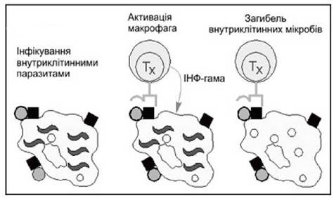
Fig. 1. Activation of macrophages by T helper cells.
Note: circles represent surface microbial antigens; squares represent MHC class II molecules; wavy lines represent intracellular parasites.
There are two subsets of CD4+ T helper cells—Type 1 and Type 2 T helpers. While they share identical antigenic structures, they differ in the profile of cytokines they synthesize in response to antigenic stimulation. This profile dictates whether a cellular or HUMORAL IMMUNE RESPONSE will be mounted.
Type 1 T helper cells (Th1) express the differentiation antigens CD3, CD4, CD29, and CD45Ra. They act as activators of cell-mediated immunity, natural killer cells, and monocytes. When a naive T cell recognizes an antigen presented by a macrophage, it transforms into a Type 1 T helper cell. These cells acquire the CD25 and CD45RB differentiation markers. Their primary function is to enhance macrophage activity aimed at destroying the captured antigen or converting it into an immunogenic form.
By producing interleukins-2, 3, 12, IFN-γ, TNF-β, and GM-CSF, they activate cytotoxic T lymphocytes, natural killer cells, macrophages, and delayed-type hypersensitivity T effectors. Th1 cells provide immunity against Viruses, intracellular Bacteria, and oncogenic cells. Th1 activity is suppressed by interleukin-10.
Type 2 T helper cells (Th2) express the differentiation antigens CD3, CD4, CD29, and CD45Ra and are responsible for cooperation with B cells. Recognition of an antigen displayed on the surface of a B lymphocyte triggers the T cell to transform into a Type 2 T helper cell, which enhances antibody production. These cells acquire the CD25 and CD45RB differentiation markers. By producing interleukins 4, 5, 6, 10, and 13, they activate the humoral immune response, B lymphocytes, and allergic inflammation. By stimulating plasma cells to produce IgM, IgG4, and IgA immunoglobulins, Th2 cells provide immunity against conventional (extracellular) bacteria and their toxins. Activation of eosinophils and mast cells, along with the stimulation of immunoglobulin E (IgE) synthesis, leads to The Development of allergies. Th2 activity is suppressed by IFN-γ.
T lymphocytes carrying CD8 antigens on their surface exhibit suppressor activity (targeting B lymphocytes and their immunoglobulin production) as well as cytotoxic activity.
CD8 T suppressor cells inhibit the development of immune responses to both self and foreign antigens, thereby ensuring immunological tolerance.
Cytotoxic CD8 T lymphocytes (CD8+ CTLs, T killers) are effectors of the cell-mediated immune response responsible for the destruction of foreign cells.
A distinctive feature of the T-cell receptor is its ability to recognize foreign antigens only in a complex with self-cellular antigens on the surface of accessory antigen-presenting cells (dendritic cells or macrophages). Unlike B lymphocytes, which can recognize soluble antigens and bind protein, polysaccharide, and lipoprotein soluble antigens, T lymphocytes can only recognize short peptide fragments of protein antigens presented on the membrane of other cells in complex with self-antigens of the major histocompatibility complex. CD4 T lymphocytes recognize antigenic Peptides in complex with MHC class II Histocompatibility Antigens, whereas CD8 T lymphocytes recognize antigenic peptides in complex with MHC class I histocompatibility antigens.
Natural killer cells (CD3-CD16+CD56+, NK cells) are a subset of lymphocytes distinguished from T lymphocytes by the absence of the T-cell receptor (TCR-). Activated NK cells may express CD25, HLA-DR, Integrins, CD69, the transferrin receptor CD71, and NK receptors on their surface. Their morphological features—large size and the presence of cytoplasmic granules—are the basis for their alternative name: large granular lymphocytes (LGLs). Unlike T cells, the lytic activity of NK cells manifests upon primary contact without prior sensitization. Their primary functional characteristic is The ability to kill certain tumor cells. In peripheral blood, NK cells account for 5 to 20% of circulating lymphocytes.
NK cells develop independently of T and B lymphocytes and lack the surface markers characteristic of T and B cells. Their surface phenotype is TCR-, CD3-CD16+CD56+, yet they share certain signaling molecules with T lymphocytes, such as CD2, individual components of CD3, and the α-chain of CD8.
Natural regulatory T cells—Tregs (CD3+CD4+CD25+)—are a subset of CD4+ T lymphocytes capable of antigen-specifically suppressing T-cell immune responses. These cells control immune responses during Organ and tissue transplantation. Natural Tregs are capable of responding to both self and microbial agents.
No specific surface markers for Tregs have been described. Tregs can be distinguished from conventional activated T lymphocytes by additional phenotypic features. Activated T cells express CD40L (CD154), a molecule required for contact with antigen-presenting cells, while lacking the CD45RO isoform. Natural Tregs in peripheral blood display the following phenotype: CD4+, CD25+, CD45RO+, CD62L+, CD122, CD152+, and GITR (glucocorticoid-induced TNF-R family).
Mononuclear phagocytes (CD14, CD64). The mononuclear phagocyte system includes bone marrow precursors originating from a common stem cell (the monoblast and promonocyte), circulating blood monocytes, and mature tissue macrophages. Mononuclear phagocytes provide innate, non-specific host defense through their phagocytic function.
The main functions of macrophages include the phagocytosis of particulate matter and microorganisms (serving as a crucial cellular factor of natural resistance—"professional scavengers"), the secretion of BIOLOGICALLY ACTIVE SUBSTANCES, and antigen presentation. The biological function of macrophages lies in phagocytosing and presenting antigens to lymphocytes, participating in the induction of inflammation, cytotoxic anti-tumor immunity, regeneration and involution processes, as well as humoral and cellular immunity.
Molecules secreted by macrophages perform effector and regulatory functions. During the development of a specific immune response, macrophages function as antigen-presenting cells.
To achieve this, antigens captured by macrophages undergo processing within phagolysosomes. Peptide fragments of the antigen, generated via Limited proteolysis, form complexes with MHC class II molecules and are displayed on the macrophage membrane in a form recognizable by T lymphocytes.
Macrophages constantly mature from circulating blood monocytes of bone marrow origin. Upon leaving the bloodstream, maturing macrophages migrate into various body tissues. In the Lungs, they are represented by alveolar macrophages. A large number of macrophages reside in connective tissue, lymph nodes, and mucosal-associated lymphoid tissue, particularly within the respiratory tract. The renewal of tissue macrophages occurs primarily through the recruitment of monocytes from the blood.
Dendritic cells and Langerhans cells are of bone marrow origin. They are subdivided into follicular and interdigitating dendritic cells. The former are found in the B-cell zones of LYMPH NODES AND the spleen; they express receptors for the Fc fragment of immunoglobulins on their surface while lacking MHC class II antigens, and they present antigens to B lymphocytes. Interdigitating dendritic cells reside in the T-cell regions of lymph nodes and the spleen; they express MHC class II antigens on their surface but lack Fc receptors, and they participate in antigen presentation to T lymphocytes.
Granulocytes. Other Blood Leukocytes, namely granulocytes or polymorphonuclear leukocytes, can also participate in the effector phase of a specific immune response. These cells constitute the first line of non-specific antimicrobial defense. They are the first to be mobilized to the site of inflammation or infection, and pathogen elimination depends heavily on their phagocytic activity. Their mobilization from the bloodstream is sharply enhanced by cytokines of macrophage origin (interleukin-8) or the C5a fraction of the activated complement system. Other macrophage products (such as tumor necrosis factor) further activate granulocyte functions.
Last update: 13/08/2026
Editorial and Educational Adaptation: This material has been compiled based on the primary/original source text. The project team performed an editorial review, corrected technical inaccuracies, structured sections, and adapted the content for an educational format.
What was processed:
- elimination of formatting defects (OCR errors, structural breaks, corrupted characters);
- editorial organization of content;
- standardization of terminology in accordance with academic sources;
- verification of factual statements against the original source text.
All mentions of the author, publication year, and origin of the primary text have been preserved in accordance with the source.