Review of Medical Physiology - William F. Ganong 2002
Circulation
Body Fluids
Blood - Leukocytes
Normally, a microliter of human Blood contains 4,000–11,000 leukocytes (Table 27-1). The most abundant among them are granulocytes (polymorphonuclear leukocytes [PMNs]).
Immature granulocytes have a indented Nucleus that becomes multilobular as the Cells mature (Fig. 27-2). Most of them contain neutrophilic granules (neutrophils), whereas some contain granules that take up acidic Dyes (eosinophils), and a fraction contain basophilic granules (basophils). The other two Cell types normally found in peripheral blood are lymphocytes, which have a large round nucleus and scant Cytoplasm, and monocytes, which feature agranular cytoplasm and a Kidney-shaped nucleus (see Fig. 27-2). The interplay of these cells provides an effective defense mechanism against tumors, Viruses, Bacteria, and parasites.
Granulocytes
All granulocytes contain cytoplasmic granules filled with BIOLOGICALLY ACTIVE SUBSTANCES involved in inflammatory and allergic reactions. The mean half-life of a neutrophil in peripheral blood is approximately 6 hours. Consequently, maintaining a normal count of these cells requires The production of 100 billion neutrophils per day. Many neutrophils migrate into Tissues. They are attracted to the endothelial surface by selectins, rolling along this surface. Subsequently, a firm bond forms between the endothelium and leukocytes via neutrophil adhesion molecules belonging to the integrin family. The next step involves neutrophils squeezing through the Capillary Wall between endothelial cells, a process known as diapedesis. A significant portion of the cells leaving the blood enters the gastrointestinal tract and is eliminated from the body. Bacterial invasion triggers an inflammatory response. The Bone Marrow becomes active, producing and releasing large numbers of neutrophils. The interaction of bacterial products, plasma factors, and Blood Cells generates compounds that attract neutrophils to the infected site (chemotaxis). Chemotactic factors, which belong to the large and widespread chemokine family (see below), include Complement components (C5a), Leukotrienes, and Polypeptides from lymphocytes, mast cells, and basophils. The stimulatory effect of C5a on chemotactic activity is enhanced by Gc-globulin. This protein is located on the neutrophil membrane and is involved in the binding and transport of vitamin D in plasma (see Chapter 21). Other plasma factors act on bacteria to make them palatable to phagocytes (opsonization). The main opsonins coating bacteria are specific classes of IMMUNOGLOBULINS (IgG) and complement Proteins (see below). These coated bacteria subsequently bind to receptors on the neutrophil membrane, triggering increased cellular motility, exocytosis, and the so-called respiratory burst. These reactions are mediated by a heterotrimeric G protein. Enhanced motility leads to the rapid Digestion of bacteria via endocytosis (phagocytosis). During exocytosis, compounds are released from neutrophil granules into the phagocytic vacuole containing the bacteria, as well as partially into the interstitial space (degranulation). The granules contain various proteases and antimicrobial proteins called defensins. Mammals possess Two Types of defensins, alpha (a) and beta (ß), whereas other types have been identified in invertebrates and plants. In addition, the membrane-bound enzyme NADPH oxidase is activated, leading to the generation of toxic oxygen derivatives. The combination of these toxic oxygen metabolites and proteolytic granule Enzymes makes the neutrophil a highly efficient microbicidal weapon.
Class="center">Table 27-1. Normal values of cellular elements in human blood
Cells |
Cells/µL, mean value |
Approximate normal range |
Percentage of total leukocytes |
Total leukocytes |
9000 |
4000-11000 |
|
Granulocytes Neutrophils |
5400 |
3000-6000 |
50-70 |
Eosinophils |
275 |
150-300 |
1-4 |
Basophils |
35 |
0-100 |
0.4 |
Lymphocytes |
2750 |
1500-4000 |
20-40 |
Monocytes |
540 |
300-600 |
2-8 |
Erythrocytes Females |
4.8 X 10⁶ |
||
Males |
5.4 X 10⁶ |
||
Platelets |
300 000 |
200 000-500 000 |
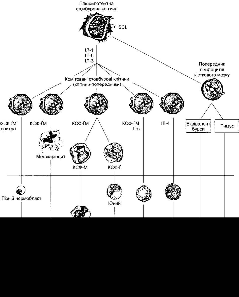
Fig. 27-2. Development of various formed elements of blood from bone marrow cells. Cells below the horizontal line are normally found in peripheral blood. Major sites of action are indicated for Erythropoietin (erythro) and various colony-stimulating factors (CSFs) that ensure Cell Differentiation; G, granulocytes; M, macrophages; IL, interleukin; see Tables 27-2 and 27-3.
Activation of NADPH oxidase is accompanied by a marked increase in O2 uptake and METABOLISM in the neutrophil (respiratory burst) and the generation of O2 through the following reactions:

Note that O2 is a free radical formed by The addition of a single electron to O2, sometimes denoted by adding a dot to form the O2 symbol.
Two O2 molecules spontaneously react with two H+ ions to yield H2O2 in a reaction catalyzed by the cytoplasmic form of superoxide dismutase (SOD):

Thus, the electron (e ) stoichiometry is as follows:

Both O2 and H2O2 are oxidants with strong bactericidal activity, although H2O2 is converted to H2O by the enzyme catalase. The cytoplasmic form of SOD contains Zn and Cu and is identified in various Tissues of the human body. In familial AMYOTROPHIC LATERAL SCLEROSIS (ALS) (see Chapter 16), a defective form of the enzyme resulting from a genetic mutation is observed. Consequently, during this progressive, fatal disease, O2 may accumulate in motor Neurons, leading to their death. Two Other forms of the enzyme encoded by at least one different Gene have been identified in humans.
Neutrophils also contain the enzyme myeloperoxidase, which catalyzes The conversion of Cl, Br, I, and SCN into their respective acids (HOCl, HOBr, etc.). These acids are also potent oxidants. Because Cl is the most abundant halide in Body Fluids, HOCl is the primary product.
In addition to myeloperoxidase and defensins, neutrophil granules contain Elastase, two Collagen-degrading metalloproteinases, and various proteases that assist in destroying microorganisms. These enzymes act in concert with O2, H2O2, and HOCl generated by the action of NADPH oxidase and myeloperoxidase to form a killing zone around the activated neutrophil. While this zone is highly effective at destroying microorganisms, in certain diseases such as rheumatoid Arthritis, neutrophils can cause localized tissue destruction in the body.
Cellular movements during phagocytosis, as well as migration to the site of infection, are mediated by microtubules and microfilaments (see Chapter 1). Normal microfilament function involves the interaction of the Actin they contain with Myosin I on the inner surface of The Cell membrane (see Chapter 1). Neutrophils also release thromboxanes, which are vasoconstrictors, alongside platelet-activating factors, leukotrienes that increase vascular permeability and attract other neutrophils to the site of inflammation, and other Prostaglandins that exhibit modest anti-inflammatory activity.
Similar to neutrophils, eosinophils have a short half-life in peripheral blood, are attracted to endothelial cell surfaces by selectins, bind to Integrins that anchor them to the vascular wall, and migrate into tissues via diapedesis. Like neutrophils, they release proteins, cytokines, and chemokines that drive inflammation and are capable of destroying microorganisms. However, there is some Specificity in the selectins and integrins mediating cell interactions and in the molecules released for microbial killing. Eosinophil maturation and activation in tissues are stimulated in part by IL-3, IL-5, and GM-CSF (see below). These cells are particularly abundant in the gastrointestinal mucosa, where they provide defense against parasites, as well as in the respiratory and urinary mucosal tracts. The number of circulating eosinophils increases in allergic conditions such as asthma, as well as in various respiratory and gastrointestinal pathologies.
Basophils also infiltrate tissues and release proteins and cytokines. They are similar, though not identical, to mast cells (tissue basophils) and, like them, contain histamine and heparin (see below). Basophils release histamine and other inflammatory Transmitters upon activation of the histamine-releasing factor secreted by T lymphocytes (see below), which play a major role in immediate-type hypersensitivity reactions. THE SPECTRUM OF such reactions ranges from mild urticaria to anaphylactic Shock.
Mast cells
Mast cells are granular, motile cells found in regions rich in Connective Tissue, particularly beneath epithelial surfaces. Their granules contain histamine, heparin, and numerous proteases. These cells also synthesize and release leukotrienes and prostaglandins. Their cell membranes bear IgE receptors, and, much like basophils, degranulation occurs following the binding of IgE-coated antigen to their surface. These cells participate in inflammatory reactions initiated by IgE and IgG (see below). During inflammation, they destroy invading pathogens. In addition to participating in adaptive immune responses, these cells release TNF-a in response to bacterial products via antibody-independent mechanisms. Consequently, they take part in non-specific immune responses directed against infectious agents (see below). Marked degranulation of mast cells underlies the clinical manifestations of allergies, including anaphylactic reactions.
Monocytes
Monocytes enter the blood from the bone marrow and circulate for about 72 hours. Subsequently, they migrate into tissues and differentiate into tissue macrophages (Fig. 27-3).
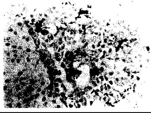
Fig. 27-3. Tissue macrophages in the Pituitary Gland. Macrophages, indicated by arrows, were stained using an immunohistochemical method with Monoclonal Antibodies specific for these cells; AL - anterior lobe; IL - intermediate lobe (courtesy of S. Gordon)
Their lifespan in tissues is unknown; however, data from human bone marrow transplantation indicate that they may survive for about three months. Evidence suggests they do not re-enter the Circulatory system. Some of them terminate their existence as multinucleated giant cells, which are observed in chronic inflammatory processes such as tuberculosis. Tissue macrophages include Kupffer cells in the Liver, alveolar macrophages (see Chapter 34), and microglia in the Brain; all of them originate from the circulatory system. They were formerly referred to as the reticuloendothelial system, but the broader term tissue macrophage system is more accurate.
T lymphocytes secrete macrophages that activate lymphokines. Activated macrophages migrate in response to chemotactic stimuli, where they engulf and destroy bacteria using mechanisms similar to those occurring in neutrophils. Macrophages play a leading role in Immunity (see below). They also secrete about 100 diverse compounds, including factors affecting lymphocytes and other cells, group E prostaglandins, and blood clotting factors.
Granulocyte-Macrophage Colony-Stimulating Factors
The production of erythrocytes and leukocytes, which is tightly regulated in a healthy Organism, is rapidly and significantly enhanced during infections. The proliferation and renewal of pluripotent cells in mice and presumably in humans depend on the production by cells of a protein encoded by the scl (stem cell leukemia) gene. Other factors are also involved. The proliferation and maturation of cells entering the blood from the bone marrow are regulated by growth factor Glycoproteins or Hormones that induce cell proliferation and maturation from one or more committed cell lineages (see Fig. 27-2, Table 27-2). The regulation of erythrocyte production by erythropoietin is described in Chapter 24. Three additional factors are called colony-stimulating factors (CSFs) because they induce a single corresponding cell to proliferate on soft Agar, forming colonies in this medium. Factors stimulating The formation of committed stem cells include granulocyte-macrophage CSF (GM-CSF), granulocyte CSF (G-CSF), and macrophage CSF (M-CSF). The interleukins IL-1, IL-6, and subsequently IL-3 (see Table 27-2) act sequentially, leading to the transformation of pluripotent uncommitted stem cells into committed precursor cells (see Fig. 27-2). Interleukin IL-3 is also known as multi-CSF. Each CSF performs a primary function; however, in addition to this, all CSFs and interleukins exert other effects. They can activate and maintain mature blood cells. Interestingly, the genes encoding most of the described factors are located closely together on a long segment of chromosome 5, and their origin may be related to the duplication of a precursor gene. It is also interesting that normal mice exhibit basal hemopoiesis during which the gene encoding GM-CSF is suppressed. This implies that the loss of one factor can be compensated for by others. Conversely, the absence of GM-CSF leads to the accumulation of surfactant in the Lungs (see Chapter 34).
Table 27-2. Factors Regulating Hemopoiesis
Name |
Cellular Sources |
Cell Type Produced in Increased Numbers |
SCL |
? |
Pluripotent cells |
Erythropoietin |
Kidney cells, Kupffer cells |
Erythrocytes |
G-CSF |
Monocytes, fibroblasts, endothelial cells |
Neutrophils |
M-CSF |
Monocytes, fibroblasts, endothelial cells |
Monocytes |
GM-CSF |
T cells, monocytes, fibroblasts, endothelial cells |
Neutrophils, monocytes, eosinophils, megakaryocytes, erythrocytes |
IL-1 |
Macrophages, fibroblasts, endothelial cells |
Neutrophils, monocytes, eosinophils, basophils, megakaryocytes, erythrocytes |
IL-3 |
T cells |
|
IL-4 |
T cells |
Basophils |
IL-5 |
T cells |
Eosinophils |
IL-6 |
Macrophages, fibroblasts, endothelial cells |
Neutrophils, monocytes, eosinophils, basophils, megakaryocytes, erythrocytes |
As noted in Chapter 24, erythropoietin is produced in part by kidney cells and acts as a circulating hormone. Other factors are produced by T-cell-activated macrophages, fibroblasts, and endothelial cells. For the most part, these factors act locally within the bone marrow.
Impaired Phagocytic Function
More than 15 primary neutrophil defects have been described, along with at least 30 other conditions accompanied by secondary neutrophil dysfunction. Patients with such disorders are prone to infections, which follow a mild course if only neutrophil function is impaired; however, when both the monocyte-tissue macrophage system and neutrophils are affected, the diseases are severe. In one syndrome (impaired neutrophil motility), normal actin polymerization fails to occur, and cell movement becomes sluggish; another condition exhibits congenital leukocyte integrin deficiency. In more serious disorders (chronic granulomatous disease), an inability to generate O2 is documented in both neutrophils and monocytes, rendering the cells incapable of destroying phagocytized bacteria. Marked congenital glucose-6-phosphate dehydrogenase deficiency leads to multiple infections because the production of NADP, required for O2 generation, becomes impossible. In congenital myeloperoxidase deficiency, the efficiency of microbial killing is reduced due to the inability to generate hypohalite ions; however, it is not completely abolished since other bactericidal mechanisms remain intact.
Lymphocytes
Lymphocytes are key elements in the formation of immunity (see below). After birth, some lymphocytes are produced in the bone marrow, but the majority are formed in the Lymph Nodes, Thymus, and Spleen from precursor cells. Precursor cells enter these Organs from the bone marrow after prior differentiation in the thymus and sites equivalent to the bursa of Fabricius (see below) to become precursors of T AND B cells (Fig. 27-4). Lymphocytes enter the blood primarily via Lymphatic vessels. Under normal conditions, only 2% of the body's lymphocytes are found in the peripheral blood. Most other lymphocytes reside in Lymphoid organs. It has been estimated that in humans, 3.5 x 10¹⁰ lymphocytes enter the blood daily solely through the Thoracic duct, although this number includes lymphocytes that recirculate through the thoracic duct. The Effects of Adrenal hormones on lymphoid organs, circulating lymphocytes, and granulocytes are discussed in Chapter 20.
Last update: 10/08/2026
Editorial and Educational Adaptation: This material has been compiled based on the primary/original source text. The project team performed an editorial review, corrected technical inaccuracies, structured sections, and adapted the content for an educational format.
What was processed:
- elimination of formatting defects (OCR errors, structural breaks, corrupted characters);
- editorial organization of content;
- standardization of terminology in accordance with academic sources;
- verification of factual statements against the original source text.
All mentions of the author, publication year, and origin of the primary text have been preserved in accordance with the source.