Review of Medical Physiology - William F. Ganong 2002
Circulation
Body Fluids
Blood and Immunity
Innate Immunity is observed in both insects and invertebrates. The core of this system consists of receptors that bind to specific sugar, lipid, and Amino acid sequences characteristic of common Bacteria, thereby triggering various defense mechanisms. These receptors are germline-encoded, and their basic Structure remains unchanged following antigen contact. Activation of these defense mechanisms results in the release of interferons, phagocytosis, The production of antimicrobial Peptides, and the activation of The Complement System and several proteolytic cascade systems. Even plants exhibit the release of antimicrobial peptides in response to infection.
Class="center">
Figure 27-4. Anatomy of a normal Lymph node (reproduced with permission from McPhee SJ et al. [editors]: Pathophysiology of Disease, 3rd ed. McGraw-Hill, 2000).
In vertebrates, innate immunity is complemented by adaptive immunity—a system in which T AND B lymphocytes are activated by specific Antigens. This activation leads to the proliferation of Cell clones that attack foreign Proteins and, after the invasion subsides, persist as a small pool of memory Cells. This ensures a rapid and robust immune attack upon re-exposure to the same antigen. The genetic changes underlying The Emergence of adaptive immunity occurred 450 million years ago in the ancestors of jawed vertebrates and likely involved the insertion of Transposons into The Genome, enabling the Generation of the vast repertoire of T lymphocytes found in the Organism.
In vertebrates, including humans, innate immunity forms the first line of defense against infections and also triggers the cascade of adaptive immune responses (Fig. 27-5). The mechanisms of both innate and adaptive immunity can also be directed against tumors and foreign tissue grafts.
Upon activation, immunocompetent cells interact via cytokines to destroy Viruses, bacteria, and other foreign cells by releasing additional cytokines and activating the complement system.
Immunology, which has seen tremendous progress in recent years, is a vast and complex discipline. Therefore, this textbook outlines only its fundamental principles.

Figure 27-5. Pathway by which bacteria, viruses, and tumors activate innate immunity and initiate adaptive immune responses. APC = antigen-presenting cell; M = monocyte; N = neutrophil; Th1 and Th2 = T-helper cells type 1 and 2, respectively.
Cytokines
Cytokines are hormone-like molecules that regulate immune responses in a paracrine manner. They are secreted not only by lymphocytes and macrophages, but also by endothelial cells, Neurons, glial cells, and other cell types (Table 27-3). Most cytokine names reflect their function, such as B-Cell Differentiation factor or B-cell growth factor 2. However, by convention, once the Amino Acid Sequence of a human factor is determined, its name is changed to interleukin. For example, B-cell differentiation factor was renamed interleukin-4. In a field advancing at such a rapid pace, compiling an exhaustive list of factors is challenging, but the major cytokines are summarized in Table 27-3.
Most cytokine and hematopoietic growth factor receptors (see above), like the receptors for prolactin (see Chapter 23) and Growth Hormone (see Chapter 22), belong to the cytokine receptor superfamily, which comprises three subfamilies (Fig. 27-6). Subfamily 1 members, which include the IL-4 and IL-7 receptors, are homodimers. Subfamily 2 members, including the IL-3, IL-5, and IL-6 receptors, are heterodimers. The receptors for IL-2 and certain other cytokines are unique in that they consist of a heterodimer combined with an unrelated protein, the so-called Tac antigen. Other members of this subfamily share the same y-chain as IL-2R. The extracellular domain of the homo- and heterodimer subunits contains four conserved Cysteine residues and a Trp-Ser-X-Trp-Ser motif. Although the intracellular domain lacks intrinsic Tyrosine kinase catalytic activity, it is capable of activating cytoplasmic tyrosine Kinases upon Ligand binding.
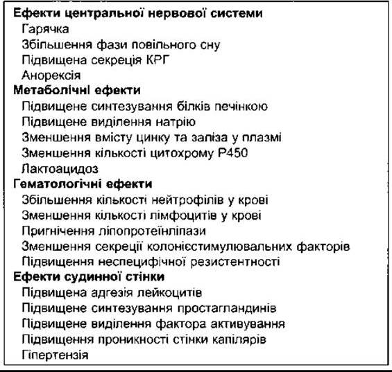
The effects of major cytokines are detailed in Table 27-3. Some exert both systemic and local paracrine effects. For instance, note the systemic effects of IL-1a and IL-1ß (Table 27-4). Although their amino acid sequences share little Homology, their biological effects are similar because they bind to the same receptors. They induce fever, which can also be caused by IL-6, TNFa, and TNFß. TNFs also share other systemic effects with IL-1. Most of these manifestations are typically observed in humans during the Cytology/cytology/16.html">Early stages of infectious diseases.
Another cytokine superfamily is the chemokine family. Chemokines are substances that attract neutrophils (see above) and other leukocytes to sites of inflammation or immune reactions. Over 40 chemokines are currently known, and it is well established that they also play a role in cell growth and angiogenesis. Chemokine receptors are serpentine (G-protein-coupled) receptors that signal through heterotrimeric G proteins, inducing pseudopodia formation and directed cell migration toward the chemokine source.
The Complement System
Cell destruction during innate and immune responses is mediated in part by a system of Plasma Proteins known as the complement system. These proteins are designated C1 through C9. Protein C1 consists of three subunits—C1q, C1r, and C1s—bringing the total number of primary proteins in the system to 11. C1 binds to antigen-bound IMMUNOGLOBULINS, initiating the reaction sequence that activates C3. This pathway of complement activation is known as the classical pathway. Activation can also proceed via an alternative pathway, also known as the properdin pathway.
Table 27-3. Major properties of human interleukins and other immunoregulatory cytokines1
Major cellular sources |
Major effects2 |
|
Interleukins IL-1a and ß |
Macrophages, other APCs, other somatic cells |
Costimulation of APCs and T cells B-cell proliferation and Ig production Phagocyte activation Inflammation and fever (see Table 27-5) |
IL-2 |
Activated Th1, Tc, and NK cells |
Proliferation of activated T cells B-cell proliferation and IgG expression |
IL-3 |
Th lymphocytes |
Growth of early hematopoietic progenitors |
IL-4 |
Th2 cells, mast cells |
B-cell proliferation, IgE and MHC class II molecule expression Proliferation and function of Th2 and T cells Growth and function of eosinophils and mast cells Suppression of inflammatory cytokine production |
IL-5 |
Th2 cells, mast cells |
Growth and function of eosinophils |
IL-6 |
Activated Th2 cells, APCs, other somatic cells |
Synergistic effects with IL-1 or TNF in T-cell stimulation B-cell proliferation and Ig production Thrombopoiesis |
IL-7 |
Thymic and Bone Marrow stromal cells |
T- and B-cell lymphopoiesis Tc-cell Functions |
IL-8 |
Macrophages, other somatic cells |
Neutrophil aggregation and activation |
IL-9 |
Cultured T cells |
Various hemato- and thymopoietic effects |
IL-10 |
Activated Th2, CD8T, and B lymphocytes, macrophages |
Suppression of Th1, NK cell, and APC cytokine production Enhanced B-cell proliferation and humoral response Suppression of cell-mediated immunity; mast cell growth |
IL-11 |
Stromal cells |
Synergistic effects on hemato- and thrombopoiesis |
IL-12 |
B cells, macrophages |
Proliferation and function of activated Tc and NK cells IFN-y production Induction of Th1 cells; suppression of Th2 cell functions Enhancement of cell-mediated immune responses |
IL-13 |
Th2 cells |
Effects similar to those of IL-4 |
Other cytokines TNF-a |
Activated macrophages, other somatic cells |
Effects similar to those of IL-1 Vascular thrombosis and tumor necrosis |
TNF-ß |
Activated Th1 cells |
Effects similar to those of IL-1 Vascular thrombosis and tumor necrosis |
IFN-a and ß |
Macrophages, neutrophils, other somatic cells |
Antiviral effects Induction of MHC class I molecules on all somatic cells Activation of macrophages and NK cells |
IFN-y |
Activated Th1 cells and NK cells |
Induction of MHC class I molecules on all somatic cells Induction of MHC class II molecules on APCs and somatic cells Activation of macrophages, neutrophils, and NK cells Enhancement of cell-mediated immunity (suppression of Th2 cells) Induction of high endothelial venules Antiviral effects |
TGF-ß |
Activated T lymphocytes, platelets, macrophages, other somatic cells |
Anti-inflammatory (suppression of cytokine production and MHC class II expression) Inhibition of macrophage and lymphocyte proliferation Enhanced IgA expression by B cells Enhanced fibroblast proliferation and wound healing |
1 Reproduced with permission from Stites DP, Terr AI, Parslow TG [editors]: Medical Immunology, 9th ed. McGraw-Hill, 1997.
2 All listed effects are stimulatory unless indicated otherwise.
The key driver of this activation pathway is a circulating protein, factor 1, which recognizes repetitive structures on The Cell membrane—specifically polyglucose or polyfructose. Such repeating sequences are found in bacteria and viruses but are absent in mammalian cells. The interaction between factor 1 and the pathogen cell surface initiates the activation of C3 and C5. Properdin is another circulating Blood protein that stabilizes the enzymatic activation complex. Both the classical and alternative pathways converge to form C3 convertase, which subsequently cleaves C3 into its active form, C3b.
Table 27-4. Systemic effects of recombinant IL-1
Protein C3b promotes bacterial opsonization, thereby enhancing phagocytosis. It also triggers reactions that activate downstream Components of the complement cascade. Upon activation, the terminal products C5–C9 generate chemotactic factors and stimulate histamine release. Much like cytotoxic T cells, they also induce the lysis of bacteria, certain viruses, and foreign cells by inserting membrane-puncturing pores known as perforins. This leads to unregulated ion flux and ultimately destroys the cell via osmotic lysis.
Innate Immunity
Cells involved in innate immune responses include neutrophils, macrophages, and natural cytotoxic cells, or natural killer (NK) cells—large lymphocytes that are not T cells yet possess cytotoxic activity. All of these cells respond to Lipids and CARBOHYDRATES specific to bacterial cell walls, as well as to other substances characteristic of tumors and grafts. The primary effect is mediated by the complement system and other systems, in which target cells are destroyed predominantly via osmotic lysis or apoptosis. Cytokines released by these cells also activate cells involved in acquired immune responses.

Fig. 27-6. Structural elements shared by members of one of the cytokine receptor superfamilies. Note that all subunits of subfamily 3, except the a-subunit, possess four conserved cysteine residues (open circles at the top) and a Trp-Ser-X-Trp-Ser motif (black). Many subunits also contain a critical regulatory domain in their cytoplasmic regions (light shading). IL-6, LIF, OSM, and CNTF contain gp130 instead of the ß-subunit; CNTF (ciliary neurotrophic factor); LIF (leukemia inhibitory factor); OSM (oncostatin M) (Modified from D’Andrea AD: Cytokine receptors in congenital hematopoietic disease. N Engl J Med 1994;330:839).
An important link in the innate immunity of Drosophila is the Toll receptor protein, which serves to bind fungal antigens and activate genes encoding antifungal proteins. Five Toll-like receptors (TLRs) have now been identified in humans. One of them, TLR4, binds bacterial lipopolysaccharides along with a protein known as CD14. These reactions initiate intracellular processes that activate the METABOLISM/31.html">Transcription of genes for numerous proteins involved in innate immune responses. This is clinically significant because bacterial lipopolysaccharides produced by Gram-negative pathogens can induce septic Shock.
Acquired Immunity
As noted above, the hallmark of acquired immunity is the ability of lymphocytes to secrete Antibodies specific to each of the millions of foreign agents that enter the body. Antigens that stimulate antibody production are typically proteins and Polypeptides, although antibodies can also be formed against Nucleic Acids and lipids if they are part of Nucleoproteins or Lipoproteins. Antibodies may even be directed against small molecules when they are coupled to carrier proteins. Acquired immunity consists of two components: humoral immunity and cellular immunity. Humoral immunity is mediated by immunoglobulin antibodies circulating in the blood, which belong to the y-globulin fraction of plasma proteins. Immunoglobulins produced by B lymphocytes activate the complement system, attack, and neutralize antigens. Humoral immunity serves as the primary defense mechanism against bacterial infections. In cellular immunity, T lymphocytes are the key players. These processes are observed during delayed-type hypersensitivity reactions and foreign tissue graft rejection. Cytotoxic T lymphocytes attack and destroy cells bearing the antigen that activated them. Cell destruction occurs via the delivery of perforins (see above) and the induction of apoptosis. Cellular immunity is the principal defense mechanism against Infections caused by viruses, Fungi, and certain bacteria, such as the tuberculosis bacillus. It also plays a vital role in tumor surveillance.
Development of the Immune System
During the fetal period, lymphocyte precursors emigrate from the bone marrow. Those that populate the Thymus (Fig. 27-7) are transformed, under the Influence of the local tissue microenvironment, into lymphocytes responsible for cellular immunity (T lymphocytes). In birds, precursors that colonize the bursa of Fabricius—a lymphoid structure near the cloaca—differentiate into lymphocytes responsible for humoral immunity (B lymphocytes). Mammals lack a bursa, and transformation into B lymphocytes occurs in bursal equivalents, such as the fetal Liver and, postnatally, the bone marrow. Following their residence in the liver or thymus, a significant proportion of T and B lymphocytes migrate to Lymph Nodes AND the bone marrow. While the processes of lymphocyte precursor maturation in the thymus and bursal equivalents, as well as migration to lymph nodes and other Tissues, occur during fetal and neonatal life, a slow generation of new lymphocytes from stem cells continues into adulthood. Morphologically, T and B lymphocytes are indistinguishable; they can be identified using cell-surface markers. B cells differentiate into plasma cells and memory B cells. There are three MAIN TYPES OF T lymphocytes: cytotoxic T cells, helper T cells, and memory T cells. Additionally, There are two subsets of helper cells: type 1 T helpers (Th1 cells) secrete IL-2 and y-interferon and are primarily involved in cellular immunity; type 2 T helpers (Th2 cells) secrete IL-4 and IL-5 and primarily interact with B cells in humoral immunity. Cytotoxic T cells destroy transplanted and other foreign cells, with their development regulated by helper T cells. Most cytotoxic T cells bear CD8 Glycoproteins on their surface, whereas helper T cells bear the CD4 glycoprotein. These proteins are closely associated with T-cell receptors and can function as coreceptors. Based on the characteristics of their receptors and functions, cytotoxic T cells are divided into aß and уδ types (see below). Natural cytotoxic cells (see above) are also cytotoxic lymphocytes, although they do not belong to the T-cell Lineage. Thus, there are three Major Types of cytotoxic lymphocytes in the body: aß T cells, уδ T cells, and NK cells.

Fig. 27-7. Development of the acquired immune system.
Following contact with an antigen, a small fraction of activated B AND T cells persist as memory B and T cells. These cells rapidly transform into effector cells upon re-exposure to the same antigen. The capacity to mount an accelerated Immune Response upon secondary antigen contact is another hallmark of acquired immunity. This capacity can persist for a long time in the lymphoid tissue where the immune response originally developed, and even longer in Blood Plasma. In some cases (e.g., immunity against measles), it may last a lifetime. The mechanisms underlying the maintenance of immunological memory have been a subject of much debate. According to one theory, this persistence is driven by recurrent, subclinical exposures to the antigen that trigger weak immune responses without causing clinical disease. According to another theory, memory cells have an exceptionally long lifespan owing to periodic exposure to Neurotrophic Factors that prevent their apoptosis. As noted in Chapter 2, nerve growth factor is found in many tissues outside The Nervous system.
Antigen Recognition
The number of different antigens recognized by lymphocytes in the body is vast. This recognition capacity is innate and develops independently of prior antigen exposure. Stem cells differentiate into millions of distinct T and B lymphocytes, each capable of reacting to a specific antigen. Upon initial entry into the body, an antigen may bind directly to appropriate receptors on B cells. However, a full humoral response typically requires the interaction of B cells with helper T cells. In the case of T cells, the antigen is engulfed by an antigen-presenting cell and partially degraded. Its peptide fragment is then presented to corresponding receptors on T cells. In each instance, this stimulates Cell Division, followed by the generation of clones of cells reactive to that antigen (clonal Selection).
Antigen Presentation
Antigen-presenting cells (APCs) include specialized dendritic Cells of the lymph nodes and Spleen, as well as Langerhans dendritic cells in the Skin. Macrophages and B cells can also function as antigen-presenting cells. Within APCs, polypeptide degradation products of antigens associate with protein products of the Major Histocompatibility Complex (MHC) genes located on the cell surface. Compounds encoded by MHC genes are known as human leukocyte antigens (HLA).
MHC genes, clustered on a short segment of human chromosome 6, encode glycoproteins that are divided into two classes based on tissue distribution and function. Class I antigens consist of a 45 kDa heavy chain noncovalently associated with a ß2-microglobulin molecule encoded by genes located outside the MHC (Fig. 27-8). They are expressed on all nucleated cells. Class II antigens are heterodimers composed of a-chains of 29–34 kDa noncovalently linked to ß-chains of 25–28 kDa. They are found on antigen-presenting cells, notably B cells, and on activated T cells. Class I MHC proteins (MHC-I) preferentially bind peptide fragments derived from endogenous proteins synthesized within the cell. Peptides to which tolerance has not been established—such as those originating from viruses or Mutations—are recognized by T cells. The degradation of these proteins occurs in proteasomes, a complex of Proteolytic Enzymes encoded by genes within the MHC region, after which the peptide fragments associate with MHC proteins in The Endoplasmic reticulum. Class II MHC proteins (MHC-II) predominantly bind peptide products of exogenous antigens, such as bacteria that enter the cell via endocytosis and are degraded in late endosomes.

Fig. 27-8. STRUCTURE OF THE human histocompatibility antigen HLA-A2. The antigen-binding cleft is located at the top and is formed by portions of the a1 and a2 domains. The a3 domain and associated ß2-microglobulin (ß2m) are situated near the membrane. The C-terminal extension of the a3 domain, which serves as the transmembrane region, and the small cytoplasmic tail are not shown (Reproduced with permission from Bjorkman PJ et al: Structure of the human histocompatibility antigen HLA-A2. Nature 1987;329:506).
T-Cell Receptors
MHC protein-peptide complexes On the surface of antigen-presenting cells bind to corresponding T cells. Consequently, T-cell receptors must be capable of recognizing an exceptionally wide array of such complexes. Most receptors on circulating T cells consist of two polypeptide subunits designated a and ß. They form heterodimers that recognize MHC proteins and the peptide fragments bound to them (Fig. 27-9). These cells are termed aß T cells. Approximately 10% of circulating T cells possess receptors containing two different polypeptides designated y and δ. These cells are referred to as уδ T cells and are found in high concentrations in the mucosa of the gastrointestinal tract. These cells are known to bridge the innate and acquired immune systems through the cytokines they secrete (see Fig. 27-5).
CD8 proteins are expressed on The surface of cytotoxic T cells, which bind to MHC-I proteins, whereas CD4 proteins are found on the surface of helper T cells, which interact with MHC-II proteins (Fig. 27-10). CD8 and CD4 proteins enhance the binding of MHC molecules to T-cell receptors and facilitate lymphocyte development, although the precise mechanisms of these effects remain unclear. Activated CD8 cytotoxic T cells directly destroy target cells, whereas activated CD4 helper T cells secrete cytokines to activate other lymphocytes.
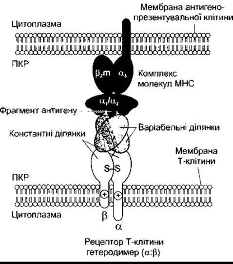
Fig. 27-9. Interaction between an antigen-presenting cell (top) and a T lymphocyte (bottom). MHC proteins (here MHC class I) and their bound peptide antigen fragment interact with the a and ß subunits, which together form the T-cell receptor.
Cellular receptors are surrounded by adhesion molecules and accessory proteins that bind to complementary molecules on antigen-presenting cells during transient cell-cell contact, forming an "immunological synapse" that triggers T-cell activation. It is currently widely accepted that full cell activation requires two signals. The first signal is generated by the binding of the processed antigen to the T-cell receptor, whereas the second results from the interaction of surrounding synaptic proteins. If the first signal is delivered in the absence of the second, the T cell becomes anergic and unable to respond.
B cells
As noted above, B cells can bind directly to antigens, but for full activation and antibody production, they must interact with helper T cells. The Th2 subtype is primarily involved; helper T cells differentiate into Th2 cells under The Influence of IL-4 (see below). Conversely, IL-12 promotes differentiation toward Th1 cells, while IL-2 induces proliferation of activated T cells. The Role of various cytokines in B- and T-cell activation is illustrated in Fig. 27-11.
Activated B cells proliferate and differentiate into memory B cells (see above) and plasma cells. Plasma cells secrete large amounts of antibodies into the blood. Antibodies circulate within the globulin fraction of plasma (see below) and, like antibodies of any localization, are called immunoglobulins. Clearly, immunoglobulins are the secreted form of antigen-binding receptors on the B-cell membrane.
Immunoglobulins
Circulating antibodies protect the body by binding and neutralizing certain protein toxins, preventing the attachment of certain viruses to cells, opsonizing bacteria (see above), and activating complement (see below). The lymphocyte-plasma cell system produces five main types of immunoglobulin antibodies. The main component of each is a symmetrical unit containing four polypeptide chains (Fig. 27-12). The two long chains are called heavy chains, and the two short chains are called light chains. Two Types of light chains—κ and λ—and eight types of heavy chains have been identified. The chains are linked together by Disulfide Bonds, which provide mobility; disulfide bonds are also present within the chains. In addition, the heavy chains are flexible in a region called the hinge. Each heavy chain has a variable (V) segment, in which The amino acid sequence is highly variable; a diversity (D) segment, where the sequence is also highly diverse; a joining (J) segment, in which the sequence is moderately variable; and a constant (C) segment, where the sequence is stable. Each light chain contains V, J, and C segments. The V segments form the antigen-binding site (the Fab region of the molecule; see Fig. 27-12). The Fc region of the molecule is the effector region, mediating antibody-stimulated reactions.
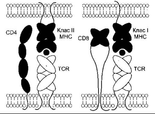
Fig. 27-10. Diagram of The structure of CD4 and CD8 and their relationship to MHC-I and MHC-II proteins. Note that CD4 is a single protein, whereas CD8 is a heterodimer. (Reprinted with permission from Leahy DJ: A structural view of CD4 and CD8. FASEB J 1995;9:17.)

Fig. 27-11. Summary of acquired immune responses. 1 - an antigen-presenting cell captures and partially digests an antigen, after which a portion of the antigen is presented along with MHC peptides (in this case, MHC II peptides on the cell membrane); 2 - formation of an "immune synapse" by an unstimulated CD4 T cell, leading to lymphocyte activation and IL-2 release; 3 - IL-2 acts in an autocrine manner, inducing cell proliferation and clone formation; 4 - an activated CD4 cell can induce B-cell activation and plasma Cell Formation, or activate cytotoxic CD8 cells. Activation of CD8 cells can also be triggered by synapse formation with an MHC I antigen-presenting cell.
Two classes of immunoglobulins contain additional polypeptide components (Table 27-5). In IgM, five main immunoglobulin units are joined around a J chain to form a pentamer. In IgA—secretory immunoglobulins—immunoglobulin units form dimers and trimers around a J chain and a polypeptide derived from epithelial cells, the secretory component (SC).
In the intestine, bacterial and viral antigens are captured by M cells (see Chapter 25) and pass through adjacent clusters of lymphoid tissue, where they stimulate lymphoblasts. These lymphoblasts then enter the Circulatory system via Lymphatic vessels and, after "maturation" in the Blood Vessels, migrate to diffuse clusters of lymphoid elements located beneath the intestinal mucosa and beneath the epithelium in the Lungs, mammary gland, urogenital tract, and FEMALE Reproductive System. There, upon re-exposure to the antigen that triggered the primary response, they secrete large amounts of IgA. Epithelial cells produce SC, which serves as a receptor and binds IgA. As a result, secretory immunoglobulins pass through the epithelial cell and are released by exocytosis. This secretory immune system is an important and effective defense mechanism.
Some immune cells contain inducible NOS (see Chapter 31); NO has been shown to enhance secretory immunity in the gastrointestinal tract (see Chapter 26).

Fig. 27-12. Structure of a typical immunoglobulin G molecule; Fab is the antigen-binding region of the molecule, and Fc is the effector region. Constant regions are colored, and variable regions are light. The constant segment of the heavy chain is divided into CH1, CH2, and CH3. On the right, lines representing intersegmental disulfide bonds are omitted to allow visualization of the JH, D, VH, JL, and VL segments.
Table 27-5. Human immunoglobulins. In all cases, the light chains are κ or λ.
Immunoglobulin |
Function |
Heavy chain |
Additional chain |
Structure |
Plasma concentration, mg/dL |
lgG |
Complement fixation |
y1, y2, y3, y4 |
Monomer |
1000 |
|
lgA |
Local defense in external secretions (tears, intestinal secretions, etc.) |
a1, a2 |
J, SC |
Monomer; dimer with J or SC chains; trimer with J chain |
200 |
lgM |
Complement fixation |
μ |
J |
Pentamer with J chain |
120 |
lgD |
Antigen recognition by B cells |
δ |
Monomer |
3 |
|
lgE |
Reaginic activity; histamine release by basophils and mast cells |
ε |
Monomer |
0.05 |
A large number of immunoglobulins can be synthesized by a single plasma cell; if fused with a tumor cell, it forms an antibody "factory." In practice, animals are immunized with a specific antigen or cell preparation. Following their sacrifice, antibody-producing cells are isolated from the spleens and then fused with myeloma cells. Myeloma is a B-cell tumor whose cells readily fuse with plasma cells to form antibody-producing hybridomas that grow and replicate well. Fused cells are isolated using standard Methods, and each gives rise to a cell clone originating from a single cell.
GENETIC BASIS OF immune system diversity
The genetic mechanisms responsible for generating the extraordinarily large number of different immunoglobulin configurations in the body's lymphocyte pool represent a fascinating biological problem. Part of this diversity is explained by the presence of two types of light chains and eight types of heavy chains. As noted above, each chain contains regions of high Variability (hypervariable regions). The variable domains of heavy chains consist of V, D, and J segments. Within the Gene family encoding these regions, there are several hundred different coding sites for the V segment, about 20 for the D segment, and 4 for the J segment. During B-cell maturation, one V, one D, and one J coding site are randomly selected and recombined to form the gene corresponding to a specific variable region. Light chains exhibit a similar V(D)J recombination of coding sites corresponding to two variable segments (V and J). In addition, J-segment variability is related to the fact that gene segments join in various and generally unpredictable ways (junctional diversity), and nucleotide insertions are sometimes observed (insertion diversity). It has been estimated that these mechanisms generate 108–1010 different immunoglobulin molecules. Additional variability is achieved through somatic mutations.
Similar gene rearrangement and joining mechanisms provide diversity for T-cell receptors. In humans, the α subunit has a V region encoded by one of 50 different genes and a J region also encoded by one of 50 different genes. The β subunits exhibit a V region encoded by one of 50 genes, a D region encoded by one of two genes, and a J region encoded by one of three genes. These variable regions allow the generation of 1015 different T-cell receptors.
Recognition of self
It has long been an intriguing question why T and B cells do not produce antibodies against or destroy the body's own cells and Organs. According current views, self-antigens appear alongside foreign ones but are subsequently eliminated in the thymus during early development (clonal deletion). The reasons why maternal rejection of the fetus as a foreign body does not occur are discussed in Chapter 23.
Autoimmune reactions
Occasionally, the elimination mechanisms for antibodies directed against self-antigens fail, resulting in various autoimmune diseases. These are classified into T-cell- or B-cell-mediated, as well as organ-specific and systemic disorders. Examples include type 1 Diabetes Mellitus (antibodies against pancreatic islet B cells), myasthenia gravis (antibodies against nicotinic cholinergic receptors), and multiple sclerosis (antibodies against myelin basic protein and certain other myelin components).
In some cases, receptor-directed antibodies are capable of activating them; for instance, TSH receptor antibodies increase thyroid activity and cause Graves' disease (see Chapter 18).
Some autoimmune responses are linked to the production of antibodies against microorganisms that cross-react with normal body constituents (molecular mimicry). Others develop through a 'bystander effect', whereby T cells residing near an inflammatory site become sensitized. This leads to their activation, which would not otherwise occur. However, much remains unclear regarding the Pathogenesis of autoimmune diseases.
Tissue Transplantation
The T-cell system is responsible for the rejection of transplanted tissue. When tissues, such as skin or Kidneys, are transferred from a donor to a recipient, the graft 'takes' and functions for a period, but necrosis and 'rejection' eventually occur as the recipient mounts immune responses against the transplant. This is typically observed even when the donor and recipient are close relatives. Rejection does not occur only in the case of identical twins.
Numerous methods have been developed to suppress transplant rejection in humans. The primary goal of Treatment is to halt rejection while preventing infectious diseases. One approach is to destroy T cells by eliminating all rapidly proliferating cells using drugs such as azathioprine, a purine antimetabolite, although this renders patients susceptible to infections and Cancer. Another method involves the administration of glucocorticoids, which suppress cytotoxic T-cell proliferation by reducing IL-2 secretion by T cells; however, this can be complicated by Osteoporosis, psychiatric disorders, and other features of Cushing's syndrome (see Chapter 20). Yet another approach is The Use of cyclosporine or tacrolimus (FK-506). Activation of T-cell receptors normally increases intracellular calcium concentration, which acts via calmodulin to activate calcineurin (Fig. 27-13). Calcineurin dephosphorylates the transcription factor NF-AT, which translocates to The Nucleus and upregulates genes encoding IL-2 and related stimulatory molecules. Nevertheless, these compounds suppress all T cell-mediated immune responses, and cyclosporine causes nephrotoxicity and cancer. A novel and promising therapeutic direction in preventing rejection is inducing T-cell unresponsiveness using drugs that block the second costimulatory signal required for normal activation (see above). Clinically effective drugs with this mode of action could be of paramount importance for transplant surgeons.
Other Clinical Correlations
As knowledge of The Immune System has advanced, over 50 immunodeficiency syndromes associated with impaired function of immunocompetent cells have been described. These conditions range in severity from a mild increase in infection frequency to severe, typically fatal cases. Figure 27-14 illustrates how a block at various stages of B- and T-cell maturation pathways leads to predictable pathological changes. Additionally, there are various types of complement deficiencies.
Malignant transformation can also occur at various stages of lymphocyte maturation. Most, if not all, cases of chronic Lymphocytic Leukemia are associated with the uncontrolled proliferation of B lymphocytes, whereas myeloma arises from the clonal tumor proliferation of mature plasma cells. Some cases of acute lymphoblastic leukemia involve the tumor transformation of T cells. Acquired Immunodeficiency Syndrome (AIDS), currently one of humanity's major challenges, is unique in that HIV (HUMAN IMMUNODEFICIENCY VIRUS), the retrovirus causing the disease, binds to CD4 and causes a drastic reduction in CD4 helper T cells. The loss of helper lymphocytes subsequently leads to impaired proliferation of CD8 and B cells, resulting in the loss of immune function and death from opportunistic infections or malignancies.
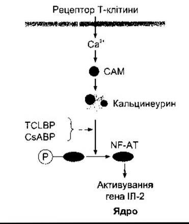
Fig. 27-13. MECHANISM OF ACTION of cyclosporine (CsA) and tacrolimus (TCL) on a lymphocyte. T-cell receptor activation triggers Ca2+ influx, which acts via calmodulin (CAM) to activate calcineurin. Calcineurin dephosphorylates the transcription factor NF-AT, which enters the nucleus and activates the IL-2 gene. Upon binding to their respective immunophilins (BP), cyclosporine and tacrolimus inhibit The Effect of calcineurin on NF-AT, thereby preventing the upregulation of IL-2 production.
Last update: 10/08/2026
Editorial and Educational Adaptation: This material has been compiled based on the primary/original source text. The project team performed an editorial review, corrected technical inaccuracies, structured sections, and adapted the content for an educational format.
What was processed:
- elimination of formatting defects (OCR errors, structural breaks, corrupted characters);
- editorial organization of content;
- standardization of terminology in accordance with academic sources;
- verification of factual statements against the original source text.
All mentions of the author, publication year, and origin of the primary text have been preserved in accordance with the source.