Review of Medical Physiology - William F. Ganong 2002
Physiology of Nerve and Muscle Cells
Excitatory Tissue: Nervous
Neurotrophic Factors
Nerve Trophic Factors
A wide variety of Proteins essential for neuronal survival and growth have been identified and characterized. Some of these—neurotrophins—are produced by Muscles or other structures innervated by the Neurons, while others are secreted by astrocytes. These proteins bind to receptors at nerve terminals, become internalized, and are transported retrogradely to the neuronal Cell body, where they promote the synthesis of proteins required for neuronal development, growth, and maintenance. Other neurotrophins are synthesized within neurons and transported anterogradely to the nerve terminals, where they are necessary for the normal functioning of the postsynaptic neuron.
Receptors
Tables 2–4 list the four definitively identified neurotrophins and their high-affinity receptors. Each of these Trk receptors undergoes dimerization, thereby initiating autophosphorylation within the receptor's cytoplasmic Tyrosine kinase domains. An additional low-affinity receptor for nerve growth factor (NGF) is also known: a 75 kDa protein designated as p75NTR. This receptor binds all four aforementioned neurotrophins with equal affinity. Evidence suggests that it can form a heterodimer with a TrkA monomer, a complex that increases the affinity and Specificity of NGF binding. However, it has been established that p75NTR homodimers capable of binding neurotrophins can themselves mediate signaling pathways leading to apoptosis—a process opposite to the conventional growth-promoting and trophic effects of neurotrophins.
Class="center">Table 2-1. Types of Nerve fibers in mammalian nerves1
Fiber type |
Function |
Fiber diameter, µm |
Conduction velocity, m/s |
Spike duration, ms |
Absolute refractory period, ms |
A α |
Proprioceptive, somatomotor |
12-20 |
70-120 |
0,4-0,5 |
0,4-1,0 |
β |
Touch, pressure |
5-12 |
30-70 |
||
γ |
Motor to Muscle spindles |
3-6 |
15-30 |
||
δ |
Pain, cold, touch |
2-5 |
12-30 |
||
B |
Preganglionic autonomic fiber |
<3 |
3-15 |
1,2 |
1,2 |
C Dorsal ROOT |
Pain, Temperature, certain mechanoreception modalities, reflex responses |
0,4-1,2 |
0,5-2 |
2 |
2 |
Sympathetic |
Postganglionic sympathetic fiber |
0,3-1,3 |
0,7-2,3 |
2 |
2 |
1 A and B fibers are myelinated, whereas C fibers are unmyelinated.
Nerve Growth Factor
The first neurotrophin to be discovered was nerve growth factor (NGF), a protein essential for the growth and maintenance of sympathetic and certain sensory neurons. This factor is present in numerous animal species, including humans, and is found in various Tissues. Particularly high concentrations have been detected in the submandibular gland of male mice. Following castration, NGF levels in males drop to those characteristic of females. The factor is composed of two α, two β, and two γ subunits. The β subunits, each with a Molecular Weight of 13,200, contain all the growth-promoting activity; the α subunits exhibit Trypsin-like activity, and the γ subunits are Serine proteases whose precise function remains unclear. The Structure of the NGF β subunit resembles that of Insulin. NGF is taken up by neurons from the extracranial Organs they innervate and is transported retrogradely from the nerve terminals to The Cell bodies. Additionally, NGF is present in the Brain, where it is apparently required for the growth and survival of cholinergic neurons in the basal Forebrain and striatum. Administration of anti-NGF Antibodies to newborn animals leads to the near-total destruction of sympathetic ganglia, resulting in immunosympathectomy. It is believed that NGF acts to suppress apoptotic pathways in these neurons.
Table 2-2. Numerical system occasionally used for classifying sensory nerve fibers
Number |
Fiber origin |
Fiber type |
Ia |
Muscle spindle, annulospiral ending |
Aα |
Ib |
Golgi tendon organ |
Aα |
II |
Muscle spindle, flower-spray ending; touch and pressure receptors |
Aβ |
III |
Pain and cold receptors, and certain touch receptors |
Aδ |
IV |
Pain, temperature, and other receptors |
Dorsal root C fiber |
Other Neurotrophins
Brain-derived neurotrophic factor (BDNF), neurotrophin-3 (NT-3), NT-4/5, and NGF support various aspects of neuronal survival and prevent apoptosis, although their Functions overlap. Disruption of the NT-3 Gene via knockout results in a marked loss of cutaneous mechanoreceptors (see Chapter 7), even in heterozygous organisms. BDNF acts rapidly and can actually depolarize neurons. BDNF deficiency in mice leads to the loss of peripheral sensory neurons, profound degenerative Changes in the vestibular ganglia, and impairment of LTP (see Chapter 4). An additional factor, NT-6, has also been described.
Other Neuronal Growth Factors
Schwann Cells and astrocytes secrete ciliary neurotrophic factor (CNTF), which promotes the survival of injured and embryonic spinal motor neurons and thus holds therapeutic potential for neurodegenerative disorders. Glial cell line-derived neurotrophic factor (GDNF) Supports Midbrain dopaminergic neurons in vitro. GDNF knockout animals appear to have morphologically normal dopaminergic neurons; however, they lack Kidneys and exhibit impaired enteric Nervous system development (see Chapter 26). Another factor that promotes neuronal growth is leukemia inhibitory factor (LIF). Furthermore, neurons, like other cells, respond to insulin-like growth factor I (IGF-I), Various Forms of transforming growth factor (TGF), fibroblast growth factor (FGF), and platelet-derived growth factor (PDGF). Consequently, The regulation of neuronal growth is a highly complex process.
Table 2-3. Comparative sensitivity of mammalian A, B, and C nerve fibers to various conduction inhibitors
Sensitivity to |
Most sensitive |
Moderately sensitive |
Least sensitive |
B |
A |
C |
|
Pressure |
A |
B |
C |
Local anesthetics |
C |
B |
A |
Table 2-4. Neurotrophins
Neurotrophin |
Receptor |
Nerve growth factor |
TrkA |
Brain-derived neurotrophic factor |
TrkB |
Neurotrophin 3 |
TrkC, to a lesser extent TrkA and TrkB |
Neurotrophin 4/5 |
TrkB |
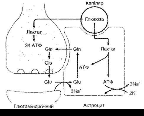
Figure 2-15. The glutamate-glutamine cycle between glutamatergic neurons and astrocytes. Glutamate (Glu) released into the synaptic cleft binds to a Na+-dependent glutamate transporter and is converted into glutamine (Gln) within the astrocyte. Glucose from capillaries enters both astrocytes and neurons. In astrocytes, glucose is metabolized to lactate with the generation of two ATP molecules; one is utilized for The conversion of glutamate to glutamine, while the other is used by the Na+-K+-ATPase to pump three Na+ ions out of the cell in exchange for two K+ ions. In neurons, glucose is metabolized via The Citric Acid Cycle to yield 34 ATP molecules.

Figure 2-14. Glial Cells of the brain (reprinted with permission from Junqueira L, Carneiro J, Kelley R: Basic Histology, 9th ed. McGraw-Hill, 1998).
Last update: 10/08/2026
Editorial and Educational Adaptation: This material has been compiled based on the primary/original source text. The project team performed an editorial review, corrected technical inaccuracies, structured sections, and adapted the content for an educational format.
What was processed:
- elimination of formatting defects (OCR errors, structural breaks, corrupted characters);
- editorial organization of content;
- standardization of terminology in accordance with academic sources;
- verification of factual statements against the original source text.
All mentions of the author, publication year, and origin of the primary text have been preserved in accordance with the source.