Review of Medical Physiology - William F. Ganong 2002
Blood Circulation
Body Fluids
Blood - Platelets
Platelets (thrombocytes) are small granular corpuscles with a diameter of 2-4 µm (see Fig. 27-2). Their Blood count is approximately 300,000/µL, and their normal half-life is about four days. Platelets are formed by the shedding of Cytoplasm from giant Bone Marrow Cells, megakaryocytes, after which they are released into the Circulation. About 60-75% of the blood platelets released from the bone marrow reside in the blood, while the remainder are localized primarily in the Spleen. Splenectomy leads to an increased platelet count (thrombocytosis). Platelets feature peripheral microtubular rings and numerous membrane invaginations with a complex system of channels communicating with the extracellular fluid. The platelet membrane contains receptors for Collagen, von Willebrand factor of the vascular wall (see below), and fibrinogen. Their cytoplasm contains Actin, Myosin, Glycogen, Lysosomes, and Two Types of granules: dense granules containing non-protein substances released upon platelet activation, including serotonin, ADP, and other adenine NUCLEOTIDES, as well as α-granules containing Proteins distinct from lysosomal Hydrolases. These proteins include clotting factors and platelet-derived growth factor (PDGF). PDGF is also produced by macrophages and endothelial cells. It is a dimer formed by two polypeptide subunits, A and B. These subunits are produced as both homodimers (AA and BB) and heterodimers (AB). PDGF promotes wound healing and is a potent mitogen for vascular smooth Muscle. Platelets, like blood vessel walls, contain von Willebrand factor, which, In addition to playing a role in adhesion, regulates the circulating level of factor VIII (see below).
Class="center">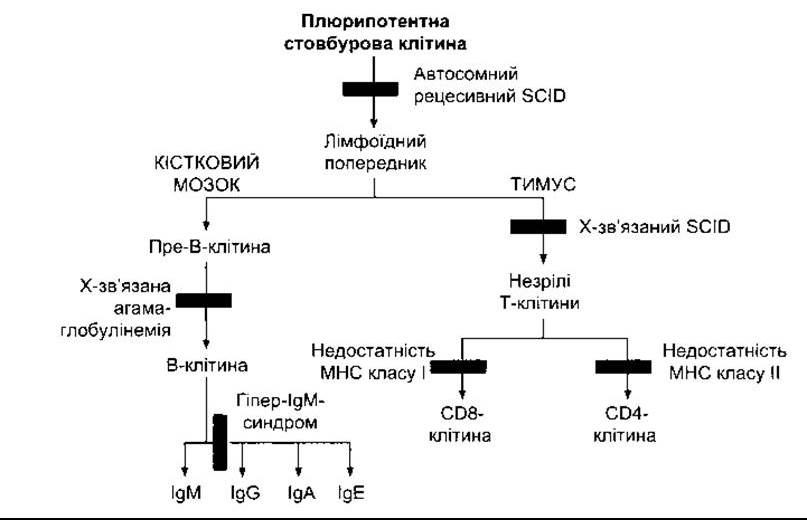
Fig. 27-14. Sites of congenital maturation block of B AND T lymphocytes in various immunodeficiency states (modified from Rosen FS, Cooper MD, Wedgwood RJP: The Primary immunodeficiencies. N Engl J Med 1995;333:431).
Upon blood vessel injury, platelets adhere to exposed collagen, Laminin, and von Willebrand factor of the wall via surface Integrins. Unlike aggregation, this platelet adhesion process does not require METABOLIC ACTIVITY OF the blood platelets. However, binding to collagen leads to platelet activation. Activation can also be triggered by ADP and Thrombin. Activated platelets change shape, form pseudopodia, release granule contents, and adhere to other platelets (platelet aggregation). Aggregation is also promoted by platelet-activating factor (PAF), a cytokine produced by neutrophils and monocytes, as well as by platelets. This substance also exhibits anti-inflammatory activity; it is a phospholipid, 1-alkyl-2-acetylglyceryl-3-phosphorylcholine, synthesized from Membrane Lipids. Its receptor is a typical serpentine G protein-coupled receptor. It activates phospholipase C, resulting in The production of diacylglycerol, which triggers the release of platelet granule contents. An increase in intracellular Ca2+ concentration and diacylglycerol leads to the activation of phospholipase A2, which causes the release of arachidonic acid from membrane Phospholipids. Arachidonic acid is converted into thromboxane A2 (SEE CHAPTER 17), which promotes further Ca2+ influx and phosphatidylinositol breakdown. Thrombin also induces Ca2+ influx and phosphatidylinositol Hydrolysis. Aspirin moderately inhibits thromboxane synthesis. Low doses of aspirin have been proven effective in preventing myocardial infarction, unstable angina, transient ischemic attacks, and stroke due to its ability to disrupt the balance between platelet thromboxane A2 and vascular wall prostacyclin (see Chapter 31).
Platelet production is regulated by colony-stimulating factors that control megakaryocyte formation (see Table 27-2), as well as thrombopoietin, a circulating protein factor. This factor promotes megakaryocyte maturation and is produced by The Liver and Kidneys. Platelets possess receptors for thrombopoietin. Consequently, when the platelet count is low, less thrombopoietin is bound, leaving more in free form to stimulate increased platelet production. Conversely, when the platelet count is high, more thrombopoietin is bound, resulting in decreased platelet production. This feedback mechanism regulates platelet production. The amino terminus of the thrombopoietin molecule exhibits platelet-stimulating activity, whereas the carboxy terminus is rich in carbohydrate residues and determines the molecule's bioavailability. The recombinant amino-terminal domain is used for therapeutic purposes. Further conjugation with polyethylene glycol (PEG) provides a significant increase in activity.
In case of platelet deficiency, clot retraction is impaired, and damaged vessels fail to constrict adequately. This leads to a clinical syndrome (thrombocytopenic purpura) accompanied by increased bruising and numerous subcutaneous hemorrhages. Purpura may also occur with a normal platelet count, sometimes due to structurally altered platelets (thrombasthenic purpura). Individuals with thrombocytosis are prone to thrombosis.
Last update: 10/08/2026
Editorial and Educational Adaptation: This material has been compiled based on the primary/original source text. The project team performed an editorial review, corrected technical inaccuracies, structured sections, and adapted the content for an educational format.
What was processed:
- elimination of formatting defects (OCR errors, structural breaks, corrupted characters);
- editorial organization of content;
- standardization of terminology in accordance with academic sources;
- verification of factual statements against the original source text.
All mentions of the author, publication year, and origin of the primary text have been preserved in accordance with the source.