Orthopedics - Oleksa A.P. 2006
Metabolic Bone Diseases
Osteoporosis
Osteoporosis is a bone disease characterized by a decrease in bone mass and microstructural deterioration of Bone tissue, leading to increased bone fragility and, consequently, a heightened risk of fractures.
According to V.V. Povoroznyuk (1993), epidemiological studies on osteoporosis across various regions of Ukraine identified contributing causal factors, its prevalence, and its distribution among different age groups, as illustrated in the diagrams (Table 2).
It has been established that osteoporosis is most frequently diagnosed in men in the southern regions of Ukraine and in women in the western regions; however, with advancing age, the frequency of its occurrence increases uniformly across all regions. Critical periods in human life associated with the onset of osteoporosis include menopause and, later, advanced age.
As of January 1, 1995, the population aged 55 and over in Ukraine accounted for 25.6% (13.2 million), while the number of women aged 50 and over—that is, in the postmenopausal period—was 9,735,000, representing 35.3% of the country's female population and 18.9% of the total population.
It was also noted that osteoporosis in women aged 30–39 develops As a result of early menopause following oophorectomy, thyroid disorders, and dietary factors. Bone mineral density is markedly reduced in individuals who have not consumed milk and dairy products as vital sources of calcium. Osteoporosis is more common in men suffering from gastric and duodenal ulcers, hepatitis, etc., as well as in those who smoke, consume alcohol, or suffer from hypogonadism.
Concurrently, studies were conducted on The impact of radionuclides on The Human Body in zones II–IV following the Chornobyl disaster. Osteotropic strontium (Sr-90), which has a half-life of 30 years, enters the body through food products. Calcium acts as an analogue of strontium in metabolic processes, meaning that the degree of Sr-90 accumulation depends on The amount of dietary calcium. This osteotropic radionuclide suppresses preosteoblasts and disrupts The Structure of hydroxyapatite crystals. This leads to an imbalance between osteoblasts and osteoclasts, thereby impairing normal bone remodeling. Studies have revealed a high frequency of osteopenic syndrome among cleanup workers of the Chornobyl catastrophe, which may be a cause of systemic osteoporosis. They exhibited decreased bone density, a high percentage of polyarthralgia, and an accelerated rate of Aging of The Musculoskeletal System (V.V. Povoroznyuk, Ye.P. Podrushnyak, E.V. Orlova et al., 1995).
From the foregoing, it follows that the causes of primary osteoporosis can vary, with the most prominent being the postmenopausal type (Type I), which occurs shortly after the decrease and cessation of estrogen levels (physiologically or surgically) in the female body, as well as senile, age-related osteoporosis (Type II).
In Ukraine, postmenopausal osteoporosis occurs in 21.5% of the population (V.V. Povoroznyuk et al., 1995). Bone tissue mineralization begins to decline after the age of 40 (by 1% annually in women and 0.5% in men).
Age-related osteoporosis develops gradually and is clinically silent. In individuals over 60 years of age, bone mass (mineral component) loss reaches 2–4% annually (Fig. 83). This diminishes bone density and mechanical strength, causing frequent fractures in elderly and senile individuals of both sexes. These can be classified as pathological fractures because they very often occur without significant physical exertion. Most common are fractures of the proximal or distal ends of the Femur (neck, intertrochanteric, and subtrochanteric fractures), the humerus, the radius at a typical site, vertebral bodies, and occasionally Ribs (Fig. 84).
Class="center">Table 2. Distribution of osteoporosis by age, sex, and region of residence
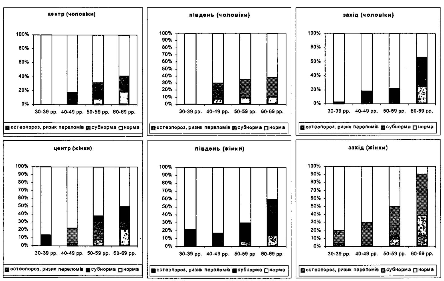
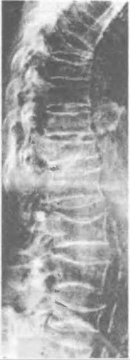
Fig. 83. Vertebral deformations caused by senile osteoporosis.
Secondary osteoporosis arises as a result of gastrointestinal diseases (PEPTIC ULCER DISEASE), Liver and Kidney disorders, Diabetes Mellitus, hyperparathyroidism, hypogonadism, Rheumatoid Polyarthritis, and The Use of corticosteroids, anticoagulants, etc.
Osteoporosis is generally considered a disease of the elderly; however, A.P. Krysyuk, T.A. Kinchaya-Polischuk, and H.V. Hayko (1997) point out that a significant number of children and adolescents suffer from osteoporosis, both congenital (in Osteogenesis Imperfecta, Renal osteodystrophy) and acquired (in diabetes mellitus, rheumatoid Arthritis, post-polio syndrome, prolonged immobilization of a limb, inflammatory Bone and joint processes, etc.). These authors propose the following Classification of osteoporosis in children:
1. By etiological factors — primary (congenital) and secondary (acquired).
2. By prevalence — systemic and local.
3. By severity — diffuse and focal.
4. By age — early childhood osteoporosis, older childhood osteoporosis, and juvenile osteoporosis.
Diagnostics and Clinical presentation.
Biochemical markers of osteoporosis include serum alkaline phosphatase assays, 24-hour urinary hydroxyproline excretion tests, and determinations of inorganic phosphorus and calcium levels in Blood and urine.
Bone alkaline phosphatase is produced by osteoblasts and serves as a marker of The rate of bone tissue formation. In osteoporosis, alkaline phosphatase activity typically falls within the normal range or is occasionally elevated.
Hydroxyproline is a breakdown product of collagens. Due to increased bone resorption and Collagen Catabolism, urinary hydroxyproline levels in a 24-hour sample are elevated. As a result of bone demineralization in osteoporosis, blood calcium levels may be normal or elevated, while phosphorus levels may be normal or reduced.
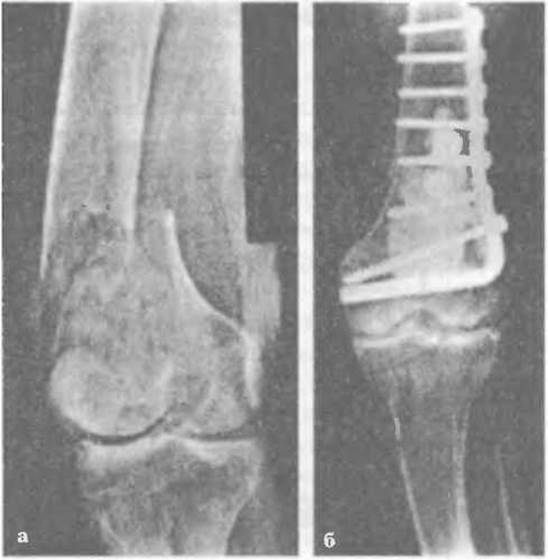
Fig. 84. Pathological fracture of the femur due to osteoporosis: a - supracondylar fracture, b - following internal fixation surgery.
As already mentioned, osteoporosis has an almost asymptomatic clinical course. Complaints of generalized fatigue can be attributed to many different conditions. Relative indicators of osteoporosis include joint and back pain, increased thoracic Kyphosis, and the eventual development of a "widow's hump"—a fixed kyphosis—sometimes accompanied by lateral curvature (Scoliosis). This is caused by microfractures of the vertebral trabeculae. Gait alterations also occur: the patient walks with short steps and the torso tilted forward.
Most commonly, signs of osteoporosis are discovered during X-ray diagnostics performed to investigate The Nature of bone fractures. Plain radiography is an accessible, inexpensive, and traditional method for examining and diagnosing osteoporosis, although it lacks high precision as it depends on the technical quality of the radiograph. Based on a lateral spinal radiograph, the degree of osteoporosis is visually assessed across three grades: mild, moderate, and severe.
It should be noted that radiographic detection of osteoporosis is only possible once 25–30% of bone mass has been lost, which indicates already significant metabolic alterations. On standard planar radiographs, osteopenia is visually apparent only when bone mineral density has decreased by 30–50%.
As a rule, both Qualitative and quantitative radiographic morphometry are performed.
Qualitative morphometry is based on evaluating the trabecular STRUCTURE OF THE vertebral endplates and the architecture of the proximal femur in comparison with normal standards. In osteoporosis, findings include reduced femoral bone density, cortical thinning, increased prominence of trabeculae along lines of vertical stress, and barely visible horizontal trabeculae in the femoral neck, HEAD, and trochanteric region.
Similar changes are observed in the vertebrae. In later Stages of the disease, wedge-shaped and "fish vertebrae" appear as a result of microfractures, and Schmorl's nodes may also be present.
Quantitative assessment of osteoporosis is based on radiographic measurements of three vertebral body heights—anterior, middle, and posterior (Fig. 85)—and their ratios for each vertebra from the fourth thoracic (Th4) to the fifth lumbar (L5) vertebra. A reduction in these parameters by more than two standard deviations from the normal mean is considered indicative of pronounced osteoporosis, while a reduction of more than three standard deviations is interpreted as an osteoporotic vertebral compression fracture.
Radiographs also allow for the Determination of the disc coefficient, which is The ratio of the intervertebral disc height to the height of the adjacent vertebral bodies. In individuals aged 25–35, this coefficient is 1/7–1/6 in the upper thoracic region, 1/5–1/6 in the mid-thoracic region, and 1/3 in the lumbar spine. Deviations from these values indicate degenerative and destructive Changes in the spine.
There are numerous Methods for determining osteoporotic indices using hand or spine radiographs (Barnett-Nordin, Exton, Rokhlin, Podrushniak, etc.). For instance, Barnett-Nordin (1960) calculated an index using a formula that incorporates measurements of the width of the second metacarpal bone at its mid-shaft and the width of its medullary cavity at the same Location. Exton’s formula additionally accounts for the length of this bone. D. G. Rokhlin calculates the osteoporosis index of the thoracic and lumbar spine from lateral radiographs using a formula based on measurements of the height of the middle part of the vertebral body (from the anterior to the posterior margin), as well as formulas defining the minor (internal) and major (external) outer rectangles of the vertebral bodies.
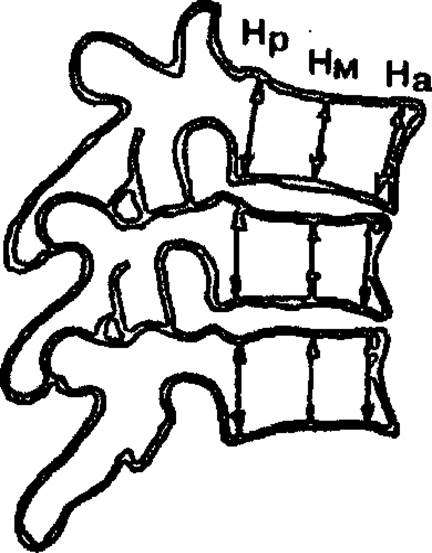
Fig. 85. Vertebral morphometry.
These methods for determining the osteoporosis index are now rarely used due to the advent of more precise and simpler techniques. These include isotope studies, direct bone biopsy with tetracycline labeling, and quantitative computed tomography (QCT), among others.
Some clinics utilize reference densitometry, which is based on assessing X-ray absorption during radiography using a standardized film, an aluminum wedge as a densitometric standard, and high-precision computerized equipment for Processing bone radiograms.
However, the use of isotope and radiographic techniques involves a certain amount of radiation exposure. Therefore, in clinical practice, ultrasonic osteometry—based on measuring the speed of ultrasound propagation through bone—is most commonly used. For this purpose, the "EOM-01" diagnostic device is employed, which can measure ultrasound transit time across any skeletal site.
The "Achilles" ultrasonic bone densitometer is also used to diagnose osteoporosis by determining the density of the calcaneus, which has a trabecular structure. Compared to histomorphometry (analysis of an iliac crest biopsy specimen), this method is non-invasive, quite accurate, and requires little scanning time (5 minutes). The device itself is compact and portable. It was utilized by V. V. Povoroznyuk and a team of researchers who conducted epidemiological studies on osteoporosis across various regions of Ukraine.
The findings of these studies are presented in Table 2 (p. 105).
Treatment and Prevention of Osteoporosis.
First and foremost, it is essential to determine the underlying cause of osteoporosis: whether it is primary or secondary.
The Therapeutic management of osteoporosis comprises both treatment and preventive measures aimed at avoiding complications such as bone fractures.
Given that the onset and progression of osteoporosis depend on numerous internal and external factors, the physician's strategy must be multifaceted.
Maximizing bone mass accumulation during growth and Puberty plays a crucial role in preventing the premature development of osteoporosis. The formation of a strong Skeleton continues up to the age of 20–25 in women and 30–35 in men, at which point peak bone mass is reached. Therefore, A balanced diet during this period—ensuring an adequate intake of calcium and vitamin D—along with regular musculoskeletal physical activity and an active lifestyle, is of utmost importance.
Physical activity is strictly necessary, as a standard daily routine is insufficient for preventing osteoporosis. According to the Wolff-Delpech law, proper Bone Formation is stimulated by physical exercises involving weight-bearing under antigravity conditions, which exerts appropriate mechanical stress on articular surfaces and improves Blood supply to the bones.
In a growing Organism, the daily calcium requirement is 1,200 mg, whereas in adulthood it is 1,000 mg. However, an increased daily dose of 1,600 mg is necessary for girls under 19 years of age, as well as for pregnant and lactating women; for older individuals, this dose may be lower, around 1,200 mg/day.
It is advisable to increase the calcium dose to 1,500 mg during the postmenopausal period; however, if estrogen replacement therapy is administered, a calcium dose of 1,000 mg/day (as in normal adulthood) is sufficient.
Calcium is obtained through dietary sources, particularly milk (100–120 mg/100 g) and dairy products (cottage cheese – 95–100 mg/100 g; processed cheese – 300 mg/100 g; hard cheese – 600–1,000 mg/100 g), as well as fish products (dried fish with bones – up to 3,000 mg/100 g; sardines – 350 mg/100 g) and nuts (hazelnuts – 290 mg/100 g; almonds – 254 mg/100 g). Among vegetables, celery is the richest in calcium (240 mg/100 g).
Calcium absorption by the body is impaired by the consumption of foods with a high oxalic acid content, such as spinach, rhubarb, gooseberries, and currants, as well as by gastrointestinal disorders (such as peptic ulcer disease) and liver diseases.
Vitamin D stimulates bone formation, promotes calcium absorption, and improves the body's calcium balance. It is prescribed not only to children for the prevention of Rickets, but also in combination with calcium supplements for patients with osteoporosis. Certain synthetic derivatives (such as 1-alphahydroxyvitamin D) can correct hyperparathyroidism, prevent bone loss, and thereby reduce the risk of fractures.
Regular Physical Exercise aimed at maintaining bone mass (such as weight training and spine-loading exercises, as well as walking) plays a crucial role in the prevention of osteoporosis. It is also essential to eliminate harmful habits, including smoking, alcohol abuse, and excessive intake of coffee and salt.
During the postmenopausal period (the first 6–10 years), women are sometimes prescribed estrogen replacement therapy. Estrogens support bone function and promote the synthesis of endogenous bioactive substances (such as cytokines, Prostaglandins, and Calcitonin) that participate in bone remodeling and fracture prevention.
Estradiol valerate is most commonly prescribed and is a key component of medications such as Progynova, Cyclo-Progynova, Klimen, and Gynodian Depot. In Progynova, estradiol valerate is the sole active ingredient. Due to potential complications associated with long-term estrogen use—such as endometrial hyperplasia, uterine bleeding, and even endometrial Cancer—estrogens are prescribed in combination with progestogens. Patients undergoing estrogen therapy must undergo regular gynecological examinations and pulmonary imaging, as the risk of cancer increases by 30%.
Cyclo-Progynova is now widely used and comes in calendar packs of 21 dragees. Treatment begins with 11 white dragees (taken one daily), each containing 0.002 g of estradiol valerate, followed by 10 light-brown dragees (also one daily), each containing 0.002 g of estradiol valerate and 0.0005 g of the progestogen norgestrel. After a 21-day treatment cycle, a 7-day break is taken before repeating the cycle. The total course of treatment lasts for 6 months.
Another medication in this category is Klimen, which is also supplied in packs of 21 dragees. The primary difference is that instead of norgestrel, the final 11 dragees contain 0.001 g of cyproterone acetate. This drug is indicated for hormone replacement therapy in menopausal disorders and androgen-deficiency states.
For Other types of osteoporosis, calcitonin or Miacalcic (synthetic salmon calcitonin) is recommended. The primary MECHANISM OF ACTION of calcitonin involves inhibiting osteoclast resorptive activity, suppressing osteolysis, and reducing the withdrawal of calcium from bones.
Miacalcic is known to significantly reduce the risk of fractures in the extremities and vertebrae while alleviating bone pain, thus acting as an analgesic. It is available in ampoules of 50 and 100 IU per 1 ml, as well as in the form of a nasal spray (providing 14 standard doses).
Miacalcic is prescribed as maintenance therapy (to preserve bone mass) at a dose of 50 IU administered subcutaneously or intramuscularly three times a week for three months. A three-month break is observed between courses.
In cases of severe pain resulting from vertebral compression fractures, Miacalcic is administered at 100 IU daily subcutaneously for one week, followed by 50 IU daily or every other day for 2–3 weeks (or 10 days if pain subsides). Alternatively, the nasal spray can be used intranasally at a dose of 50 IU twice daily for two weeks. Patients receiving Miacalcic therapy should maintain a daily calcium intake of 600–1200 mg.
Antiresorptive agents also include anabolic Steroids, which exert an antiresorptive effect and stimulate an increase in bone mass. They are prescribed for patients with senile osteoporosis. Commonly recommended agents include Retabolil, Phenobolin (turabolil, turinabol), and Nerobol (methandrostenolone).
Phenobolin in an oil solution is administered intramuscularly 3 to 4 times—once every 7–10 days at a dose of 0.025–0.05 g. Retabolil is administered at the same dose every 2–3 weeks for a total of 5–6 injections.
Methandrostenolone is prescribed for patients over the age of 55 according to a tapering schedule: 5 mg daily for 10 days, 2.5 mg daily for the next 10 days, and 1 mg daily for 5 days.
Anabolic steroids must not be prescribed to patients with prostatitis or prostate cancer.
Bone-stimulatory agents include sodium fluoride (osseine, coreberon) and osteochin (ipriflavone). Sodium fluoride stimulates osteogenesis, prompting osteoblasts to produce increased amounts of bone matrix, while its ions are incorporated into the newly forming bone during mineralization, substituting for the hydroxyl ion in the apatite crystal lattice. This process yields sparingly soluble fluorapatite, which is significantly more resistant to osteoclastic resorption.
Osteochin belongs to the flavonoid group; it improves metabolic processes within bone tissue, stimulates bone formation while reducing resorption, acts as an analgesic, and helps prevent kyphosis (vertebral body fractures) associated with osteoporosis.
The current state-of-the-art approach to treating osteoporosis involves the use of bisphosphonates to block osteoclastic activity. This drug class includes alendronate, etidronate, pamidronate, and the newer agent Fosamax (sodium alendronate) manufactured by MSD. Fosamax belongs to the aminobisphosphonate group, which is capable of preventing osteoporosis in postmenopausal women. It suppresses osteoclast activity, promotes a positive bone tissue balance, and increases bone mineralization—including in the pelvic bones and spine—thus restoring skeletal bone status closer to normal. In most women, bone density returns to pre-menopausal levels after just one month of Fosamax therapy.
For the prevention and treatment of osteoporosis, the Institute of Gerontology of the AMS of Ukraine and the Institute of Meat and Dairy Products have developed "Cosmol". This dry dairy product consists of whole milk powder, dextrin-maltose, Vitamins C, D, and E, and calcium lactate. The optimal ratio of protein, calcium, phosphorus, and lactose promotes the normalization of calcium METABOLISM and its assimilation by bone tissue. Cosmol (20 g of powder) is dissolved in a Glass of warm Water and taken daily for one month, twice a year.
Comprehensive treatment and prevention strategies for osteoporosis are applied with the ultimate goal of increasing bone mass. However, the long-term therapeutic efficacy of these interventions has not yet been fully elucidated in all cases.
Last update: 10/08/2026
Editorial and Educational Adaptation: This material has been compiled based on the primary/original source text. The project team performed an editorial review, corrected technical inaccuracies, structured sections, and adapted the content for an educational format.
What was processed:
- elimination of formatting defects (OCR errors, structural breaks, corrupted characters);
- editorial organization of content;
- standardization of terminology in accordance with academic sources;
- verification of factual statements against the original source text.
All mentions of the author, publication year, and origin of the primary text have been preserved in accordance with the source.