Orthopedics - Oleksa A.P. 2006
Joint Diseases
Arthritis of Unknown Etiology
Rheumatoid Polyarthritis
Rheumatoid Arthritis (infectious non-specific arthritis, infectarthritis, primary chronic polyarthritis, chronic evolving polyarthritis) is a severe infectious-allergic disease belonging to the group of systemic Connective Tissue disorders, primarily affecting The Musculoskeletal System.
The prevalence of rheumatoid arthritis across various Regions of the former USSR varies insignificantly, averaging about 1% of the surveyed population (Astapenko M.G., Pavlenko T.M., 1966). In Ukraine, such epidemiological studies were conducted by Oleksa A.P. (1972) in three western regions—ranging from the high-altitude Carpathians (Dolyna) to the low-lying, Water-rich Volyn (Liuboml). Adhering to Kellgren's diagnostic criteria, no difference in the incidence of rheumatoid arthritis (1.25 per 1,000) was found across these geographical zones.
A characteristic feature of rheumatoid arthritis is its progressive course, leading to severe disability, which, according to our data, ranks first (23%) among all causes of primary disability resulting from musculoskeletal disorders.
The Pathogenesis of the disease involves systemic progressive damage to connective tissue (primarily the disorganization of its ground substance), with the process most frequently localizing in the joints. However, articular-visceral forms of rheumatoid polyarthritis also occur.
The exact causes of the disease remain undetermined to this day. Most specialists support the infectious-allergic theory, which attributes the onset of the disease to a specific allergic state of the Organism caused by hypersensitization to bacterial or viral protein breakdown products, as well as their endotoxins. The disease is linked to streptococcal infection, predominantly localized in the nasopharynx and Oral Cavity (carious Teeth, acute tonsillitis, etc.). However, the focus of infection may serve merely as a triggering mechanism for sensitization and impaired immunological reactivity of the organism, manifesting as arthritis. The subsequent course of the disease is determined by the degree of the organism's reactivity, metabolic disturbances, and dysfunctions across various systems. Autoimmune and autoaggressive processes take place within the synovial membrane of the Joint Capsule and surrounding Blood Vessels, manifesting as massive cellular infiltration of the membrane and alterations in its vascular bed. This leads to microvascular dysfunction, increased vascular permeability, progressive connective tissue disorganization, and exudative-dystrophic and sclerotic tissue changes.
Clinical symptoms. The disease may begin acutely (within a single day), subacutely, or as a primary chronic condition. An acute onset of rheumatoid arthritis is typically observed in children and young women following acute tonsillitis, a cold, or similar illnesses. In men and elderly individuals, the disease most often begins gradually and imperceptibly, progressing with characteristic clinical and pathomorphological Changes in the joints.
There are three clinical-anatomical stages of rheumatoid arthritis (Skliarenko Ye.T., 1965). The First stage is the exudative stage (of acute, subacute, or chronic synovitis). In the acute course, arthritis begins with sharp pain in the joints, Swelling, and elevated body Temperature. The pain can be so severe that the patient adopts a protective posture, becoming immobilized and resistant to movement.
In the subacute process, pain develops without apparent cause in one or several (paired) joints, accompanied by localized temperature elevation, restricted movement, antalgic contracture, and smoothing of joint contours. Palpation clearly reveals free fluid within the joints. Characteristic signs include morning stiffness, subcutaneous rheumatoid nodules on the extensor surfaces near the joints, symmetrical joint involvement, and—as noted previously—the absence of involvement in the distal phalangeal joints.
Rheumatoid arthritis typically begins in one or two paired joints (most commonly with synovitis of the knee (Fig. 304) or interphalangeal joints), occasionally presenting as transient pain and stiffness in several finger JOINTS OF THE hands. Contractures are generally absent.
Class="center">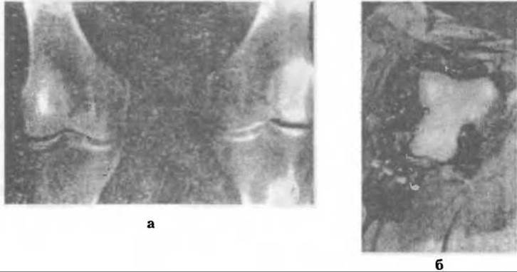
Fig. 304. Rheumatoid monoarthritis of the knee joint (a); appearance of the hypertrophied synovial membrane during surgery (b).
Macroscopically, the synovial membrane is edematous with hypertrophied villi, which sometimes doubles or more than doubles its secretory surface area. The joint cavity contains turbid exudate with flecks of floating fibrin. Within the joint, fibrin precipitates into clots and settles on the synovial membrane and articular Cartilage. During joint movement, fibrin is mechanically worn away from the cartilage and accumulates along its margins. Granulation tissue develops in the same areas, gradually creeping over the cartilage (pannus).
Microscopically, the synovial membrane exhibits edema, vascular dilatation, lymphoid and plasma Cell infiltration, as well as mucoid and fibrinoid disorganization of the connective tissue in the villi.
Radiologically, the first stage of arthritis reveals Osteoporosis of the epimetaphyseal regions of the bones (especially the hands) and widening of the joint space if there is excessive effusion (pressure) within the joint, along with Baker's Cyst (Fig. 305).
The severity and extent of connective tissue damage are reflected in laboratory parameters: the ERYTHROCYTE SEDIMENTATION RATE (ESR) increases, along with elevated levels of globulins, fibrinogen, diphenylamine (DPA) test values, hyaluronidase activity, and the appearance of C-reactive protein. If process activity is high, the Waaler-Rose test may be positive. The more acute and severe the disease course, the more pronounced these indicators become.
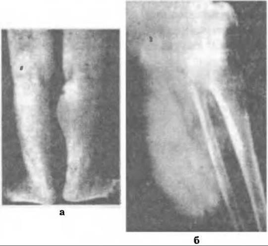
Fig. 305. Popliteal Baker's cyst in rheumatoid synovitis: a - clinical view, b - radiological view after contrast medium administration.
There are three degrees of activity in the rheumatoid process: Grade I — minimal activity; Grade II — moderate; Grade III — high. The overall activity of the process is assessed based on the general course of the disease, the quality of clinical manifestations, and blood laboratory parameters. Local activity for each individual joint is evaluated by the severity of the inflammatory-exudative clinical manifestations, alongside laboratory analyses of synovial fluid: cytological (phagocyte count, cellular composition), biochemical (protein, Enzymes, etc.), and immunological (Waaler-Rose test, latex test, etc.).
Process activity serves as an indicator of Treatment efficacy over time. Local assessment is necessary for orthopedic surgeons to establish indications for Surgical treatment and determine The Scope of surgery. Fig. 306 illustrates the potential course of rheumatoid arthritis and its outcomes: 1) possible restoration of joint function; 2) chronic relapsing course; 3) destruction of articular cartilage resulting in secondary arthrosis-arthritis or ankylosis.

Fig. 306. Potential course of rheumatoid arthritis: 1 - possible joint restoration, 2 - destruction of articular cartilage with subsequent ankylosis, 3 - destructive osteoarthritis.
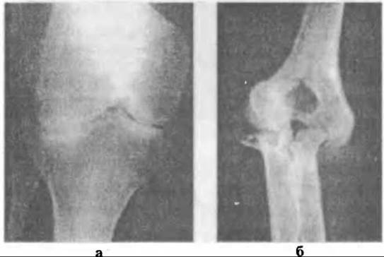
Fig. 307. Radiographs of the knee (a) and elbow (b) joints In the second stage of the rheumatoid process.
The duration of the first stage varies. In primary arthritis, it can last for years, whereas in an acute disease course, the process progresses so rapidly that destructive changes in the articular cartilage (Second Stage) develop within a few months (4–6).
The second stage is the proliferative-destructive stage (Fig. 307). In this stage, pain and joint effusion decrease, and proliferative reactions and sclerotic changes predominate over exudation. Fibrin that had precipitated from the exudate into the recesses (pouches) of the joints as plaque and clots becomes organized, leading to adhesive arthritis which, As a result of scarring, causes arthrogenic scar contracture.
Regarding the destruction of the articular cartilage, which is the primary feature of the second stage, various explanations of its Etiology have been proposed. It is generally accepted that articular cartilage damage in rheumatoid arthritis is secondary. Due to subchondral rheumatoid granulomas developing near the epiphyseal vessels and their subsequent proliferation, the Nutrition of the deep layers of the articular cartilage is disrupted. Pathologically altered synovial fluid, a fibrinous film, and pannus on the cartilage compromise the nutrition of its superficial layers. Degenerative changes, microcracks, and destruction occur within the cartilage, which are further aggravated by lysosomal Enzymes of the synovial fluid and mechanical factors. Erosions of varying size and depth appear; these are less pronounced in areas of the cartilage where fibrinous deposits and pannus are mechanically worn away during joint movement, and more prominent in weight-bearing areas.
It is very common to perform surgery on joints where pathological changes are initially diagnosed as the first stage of the disease, only to discover during the Procedure that these changes correspond to the second stage, having remained invisible on standard radiographs. Therefore, to accurately determine the stage, it is advisable to use arthroscopy or contrast radiography of the joints.
Clinically, along with Complaints of pain, there are classic joint deformities against a Background of Muscle atrophy. In most cases, the limbs are held in a forced position with pronounced contractures, subluxations, and restricted mobility, with the degree of functional impairment directly determining the extent of disability. There are three degrees of functional Impairment of the joints (FIJ): Grade I — professional capacity is preserved; Grade II — working capacity is lost and self-care is limited; Grade III — inability to care for oneself (patients require outside assistance).
Rheumatoid arthritis is dominated by flexion and flexion-adduction contractures that originate as antalgic and progress to arthrogenic. In the fingers, classic flexion and extension contractures may develop (such as "swan-neck" or "boutonnière" deformities) with subluxations and dislocations of the Phalanges (Fig. 308). As a result of rheumatoid tenosynovitis, changes occur in the tendons and their sheaths, potentially leading to pathological tendon ruptures. If the contractures in the limb joints still allow for various movements, such limb positions are termed concordant, whereas if they do not, they are discordant. Patients with discordant (fixed) positions of the lower extremities are bedridden.

Fig. 308. Rheumatoid hand: a - clinical view, b - radiological view.
Under METABOLISM/18.html">The Influence of treatment, The activity of the rheumatoid process gradually subsides, blood parameters improve, and remission ensues, the duration of which is difficult to predict.
The Third Stage is fibrous-ankylosing arthrosteitis. It is characterized by persistent, irreversible changes in the joints. Fibrous changes occur in the periarticular Tissues (ligaments, joint capsule), alongside fibrous adhesions within the joint cavity, leading to fibrous and eventually bony ankylosis (Fig. 309). Sometimes the process stops at severe secondary deforming arthrosteitis with marked pain and functional impairment, although the inflammatory process is very mild and joint effusion is generally absent. Persistent concordant and discordant limb positions develop, resulting in severe disability.
In rheumatoid polyarthritis, which exhibits a chronic course with remissions and frequent exacerbations, more and more joints are progressively involved. Consequently, various morphological changes, clinical and radiological stages, local activity, and degrees of functional impairment can be observed simultaneously in different joints.
Due to the systemic involvement of connective tissue, rheumatoid arthritis is accompanied by lesions of Internal Organs and all body systems. Therefore, predominantly articular, articular-visceral, or predominantly visceral forms are distinguished. The Cardiovascular system, Liver, digestive tract, Kidneys, and other organs are affected. Rheumatoid arthritis may also be combined with other conditions (systemic connective tissue diseases, age-related, allergic, or Metabolic Disorders).
Treatment. Patients with rheumatoid arthritis are treated conservatively, as a rule, by rheumatologists. However, given the specific nature and progressive dynamics of the disease leading to disability, successful treatment requires the active participation of an orthopedic surgeon, physiotherapist, adaptive physical education (APE) specialists, and massage therapists. Furthermore, comprehensive treatment must be administered continuously across inpatient, outpatient, and spa settings.
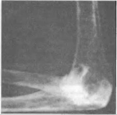
Fig. 309. Radiograph of the elbow joint in the ankylosis stage.
Conservative treatment includes comprehensive drug therapy and measures aimed at preventing muscle hypotrophy, deformities, and joint contractures. Pharmacological agents used in the management of rheumatoid arthritis are categorized as disease-modifying antirheumatic drugs (DMARDs), antirheumatic agents, and anti-inflammatory drugs.
Disease-modifying antirheumatic drugs (gold preparations, hydroxychloroquine, chloroquine, cytostatic and non-hormonal immunosuppressants, corticosteroids, etc.) are aimed at improving the body's immunological processes and halting the progression of rheumatoid arthritis, whereas anti-inflammatory drugs exert a local effect.
Since immunological reactions primarily take place in the synovial membrane, the intra-articular administration of corticosteroids (triamcinolone acetonide, hydrocortisone, etc.) and cytostatics (cyclophosphamide, thiophosphamide, etc.) is widely employed. Positive results are achieved by introducing into the joint cavity agents such as osmic acid, varizole, or varicocide, which are capable of inducing destruction and scarring of the synovial membrane (chemical synovectomy or synoviorthesis). Radionuclides such as gold-198 and yttrium-90 are administered for the same purpose.
Chemical and radionuclide synovectomy is most effective in the first stage of the rheumatoid process with high local activity, particularly in mono- and oligoarthritis.
Orthopedic treatment as part of conservative management is of great importance, as it aims to prevent joint stiffness and permanent contractures. During periods of acute pain, protective antalgic contractures develop, placing limb segments in functionally unfavorable positions. The application of splints and braces to the limbs, especially at night, reduces pain and prevents fixed contractures. During the day, the patient should engage in therapeutic exercise. For the same purpose, they must rest on a firm, flat bed. To eliminate established contractures, serial corrective casts, traction, etc., are utilized. In the presence of persistent contractures, after the inflammatory activity has subsided, the Volkov-Oganesyan apparatus is employed, yielding favorable outcomes.
To preserve joint mobility and prevent rapidly developing muscle hypotrophy, active Therapeutic Exercises and massage are prescribed from the onset of the disease. To consolidate the results of inpatient treatment, patients are recommended to undergo physical therapy and balneotherapy in a balneological clinic or health resort. Anti-relapse therapy is administered in an outpatient Setting.
Surgical treatment Methods. Despite comprehensive conservative treatment, in one-third of patients, the rheumatoid process in the joints progresses, leading to severe deformities and disability.
Historically, surgical interventions (corrective osteotomy, arthroplasty) were rarely used and aimed primarily at improving the static-dynamic function of the limbs in Stage III of the disease and facilitating the daily lives of disabled individuals. Today, theoretically grounded early surgical approaches for rheumatoid arthritis are widely applied before destructive changes occur in the articular cartilage.
In the first stage, when the rheumatoid process is limited to involvement of the synovial membrane alone and clinically manifests as acute, subacute, or chronic synovitis, a timely synovectomy (Fig. 310) can prevent subsequent joint destruction. Most specialists (Pavlov V.P., 1970; Sklyarenko E.T., 1971; Oleksa A.P., 1972, etc.) view these operations as prophylactic, since they can halt the destructive process and reduce inflammation not only in the operated joint but also in others. This is particularly evident following synovectomy of the knee joint, where the surface area of the synovial lining is largest.
Synovectomy is indicated for progressive rheumatoid arthritis that does not respond to conservative treatment—within three months according to Vainio (1967) or 18 months according to Euving (1971)—when intra-articular hydrocortisone injections or spa therapy yield no effect (Sklyarenko E.T., 1971).
Clinical experience demonstrates that synovectomy should be performed if the rheumatoid process is progressive and frequently recurrent, specifically after the third exacerbation. Some surgeons prefer operating during the inactive phase of the process, whereas others (Oleksa A.P., 1972) perform synovectomy regardless of disease activity, provided preoperative drug therapy and subsequent conservative management are administered. Given that synovectomy is a rather complex procedure, it is preferable to perform the simpler subtotal syncapsulectomy. It is essential to remove all pathologically altered areas of the synovial recesses.
To prevent intra-articular adhesions following synovectomy and syncapsulectomy, we employ joint distension with a novocaine solution every other day for 2–3 times, or interposition of a preserved membrane (Oleksa A.P., 1972) used to line the recesses of the knee joint. This technique leads to the best functional outcomes. The membranes were produced according to G.M. Dzyk’s method in tissue preservation laboratories.
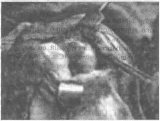
Fig. 310. Segmental arthroplasty in the area of femoral condyle erosions.
In the second stage of the disease, the scope of surgical intervention is significantly larger. In addition to synovectomy of the knee joint, surgeons have to perform debridement, which involves removing hypertrophied fat pads, clearing the cartilage of pannus and granulations, shaping bone erosions, and resecting marginal osteophytes.
When there is minor cartilage destruction on one of the femoral condyles following arthrolysis, it is advisable to reshape the surface and perform segmental arthroplasty—that is, cover this area with a biological membrane by suturing it under tension from the margins. To eliminate joint contractures, capsulotomy is performed.
If the destruction of the articular surfaces is extensive, Ye. T. Sklyarenko performed arthroplasty involving resection of the articular ends combined with covering them with a preserved biological membrane. In the 1970s, to maintain knee stability during surgery, we considered it appropriate to perform conservative resection of the articular ends while preserving the ligamentous apparatus. Hemiarthroplasty of the femoral condyles combined with shaping of the tibial articular surface was also considered justified at that time.
In the third stage of the disease, arthroplasty of various joints was used, but the functional outcomes failed to satisfy either patients or physicians due to pain, contractures, and joint instability. Today, joint replacement (arthroplasty) has taken its rightful place and is widely used—especially by foreign surgeons—even in the second stage of the disease. Numerous endoprosthesis designs have been proposed for the hip (Sivash, Virabov, Movshovych, Gerchev, Miller, Weller, McKee-Farrar), knee (Sivash, Swanson, "ESKA-medical", etc.), shoulder (Neer, "Howmedica"), elbow (Golyakhovsky, Mayo-Kondrat), as well as for the joints of the hand and fingers (I. H. Hryshyn, Swanson, etc.) (Figs. 311, 312).
The goal of surgical treatment in patients with rheumatoid polyarthritis is to restore joint function and limb congruity as much as possible, enabling patients to care for themselves and engage in manageable socially useful work.
To restore the gripping function of the fingers, particularly in cases of "boutonnière" deformity, Matev's operation or its modification is performed. The procedure involves a linear Skin and subcutaneous incision along the dorsal surface of the middle phalanx, exposing the split tendon of the superficial extensor and the insertion site of the deep extensor on the distal phalanx.
Next, the deep flexor is incised near the bone, along with both bands of the superficial extensor, so that the lateral band fills and replaces the defect of the deep extensor after flexion of the distal phalanx. Subsequently, the distal end of the medial band is sutured to the distal end of the lateral band (Fig. 313).
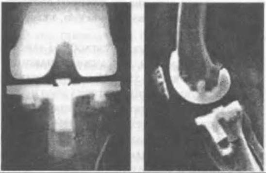
Fig. 311. Radiograph of the knee after joint replacement.
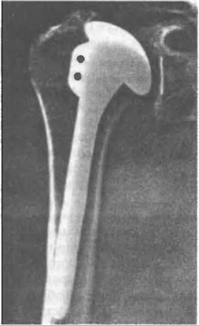
Fig. 312. Radiograph of the shoulder joint after joint replacement.
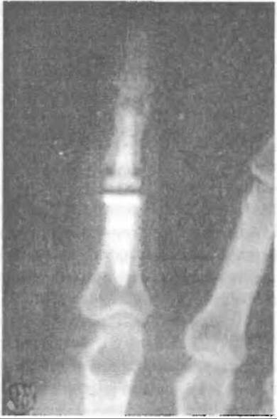
Fig. 313. Radiograph of an endoprosthetic finger.
The wound is closed, and the semi-flexed finger is immobilized with a plaster splint extending to the upper third of the forearm. Three weeks later, the plaster cast is removed, and a comprehensive rehabilitation program is initiated.
Last update: 10/08/2026
Editorial and Educational Adaptation: This material has been compiled based on the primary/original source text. The project team performed an editorial review, corrected technical inaccuracies, structured sections, and adapted the content for an educational format.
What was processed:
- elimination of formatting defects (OCR errors, structural breaks, corrupted characters);
- editorial organization of content;
- standardization of terminology in accordance with academic sources;
- verification of factual statements against the original source text.
All mentions of the author, publication year, and origin of the primary text have been preserved in accordance with the source.