Orthopedics - Oleksa A.P. 2006
Examination of the Orthopedic Patient
Palpation
Palpation is a widely used examination method in orthopedic trauma patients. It is performed using the pad of one or two to three fingers, with one or both hands (bimanually). Palpation is typically comparative, meaning that palpation findings from specific symmetrical areas of the body are compared.
Palpation helps identify various signs and symptoms characteristic of musculoskeletal disorders. In some cases, palpation is the only reliable diagnostic tool, such as for crepitant tenosynovitis, subcutaneous emphysema, or infiltrates. Palpation is used to determine the presence and character of the pulse, Skin moisture or dryness, Temperature, turgor and mobility, as well as soft tissue infiltration. This information is crucial for diagnosing inflammatory processes, tumors, etc. Palpation is also used to assess the condition of periarticular Tissues and the Joint Capsule, as well as the size, shape, and consistency of various swellings, exostoses, and tumors.
Palpation is used to determine the area or point of maximum superficial tenderness. This allows for the identification of a bone fracture line, as well as the degree and direction of fragment displacement. Deep tenderness is diffuse, making it difficult to localize precisely, such as in compression fractures of vertebral bodies.
Tapping the heel of an extended leg with the fingers can reveal referred pain in the affected limb segment (knee, hip joints, etc.). Rib fractures are diagnosed using both palpation and bimanual compression of the chest in the anteroposterior and lateral directions.
Bimanual digital examination is used to detect the fluctuation sign in soft tissue hematomas or abscesses, as well as to determine the presence of Blood in the joint cavity in hemarthrosis, effusion in synovitis (the patellar tap or ballottement sign) (Fig. 6, a, b), and pathological mobility in joints and bone fractures.
Class="center">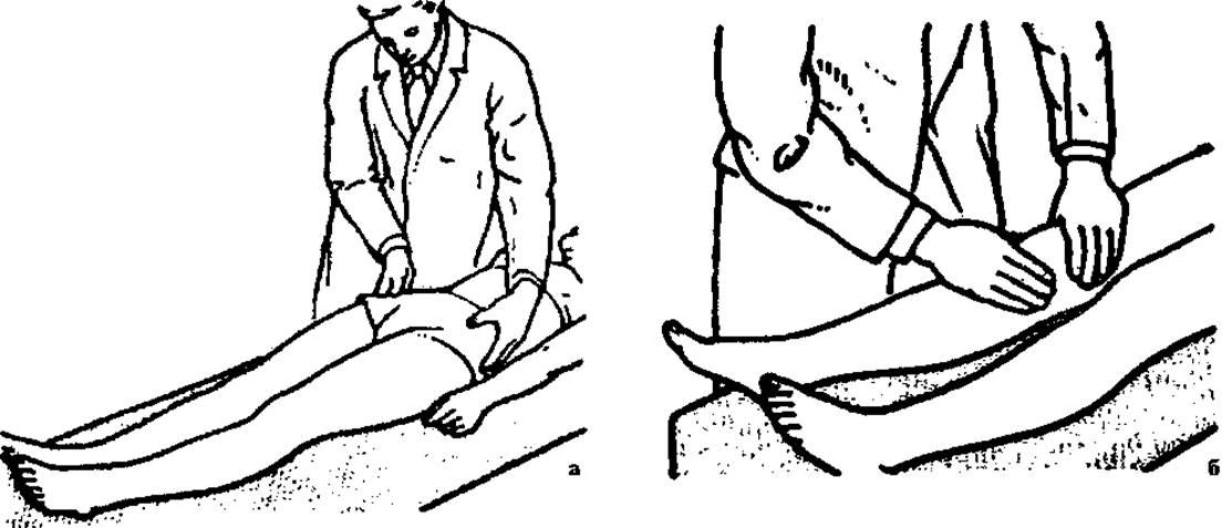
Fig. 6. Bimanual palpation of the greater trochanters relative to the pelvis (a) and the knee (b).
By using bony prominences in certain areas of the body as landmarks during palpation, the correct mutual positioning of these structures is determined using various auxiliary lines and geometric figures.
The Nélaton's line (Roser–Nélaton line) (Fig. 7, a) connects the ischial tuberosity with the anterior superior iliac spine and serves to detect pathology in the hip joint region. Normally, when the thigh is flexed at an angle of 130–140°, the tip of the greater trochanter is palpable along this line. In hip dislocations and femoral neck fractures, the tip of the greater trochanter is located above or below the Nélaton's line.
Schoemaker's line (Fig. 7, b) connects the tip of the greater trochanter with the anterior superior iliac spine and extends onto the anterior abdominal wall. When these lines are drawn on both sides, they intersect on the midline of the abdomen, typically above the umbilicus. If There is a pathology in the hip joint accompanied by upward Displacement of the greater trochanter, Schoemaker's line will intersect with the corresponding line from the opposite side of the abdomen below the umbilicus.
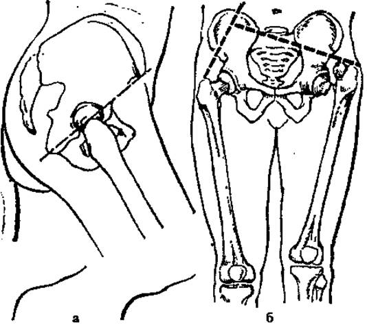
Fig. 7. Determination of lines: a — Nélaton's, b — Schoemaker's.
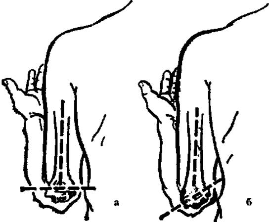
Fig. 8. Marx's line: a — normal, b — pathological.
Marx's line (Fig. 8) connects both epicondyles of the humerus and normally lies perpendicular to the longitudinal axis of the bone.
Hueter's triangle (Fig. 9) is formed when the elbow is flexed by three bony prominences: both epicondyles of the humerus and the most prominent part of the olecranon. Normally, this triangle is isosceles, with its apex being the olecranon (Fig. 9, a). When the elbow is fully extended, Hueter's line is formed, connecting the aforementioned bony structures.
Marx's line and Hueter's triangle are used for the differential clinical Diagnosis of forearm dislocation and supracondylar humeral fracture. In a fracture, this line deviates and may form various angles with the axis of the humerus (Fig. 9, b), while Hueter's triangle remains normal. Conversely, in a forearm bone dislocation, Marx's line remains normal, whereas Hueter's triangle is disrupted.
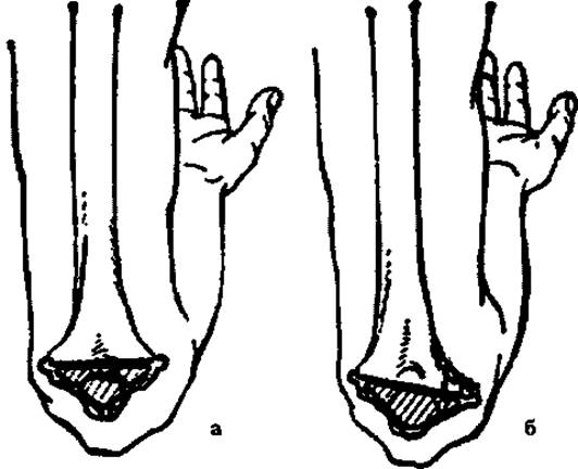
Fig. 9. Hueter's triangle: a — normal, b — pathological.
Bryant's triangle (Fig. 10, a) is normally isosceles and is formed by a line drawn along the axis of the thigh (extended to 180°) upward through the trochanteric region to its intersection with a perpendicular line starting from the anterior superior iliac spine and connecting it to the tip of the greater trochanter.
The isosceles nature of this triangle can be disrupted in hip dislocations (Fig. 10, b), femoral neck fractures due to displacement of the greater trochanter, fractures of the iliac wing, etc.
Last update: 10/08/2026
Editorial and Educational Adaptation: This material has been compiled based on the primary/original source text. The project team performed an editorial review, corrected technical inaccuracies, structured sections, and adapted the content for an educational format.
What was processed:
- elimination of formatting defects (OCR errors, structural breaks, corrupted characters);
- editorial organization of content;
- standardization of terminology in accordance with academic sources;
- verification of factual statements against the original source text.
All mentions of the author, publication year, and origin of the primary text have been preserved in accordance with the source.