Human Anatomy and Physiology (with Age-Related Features of the Child's Body) - Sapin M.R., Sivoglazov V.I. 2002
Musculoskeletal System
Muscular System
Muscles and Fasciae of Body Regions
Skeletal Muscles are subdivided into several major groups. These include the Muscles of the Trunk, HEAD and Neck, as well as the upper and lower extremities (Table 6).
Class="center">Table 6 Muscle groups of The Human Body
|
Muscles of the trunk |
Muscles of the extremities |
|
Muscles of the back |
Muscles of the upper extremity |
|
Superficial back muscles |
Muscles of the shoulder girdle |
|
Muscles of the free upper extremity |
|
|
Deep back muscles |
|
|
Suboccipital muscles |
Muscles of the arm |
|
Muscles of the chest |
Muscles of the forearm |
|
Superficial chest muscles |
|
|
Deep chest muscles |
Muscles of the lower extremity |
|
Muscles of the lateral abdominal walls |
Muscles of the free lower extremity |
|
Muscles of the anterior abdominal wall |
Muscles of the thigh |
|
Muscles of the Perineum |
Muscles of the FOOT |
|
Muscles of the pelvic Diaphragm |
|
|
Muscles of the urogenital diaphragm |
|
|
Muscles of the head |
|
|
Facial muscles (Muscles of facial expression) |
|
|
Masticatory muscles |
|
|
Muscles of the neck |
|
|
Infrahyoid muscles |
|
|
Deep neck muscles |
In different Regions of the body, muscles exhibit Structural and functional features tailored to their Location. The trunk walls are formed by ribbon-like and flat muscles featuring wide, thin tendons known as aponeuroses. In the head region, the masticatory muscles originate from the bones forming the cranial base and insert into the Mandible. The facial muscles blend into the Skin of the face. The extremities typically contain fusiform (spindle-shaped) muscles with narrow, long tendons.
Muscles of the trunk
Muscles and fascia of the back. The back muscles are paired and located on the posterior surface of the trunk, extending along the entire Vertebral Column. They are subdivided into superficial and deep groups (Fig. 29). The superficial muscles include the thin, broad trapezius and latissimus dorsi.
The trapezius muscle originates from the Occipital bone, nuchal ligament, and the spinous processes of all thoracic vertebrae. It inserts into the acromial end of the clavicle, the acromion, and the spine of the scapula. When the entire muscle contracts, the scapula is retracted toward THE VERTEBRAL COLUMN. Its upper fibers pull the scapula upward and medially, while the lower fibers pull it downward and medially.

Fig. 29. Muscles of the back:
1 — trapezius muscle, 2 — splenius capitis muscle, 3 — Major and minor rhomboid muscles, 4 — serratus posterior inferior muscle, 5 — thoracolumbar fascia, 6 — latissimus dorsi muscle
The latissimus dorsi originates via a broad tendon (aponeurosis) from the spinous processes of the lower six thoracic vertebrae, all lumbar vertebrae, and the iliac crest. Ascending laterally and superiorly, the narrow tendon of the muscle inserts into the crest of the lesser tubercle of the humerus.
Deep to the trapezius lie the major and minor rhomboid muscles and the levator scapulae muscle.
The rhomboid muscles originate from the spinous processes of the lower cervical vertebrae (minor rhomboid) and the upper four thoracic vertebrae (major rhomboid), and insert into the medial border of the scapula. Both muscles retract the scapula toward the spine and elevate it.
The levator scapulae muscle is located in the posterior region of the neck; it originates from the transverse processes of the upper four cervical vertebrae and inserts into the superior angle of the scapula, which it elevates upon contraction.
The next layer contains the serratus posterior superior and serratus posterior inferior muscles, which attach to the Ribs.
The serratus posterior superior muscle is situated beneath the rhomboid muscles. It originates from the spinous processes of the lower two cervical and upper two thoracic vertebrae, courses inferolaterally, and inserts into ribs II–V.
The serratus posterior inferior muscle lies beneath the latissimus dorsi; it originates from the spinous processes of the lower two thoracic and upper two lumbar vertebrae and inserts into the lower four ribs. The serratus posterior superior elevates the ribs, whereas the inferior depressor pulls them downward, thereby increasing the volume of the thoracic cavity.
The deep back muscles extend along the entire length of the spine from the sacrum to the occipital bone. In this group, the erector spinae muscle, along with the splenius capitis and splenius cervicis, lies more superficially. Deeper, lying directly against the spine, are the short transversospinales, interspinales, and intertransversarii muscles, and in the upper neck region, the suboccipital muscles.
The erector spinae is a thick, powerful muscle originating from the posterior surface of the sacrum, the spinous processes of the lumbar and lower thoracic vertebrae, the posterior part of the iliac crest, and the thoracolumbar fascia. The muscle courses upward and inserts via individual bundles into the spinous and transverse processes of the overlying lumbar, thoracic, and cervical vertebrae, the angles of the ribs, and the mastoid process of the Temporal bone.
The transversospinales muscle is located superficial to the erector spinae and consists of short muscular bundles originating from the transverse processes of the vertebrae and running obliquely upward in a medial direction. The more superficial bundles span 5–6 vertebrae, while the deepest ones span a single vertebra. Bilateral contraction of the transversospinales extends the vertebral column, whereas unilateral contraction flexes it laterally and rotates it.
The deepest muscles, which connect the corresponding processes of adjacent vertebrae, are the interspinales and intertransversarii muscles.
At the level of the atlanto-occipital joints lie the paired suboccipital muscles, which include the rectus capitis posterior major and minor, as well as the obliquus capitis superior and inferior. These muscles participate in the extension (tilting backward) and lateral Rotation of the head.
In the posterior neck region, lateral to the deep extensor muscles, lie the splenius cervicis and splenius capitis muscles, which act as neck and head extensors. These muscles originate from the spinous processes of the lower cervical and upper thoracic vertebrae and insert into the transverse processes of the cervical vertebrae and the occipital bone.
The superficial Fascia of the back covers the trapezius and latissimus dorsi muscles. Deeper lies the thoracolumbar fascia, which separates the deep muscles from the superficial ones.
Muscles and fascia of the chest. The chest muscles are categorized into superficial muscles attaching to the BONES OF THE shoulder girdle (pectoralis major, pectoralis minor, serratus anterior, and subclavius) and deep, or intrinsic, chest muscles (external and internal intercostal muscles). The diaphragm is also considered a chest muscle. The pectoral and subclavius muscles are located on the anterior surface of the chest inferior to the clavicle, while the serratus anterior is positioned on its lateral wall (Fig. 30).
The pectoralis major muscle is triangular in shape and originates from the outer surface of the clavicle, Sternum, and cartilages of ribs II–VII. It inserts via a narrow tendon into the crest of the greater tubercle of the humerus. The muscle adducts the arm and medially rotates it. When the arm is fixed, it elevates the ribs and expands the thoracic cavity.
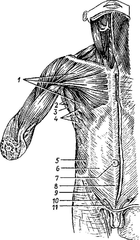
Fig. 30. SUPERFICIAL MUSCLES OF the chest and abdomen:
1 — pectoralis major muscle, 2 — axillary fossa, 3 — latissimus dorsi muscle, 4 — serratus anterior muscle, 5 — external oblique muscle of the abdomen, 6 — aponeurosis of the external oblique muscle of the abdomen, 7 — umbilical ring, 8 — linea alba (white line) of the abdomen, 9 — inguinal ligament, 10 — superficial inguinal ring, 11 — Spermatic Cord
The pectoralis minor muscle lies beneath the pectoralis major. It originates from ribs II–V, courses superiorly and laterally, and inserts into the coracoid process of the scapula. The muscle pulls the scapula anteriorly and inferiorly; when the scapula is fixed, it elevates the ribs, participating in the act of inspiration.
The subclavius muscle is located between the clavicle and the first rib, drawing the clavicle inferiorly and medially.
The serratus anterior muscle originates by fleshy slips from the upper nine ribs, passes posteriorly and medially, and inserts into the medial border of the scapula, extending down to its inferior angle. The muscle pulls the scapula anteriorly, rotating its inferior angle laterally. With a fixed scapula, the muscle elevates the ribs, participating in the act of inspiration.
The external and internal intercostal muscles are arranged in two layers within the intercostal spaces. The external muscles elevate the ribs (inspiration), while the internal muscles depress the ribs (expiration).
The diaphragm, or thoracoabdominal partition, separates the thoracic and abdominal cavities and is dome-shaped, with its convexity facing upward (into the thoracic cavity). Its muscular bundles originate along the lower boundary of The thoracic cage: on the sternum (sternal part), ribs (costal part), and lumbar vertebrae (lumbar part). These muscular bundles then transition into a central flat tendinous aponeurosis occupying the middle part of the diaphragm, known as its central tendon (Fig. 31). The lumbar part of the diaphragm forms two crura: right and left. The medial PARTS OF THE crura bound two openings: the posterior for the aorta and the anterior for the Esophagus. The central tendon contains an opening for the INFERIOR VENA CAVA. The diaphragm is a respiratory muscle; upon contraction, it flattens and descends, increasing the volume of the thoracic cavity (inspiration). Upon relaxation, the diaphragm ascends, thereby decreasing the volume of the thoracic cavity (expiration).
Fasciae cover the muscles of the chest. The superficial pectoral fascia overlies the pectoralis major and serratus anterior muscles. The deep fascia forms a sheath for the pectoralis minor and subclavius muscles and is also adherent to the external intercostal muscles. The inner surface of the thoracic walls is lined by the endothoracic fascia, which continues inferiorly onto the diaphragm.
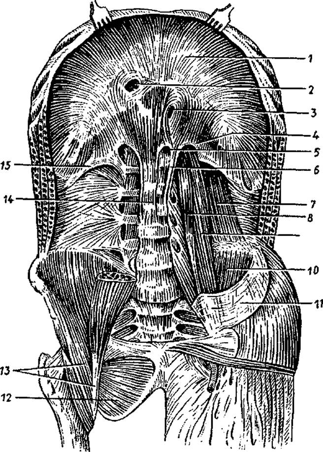
Fig. 31. Diaphragm and muscles of the posterior abdominal wall:
1 — central tendon, 2 — opening for the inferior vena cava, 3 — esophageal hiatus, 4 — medial lumbocostal arch (arcuate ligament), 5 — aortic hiatus, 6 — left crus of the diaphragm, 7 — quadratus lumborum muscle, 8 — psoas minor muscle, 9 — psoas major muscle, 10 — iliacus muscle, 11 — iliac fascia, 12 — obturator externus muscle, 13 — iliopsoas muscle, 14 — right crus of the diaphragm, 15 — lateral lumbocostal arch (arcuate ligament)
The diaphragm of an infant differs significantly from that of an adult. In a newborn that has not yet breathed, the diaphragm lies at the level of the 7th thoracic vertebra. In an infant that has begun to breathe, the diaphragm descends to the VIII thoracic vertebra, in a one-year-old child to the X, in a five-year-old to the IX, and by age 13 it settles at the level of the XII thoracic vertebra.
The shape of the diaphragm begins to take form in the third year of life. During respiratory movements, the diaphragm of a newborn infant moves up and down between the upper border of the IV rib and the lower border of the VI rib; in an adult, between the V and VI ribs; and in elderly individuals, up to the VII–IX ribs. The range of diaphragmatic excursion is greater in men than in women, and greater in the young than in the elderly.
MUSCLES AND FASCIAE of the abdomen. The abdomen is the part of the trunk located between the Thorax and the pelvis. The Abdominal cavity is the space whose walls are formed superiorly by the diaphragm, and inferiorly by the pelvic bones, muscles, and the pelvic floor (perineum). The posterior wall is formed by the Vertebral Column and the paired quadratus lumborum muscle. The anterior and lateral walls are likewise formed by paired Introduction/39.html">Muscles and their fasciae—specifically, the paired external and internal oblique, transverse, and rectus abdominis muscles.
The external oblique muscle of the abdomen is broad and thin, originating by slips from the lower eight ribs, from where it courses anteriorly and inferiorly. The muscle continues into a broad tendon (aponeurosis) that inserts into the iliac crest and the Pubic Symphysis. In the region between the anterior superior iliac spine and the pubic tubercle, the aponeurosis of the external oblique folds inward to form the inguinal (Poupart's) ligament. Along the midline of the anterior abdominal wall, the aponeurosis of the external oblique joins the corresponding tendon of the opposite external oblique to form the linea alba. This line extends from the xiphoid process to the pubic symphysis.
In some cases (such as increased intra-abdominal pressure caused by constipation), the Connective Tissue fibers forming the linea alba may separate, creating narrow slits. Consequently, these areas become weak points where hernias of the linea alba can develop.
Approximately at the middle of the linea alba lies the umbilical ring (navel), which is closed by connective tissue. In embryos and fetuses, Blood Vessels (umbilical vessels) pass through the umbilical ring. The umbilical ring may also serve as a site for The formation of umbilical hernias.
The internal oblique muscle of the abdomen lies beneath the external oblique. It originates from the thoracolumbar fascia, the iliac crest, and the inguinal ligament, and courses anteriorly and superiorly. The posterior fibers of the internal oblique insert into the cartilages of the lower ribs, while its broad aponeurosis contributes to the Formation of the linea alba.
The transverse muscle of the abdomen lies in the third layer, beneath the two preceding oblique muscles. It originates from the inner surface of the lower six ribs, the thoracolumbar fascia, the iliac crest, and the inguinal ligament. The muscle courses anteriorly, continuing into a broad aponeurosis that intertwines with the linea alba.
The rectus abdominis muscle is located lateral to the anterior midline (linea alba), and its fibers run vertically. The muscle originates from the xiphoid process of the sternum and the cartilages of ribs V–VII, and inserts into the pubic crest and pubic symphysis. Its muscular bundles are interrupted by three to four tendinous intersections. Both the right and left rectus abdominis muscles are each enclosed within their own sturdy tendinous sheath (rectus sheath), formed by the aponeuroses of the external and internal oblique and transverse abdominal muscles.
The rectus abdominis muscles pull the thoracic cage inferiorly and flex the trunk. The abdominal oblique muscles also flex the trunk forward, participate in its rotation to the right and left, and aid in Respiration due to their attachments to the ribs.
The quadratus lumborum muscle is located lateral to the lumbar spine (see Fig. 31). It participates in forming the posterior abdominal wall. This muscle originates from the XII rib and the transverse processes of lumbar vertebrae I–IV, and inserts into the iliac crest and the transverse processes of the lumbar vertebrae. Upon contraction, this muscle bends the spine laterally toward its own side.
By forming the walls of the abdomen, the abdominal muscles contract to increase intra-abdominal pressure, which is essential for maintaining Internal Organs in their natural positions. Intra-abdominal pressure facilitates bowel evacuation (defecation), urination, and, in women, the expulsion of the fetus from the Uterus during childbirth. Due to these Functions, the abdominal muscles constitute what is known as the abdominal press.
The abdominal fasciae cover not only individual muscles of the abdominal wall. On the exterior, beneath the subcutaneous fat, lies the superficial fascia, which covers the Superficial layer of the abdominal muscles and serves as a continuation of the superficial pectoral fascia. From the internal abdominal cavity, the abdominal walls are lined by the endoabdominal (intraperitoneal) fascia.
Due to their complex Structure, the abdominal walls feature vulnerable areas prone to hernia formation, namely the linea alba, the umbilical ring, and the Inguinal Canal.
The slit-like inguinal canal is situated just above the inguinal ligament, which forms its lower wall. The anterior wall of the inguinal canal is formed by the lower part of the external oblique aponeurosis. The upper wall is created by the lower muscle bundles of the internal oblique and transverse abdominal muscles, while the posterior wall consists of the transversalis fascia, a component of the endoabdominal fascia. The inguinal canal is approximately 5 cm long. On the abdominal cavity side, there is the deep (internal) inguinal ring, located about 2 cm above the inguinal ligament, roughly at its midpoint. The superficial inguinal ring lies above the medial part of the inguinal ligament within the cleft between the diverging fibers of the external oblique muscle. Passing through the inguinal canal is the spermatic cord in men and the round ligament of the uterus in women.
Pelvic floor muscles (perineum). The perineum, or pelvic floor, refers to the musculofascial plate that closes the outlet of the lesser pelvis, namely its inferior aperture.
The diamond-shaped perineum is bounded anteriorly by the pubic symphysis, laterally by the ischial tuberosities, and posteriorly by the coccyx. A line connecting the ischial tuberosities divides the perineum into two triangular regions.
The anterior region is termed the urogenital diaphragm, and the posterior one is the pelvic diaphragm. Through the urogenital diaphragm passes the Urethra in men, and the urethra and Vagina in women. The terminal part of the rectum passes through the pelvic diaphragm.
Both the urogenital diaphragm and the pelvic diaphragm are formed by two muscular layers and their covering fasciae.
In the superficial layer of the pelvic diaphragm, beneath the skin and superficial fascia, lies an unpaired annular muscle—the external anal sphincter, a voluntary muscle that closes the anal opening. In the second, deep layer lies the paired, triangular levator ani muscle. This muscle originates on the internal surface of the pelvic walls, descends, and blends into the terminal section of the rectum. Both levator ani muscles surround the lower part of the rectum in a funnel-like fashion. The posterior section of the pelvic diaphragm is completed by the coccygeus muscle, which originates from the ischial spine and attaches to the margin of the coccyx and the apex of the sacrum.
Flanking the anus is a fat-filled, paired depression known as the ischioanal fossa, the apex of which points toward the lesser pelvis. The lateral wall of the fossa is formed by the fascia-covered obturator internus muscle and the ischial tuberosity. The medial wall is formed by the external surface of the levator ani muscle, covered by the inferior fascia of the pelvic diaphragm.
The urogenital diaphragm likewise comprises superficial and deep muscular and fascial layers. The superficial layer contains the paired superficial transverse perineal muscles, as well as the bulbospongiosus and ischiocavernosus muscles, which facilitate the erection of the Penis or Clitoris. In men, the bulbospongiosus muscle surrounds the bulb and corpus spongiosum of the penis, while in women it surrounds the vaginal opening. The ischiocavernosus muscle originates at the ischial tuberosity and inserts into the corpus cavernosum of the penis in men or the clitoris in women. The Deep Layer of the urogenital diaphragm contains the unpaired external urethral sphincter and the paired deep transverse perineal muscle, which originates, like its superficial counterpart, on the internal surface of the ischium. The thin, flat tendons of the right and left transverse muscles blend into the perineal body (central tendon of the perineum), located between the external genitalia anteriorly and the anus posteriorly.
The muscles of the perineum are externally covered by the superficial perineal fascia, which is a continuation of the general subcutaneous fascia. Additionally, the levator ani and its sphincter are enclosed between the inferior and superior Fasciae of the pelvic diaphragm. The deep transverse perineal muscles and the urethral sphincter are covered by the inferior and superior fasciae of the urogenital diaphragm.
Between the anterior margin of the urogenital diaphragm and the pubic symphysis lies a narrow cleft through which blood Vessels and nerves pass from the pelvic cavity to the external genitalia.
Muscles and Fasciae of the Head
Based on their location and function, the muscles of the head are divided into two main groups: facial muscles (muscles of facial expression) and masticatory muscles (Fig. 32).
Facial muscles (muscles of facial expression) lie beneath the skin and are arranged as sphincters and dilators around the oral and nasal orifices, the orbits, and the external acoustic meatus. These muscles lack a superficial fascia. They originate on the bones of The Skull and insert into the connective tissue framework of the skin. Upon contraction, facial muscles shift the skin, alter its relief, and form dimples, grooves, and folds. In this manner, the muscles produce complex expressive facial movements—mimicry. Facial muscles close and open the orifices they surround, contracting in response to impulses transmitted via the Facial Nerve from the Brain. The resulting facial expressions reflect various emotional states and experiences—joy and sorrow, pain and shame, attention and hope. According to their anatomical location, the paired facial muscles are subdivided into groups: muscles of the cranial vault; muscles surrounding the palpebral fissure; muscles surrounding the nostrils (nasal apertures); muscles surrounding the oral aperture; and muscles of the auricle.
The epicranius Muscle consists of two bellies (frontal and occipital) connected by a broad epicranial aponeurosis (galea aponeurotica), which is firmly fused with the scalp. The muscle connects loosely to the periosteum of the cranial roof. The occipital belly originates from the highest nuchal line of the occipital bone, while the frontal belly blends into the skin of the forehead. Contraction of the frontal belly elevates the eyebrows and creates transverse wrinkles on the forehead (the muscle of surprise and inquisitive attention). The occipital belly pulls the entire muscle posteriorly and smooths out the forehead wrinkles.
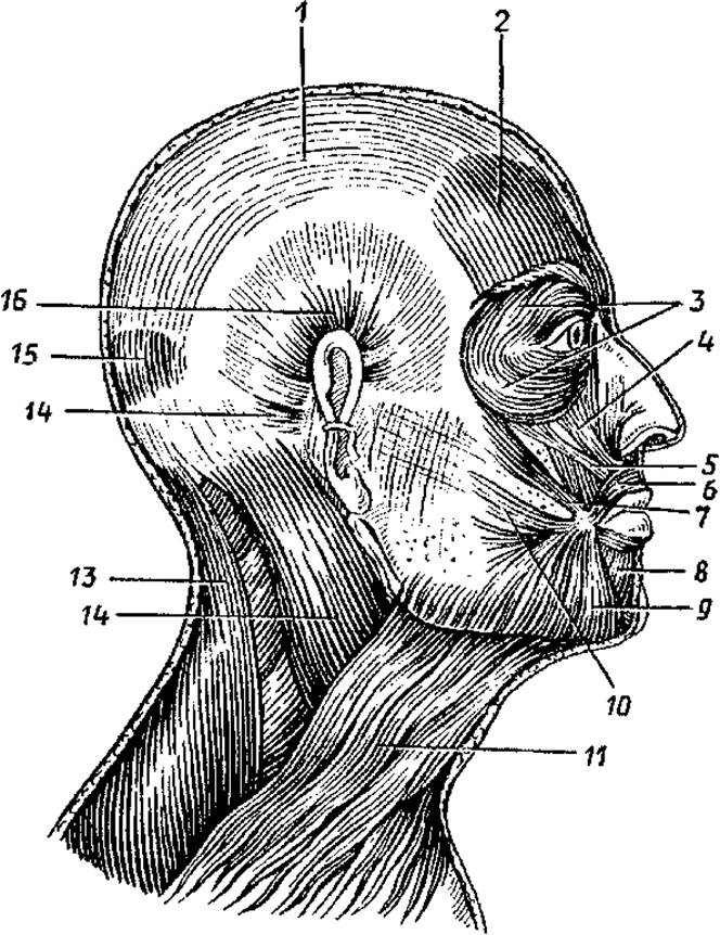
Fig. 32. Muscles of the head and neck. Right lateral view:
1 — tendon helmet (epicranial aponeurosis), 2 — frontal belly of the occipitofrontalis muscle, 3 — orbicularis oculi muscle, 4 — levator labii superioris muscle, 5 — levator anguli oris muscle, 6 — orbicularis oris muscle, 7 — zygomaticus major muscle, 8 — depressor labii inferioris muscle, 9 — depressor anguli oris muscle, 10 — risorius muscle, 11 — platysma, 12 — sternocleidomastoid muscle, 13 — trapezius muscle, 14 — auricularis posterior muscle, 15 — occipital belly of the occipitofrontalis muscle, 16 — auricularis superior muscle
The corrugator supercilii muscle is located within the thickness of the eyebrow, originating from the nasal part of the Frontal bone and inserting into the skin of the eyebrow. When contracted bilaterally, these muscles draw the eyebrows together, creating the facial expression of pain, suffering, or gloom.
The orbicularis oculi muscle (surrounding the palpebral fissure) is formed by circular bundles encircling the Orbit (orbital part), inserting into the skin of the eyelids (palpebral part), and attaching to the lacrimal sac (lacrimal part). Upon contraction, this muscle closes the eyelids, tightly squints the eyes, and facilitates tear drainage into the nasolacrimal duct.
The muscles surrounding the nasal openings originate on the anterior surface of the Maxilla and insert into the ala and the cartilaginous part of the dorsum of the Nose. These muscles narrow the nostrils.
The muscles surrounding the oral aperture are formed by bundles oriented radially relative to the oral fissure (dilators) and bundles located within the thickness of the Lips (sphincters). Certain dilators lie superior to the oral fissure (levator anguli oris, levator labii superioris, and zygomatic muscles), which produce the expression of a smile or laughter. Other muscles lie inferior to the oral fissure (depressor anguli oris, depressor labii inferioris). Located here is also the mentalis muscle, which inserts into the skin of the chin and forms a dimple.
The buccinator muscle, which forms the muscular foundation of the cheek, originates from the posterior parts of the upper and lower jaws and likewise blends into the orbicularis oris muscle. This muscle participates in suckling and in moving the food bolus toward the Pharynx; it tenses the cheek, earning it the traditional name of the "trumpeter's muscle." Posteriorly, the fascia of the buccinator muscle fuses with the adventitia of the pharynx.
Facial muscles also include rudimentary auricular muscles of the auricle (anterior, superior, and posterior auricular muscles), which are well-developed in animals, as well as the broad, flat platysma of the neck, among others.
The masticatory muscles are formed by four pairs of short, thick, powerful muscles that originate on the bones of the skull base and attach to its only movable bone, the mandible. Two of these muscles are superficial (the masseter proper and the temporalis) and two are deep, located medially to the mandibular ramus within the infratemporal fossa.
The masseter muscle originates on the zygomatic arch, runs inferiorly and posteriorly, and attaches to the outer surface of the mandibular angle. This muscle elevates the mandibular angle, participating in the acts of mastication and food grinding.
The temporalis muscle, broad superiorly and narrow inferiorly, originates on the lateral surface of the neurocranium, filling the entire temporal fossa, and attaches to the coronoid process of the mandible. This muscle acts primarily on the anterior Teeth (incisors and canines), which is why it is often referred to as the biting muscle. The posterior fibers of this muscle pull the mandible backward.
The medial pterygoid muscle originates in the fossa of the pterygoid process of the Sphenoid bone, runs inferiorly and posteriorly, and attaches to the corresponding tuberosity on the Medial surface of the mandibular angle. This muscle presses the lower molars against the upper ones and participates in mastication, much like the masseter proper.
The lateral pterygoid muscle is located in the infratemporal fossa. It originates on the posterior surface of the maxillary tuberosity and the pterygoid process of the sphenoid bone. The muscle fibers run posteriorly and attach to the condylar (articular) process of the mandible. Bilateral contraction of these muscles protrudes the mandible forward, whereas unilateral contraction pivots it to the opposite side.
The arrangement of the masticatory muscles relative to the mandible and the complex temporomandibular joints ensures efficient biting, chewing, and grinding of food. The wide variety of mandibular movements allowed by the temporomandibular joints has led to the human masticatory mechanism being described as universal.
All masticatory muscles possess their own fasciae. However, the superficial fascia of the masseter is thick and fused with the capsule of the Cytology/practical/97.html">Parotid salivary gland; consequently, it is termed the parotideomasseteric fascia in scientific literature. The outer surface of the temporalis is also covered by a dense temporal fascia, which originates on the lateral surface of the neurocranium just above the muscle origin and attaches to the zygomatic arch.
Muscles and fasciae of the neck
The posterior neck muscles are generally considered part of the back muscle group. The neck muscles proper include those situated in its anterior and lateral regions, which are subdivided into superficial and deep groups.
The superficial neck muscles comprise the platysma (which developmentally, anatomically, and functionally belongs to the facial expression muscles), the sternocleidomastoid, as well as the suprahyoid and infrahyoid muscles. The deep neck muscle group includes the scalene and prevertebral muscles.
The platysma is a thin, flat muscle located directly beneath the skin. It originates within the pectoral fascia and the subcutaneous tissue below the clavicle, ascends to blend with the masseteric fascia and the Tissues of the oral angle. Upon contraction, the muscle depresses the angle of the Mouth and pulls the skin of the neck taut, thereby protecting the superficial Veins from compression.
The sternocleidomastoid muscle originates via two heads from the sternum and clavicle and inserts into the mastoid process of the temporal bone. Bilateral contraction of these muscles tilts the head backward. Unilateral contraction tilts the head toward the contracting side while simultaneously rotating it in the opposite direction.
The suprahyoid group unites four paired muscles: the digastric, stylohyoid, geniohyoid, and mylohyoid. The digastric muscle features two bellies (anterior and posterior) connected by an intermediate tendon. The anterior belly originates in the digastric fossa of the mandible, while the posterior belly attaches to the mastoid notch of the temporal bone. The intermediate tendon, linking both bellies, is anchored to the Hyoid bone by a tendinous sling. This muscle elevates the hyoid bone, or depresses the mandible when the hyoid bone is fixed. The stylohyoid muscle originates on the styloid process of the temporal bone. The geniohyoid muscle arises from the mental spine (lingual tubercle) of the mandible. The mylohyoid muscle originates on the internal surface of the mandible. By uniting with its counterpart from the opposite side, the mylohyoid forms the floor of the Oral Cavity (the oral diaphragm), upon which the Tongue rests. All these muscles attach to the hyoid bone to fixate it; when the bone is stabilized, the geniohyoid and mylohyoid muscles act to depress the mandible.
There are also four infrahyoid muscles (the sternohyoid, omohyoid, sternothyroid, and thyrohyoid). The sternohyoid muscle originates on the sternum, attaches to the hyoid bone, and pulls it downward. The omohyoid muscle, which connects these two bones, consists of two bellies (superior and inferior) linked by an intermediate tendon. The sternothyroid muscle runs from the sternum to the outer surface of the thyroid Cartilage, depressing both the Larynx and the hyoid bone. The thyrohyoid muscle essentially acts as a continuation of the previous one; by attaching to the hyoid bone, it elevates the larynx. Together with their action on the mandible, all suprahyoid and infrahyoid muscles play a major role in stabilizing the hyoid bone and, consequently, the larynx.
The deep neck muscles comprise the anterior, middle, and posterior scalene muscles, as well as the prevertebral muscles — the longus capitis and longus colli, alongside the anterior and lateral rectus capitis muscles.
The scalene muscles originate on the transverse processes of the cervical vertebrae and attach to the ribs: the anterior and middle scalenes attach to the first rib, while the posterior attaches to the second.
A wide interscalene space exists between the anterior and middle scalene muscles, through which the Subclavian Artery and major NERVES OF THE upper limb pass. Upon contraction, the scalene muscles elevate the ribs, thus acting as accessory Muscles of inspiration.
The longus capitis, longus colli, and rectus capitis anterior muscles flex the head and the cervical spine forward. The rectus capitis lateralis tilts the head to its own side.
Fasciae of the Neck. The Cervical fascia, situated anterior to the vertebral column, is divided into three distinct layers or plates: the superficial lamina (superficial cervical fascia), the pretracheal lamina (middle cervical fascia), and the prevertebral lamina (deep prevertebral cervical fascia). The superficial lamina lies beneath the platysma, encircles the neck, and forms fascial sheaths for the sternocleidomastoid and trapezius muscles. The pretracheal lamina forms fascial compartments for all the infrahyoid muscles. This fascial plate resembles a sail stretched between the omohyoid muscles of the right and left sides, leading to its designation in medical literature as the "Richet's sail". The prevertebral lamina covers the anterior aspect of the muscles overlying the cervical vertebrae.
Review and Self-Assessment Questions:
1. What groups are the trunk muscles divided into? Where are their origins and insertions located? What functions do these muscles perform?
2. Name the back muscles that elevate and depress the ribs. Where do these muscles originate and insert?
3. Name the muscles involved in forming the anterior and posterior walls of the abdomen. Where do these muscles originate and insert?
4. List the "weak spots" in The structure of the abdominal wall. Where are these locations found? What do you know about their anatomy, and why are they referred to as "weak spots"?
5. Describe the Anatomy of the perineum, including the arrangement of its muscular layers and fasciae.
6. Describe the Functions of the facial muscles in relation to their arrangement.
7. What functions does each masticatory muscle perform by acting on the mandible?
8. What muscle groups are distinguished in the neck, and which muscles make up each group? Name the fascias of the neck associated with each of these muscle groups.
Muscles and Fascias of the Limbs
Differences in the Skeletal structure of the upper and lower limbs, along with their distinct functions, have led to The Development of specialized homologous muscles and even The Emergence of new ones.
Muscles and Fascias of the Upper Limb
The Muscles of the Upper Limb are divided into those of its girdle (Pectoral Girdle) and those of the free upper limb, which are grouped around the joints (Figs. 33 and 34).
The muscles of the upper limb girdle surround the shoulder joint. Originating on the bones of the pectoral girdle and inserting into the humerus, they ensure a wide range of Movements of the free upper limb. There are six such muscles: the deltoid, supraspinatus, infraspinatus, teres major, teres minor, and subscapularis muscles.
The deltoid muscle is massive and powerful, originating on the spine of the scapula, the acromion, and the clavicle, and inserting into the deltoid tuberosity of the humerus. The muscle covers the shoulder joint anteriorly, superiorly, and posteriorly. Its anterior part flexes the arm, the middle part abducts it from the trunk, and the posterior part extends the arm. The supraspinatus muscle originates in the supraspinous fossa of the scapula. Passing beneath the coracoacromial ligament, the tendon of the muscle inserts into the greater tubercle of the humerus, which it abducts from the trunk. The infraspinatus muscle originates in the infraspinous fossa and inserts into the greater tubercle of the humerus. Upon contraction, it laterally rotates the arm. The teres major muscle originates on the lateral border of the scapula and inserts into the crest of the lesser tubercle of the humerus. It extends the arm and medially rotates it. The teres minor muscle originates on the lateral border of the scapula and inserts into the greater tubercle of the humerus. It laterally rotates the arm. The subscapularis muscle has a broad origin on the costal surface of the scapula and inserts into the lesser tubercle of the humerus. The muscle medially rotates the arm and adducts it to the trunk, as well as pulls the capsule of the shoulder joint to protect it from impingement.
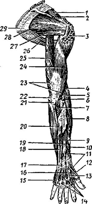
Fig. 33. Muscles of the upper limb. Posterior view:
1 — supraspinatus muscle, 2 — spine of scapula (partially removed), 3 — deltoid muscle (partially removed), 4 — brachioradialis muscle, 5 — extensor carpi radialis longus, 6 — lateral epicondyle, 7 — anconeus muscle, 8 — extensor carpi radialis brevis, 9 — extensor digitorum, 10 — abductor pollicis longus, 11 — extensor pollicis brevis, 12 — tendon of extensor pollicis longus, 13 — first dorsal interosseous muscle, 14 — tendon of extensor digitorum, 15 — tendon of extensor digiti minimi, 16 — tendon of extensor indicis, 17 — extensor retinaculum, 18 — extensor carpi ulnaris, 19 — extensor digiti minimi, 20 — flexor carpi ulnaris, 21 — olecranon, 22 — medial epicondyle, 23 — triceps brachii, 24 — lateral head of triceps brachii, 25 — long head of triceps brachii, 26 — teres major muscle, 27 — teres minor muscle, 28 — infraspinatus muscle, 29 — inferior angle of scapula
The Muscles of the Free Upper Limb include the muscles of the arm, forearm, and hand. The arm features an anterior muscle group (flexors) and a Posterior muscle group (extensors).
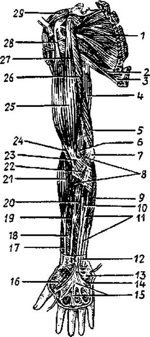
Fig. 34. Muscles of the upper limb. Anterior view:
1 — subscapularis muscle, 2 — teres major muscle, 3 — latissimus dorsi, 4 — long head of triceps brachii, 5 — medial head of triceps brachii, 6 — cubital fossa, 7 — medial epicondyle of humerus, 8 — pronator teres, 9 — flexor carpi ulnaris, 10 — palmaris longus, 11 — flexor digitorum superficialis, 12 — part of forearm fascia, 13 — palmaris brevis, 14 — hypothenar eminence, 15 — palmar aponeurosis, 16 — thenar eminence, 17 — tendon of abductor pollicis longus, 18 — flexor pollicis longus, 19 — flexor digitorum superficialis, 20 — flexor carpi radialis, 21 — brachioradialis, 22 — bicipital aponeurosis, 23 — tendon of biceps brachii, 24 — brachialis, 25 — biceps brachii, 26 — coracobrachialis, 27 — short head of biceps brachii, 28 — long head of biceps brachii, 29 — deltoid muscle
The anterior muscle group of the arm comprises the biceps brachii, coracobrachialis, and brachialis muscles.
The biceps brachii has two heads: long and short. The long head originates on the supraglenoid tubercle of the scapula, while the short head originates on its coracoid process. The common muscle belly transitions into a tendon that inserts into the radial tuberosity. The muscle flexes the arm at the shoulder joint and the forearm at the elbow joint, and also laterally rotates the forearm (supinates it). The coracobrachialis muscle originates on the coracoid process of the scapula and inserts into the humerus distal to its lesser tubercle. The muscle flexes and adducts the arm. The brachialis muscle originates on the humerus (beneath the biceps). Its tendon inserts into the ulnar tuberosity. The muscle flexes the forearm at the elbow joint.
The posterior muscle group of the arm includes the triceps brachii and anconeus muscles, which extend the forearm at the elbow joint.
The triceps brachii, possessing three heads, originates on the infraglenoid tubercle of the scapula (long head) and on the posterior surface of the humerus (medial and lateral heads). The tendon of the muscle inserts into the olecranon of the ulna. The anconeus muscle originates on the lateral epicondyle of the humerus and inserts into the olecranon of the ulna.
The muscles of the forearm are likewise subdivided into anterior and posterior groups. Within each group, the muscles are not located at a single level but form multiple layers. The muscles of the anterior group function as flexors of the forearm, wrist, and digits, as well as pronators. The majority of the muscles in this group originate on the medial epicondyle of the humerus.
The anterior forearm group includes 9 muscles: the pronator teres, brachioradialis (which insert into the radius at different levels), flexor carpi ulnaris (inserting into the Carpal Bones), flexor carpi radialis (inserting into the Metacarpal bones), palmaris longus (blending into the palmar aponeurosis), flexor digitorum superficialis and profundus, flexor pollicis longus (inserting into the Phalanges of the digits), and the deeply situated quadratus pronator (inserting into the radius).
The posterior forearm group comprises 10 muscles, which act as extensors of the wrist and digits, supinators, and extensors of the forearm. These muscles originate on the lateral epicondyle of the humerus, the posterior surfaces of the ulna and radius, and the interosseous membrane. The supinator inserts into the radius; the extensor carpi radialis longus and brevis, extensor carpi ulnaris, and abductor pollicis longus insert into the metacarpal bones; and the extensor digitorum, extensor digiti minimi, extensor pollicis longus and brevis, and extensor indicis insert into the phalanges of the digits.
The intrinsic muscles of the hand are short and located exclusively on its palmar surface, where they form three muscle groups. Laterally lie the muscles of the thenar eminence (ball of the thumb), medially lie the muscles of the hypothenar eminence (ball of the little finger), and between them is the intermediate muscle group of the hand. There are no muscles on the DORSAL SIDE OF the hand; only the long tendons of the digital extensors pass through this area, which belong to the posterior muscle group of the forearm.
The thenar eminence includes four muscles that originate on the carpal bones and the flexor retinaculum, and insert into the phalanges of the thumb. These are the abductor pollicis brevis, flexor pollicis brevis, opponens pollicis, and adductor pollicis muscles.
The hypothenar eminence is likewise formed by four muscles acting on the bones of the little finger. These are the abductor digiti minimi and the flexor digiti minimi brevis (which attach to the phalanges of the little finger), the opponens digiti minimi (attaching to the fifth metacarpal bone), and the palmaris brevis (which blends into the skin of the palm). These muscles originate from the carpal bones and the flexor retinaculum.
The middle group of hand muscles consists of four lumbricals, alongside seven interosseous muscles (three palmar and four dorsal) located in the intervals between the metacarpal bones. The lumbricals originate from the tendons of the flexor digitorum profundus and insert into the dorsal surface of the proximal phalanges of digits 2–5. The interosseous muscles originate from the metacarpal bones and insert into the proximal phalanges in such a way that the palmar interossei adduct digits 2, 4, and 5 toward the middle digit, whereas the dorsal interossei abduct digits 2, 4, and 5 away from the middle (third) digit. Although not very large in size, the hand muscles are quite numerous (19 muscles) and ensure A wide variety of delicate finger movements. Particularly important are the movements of opposition of the thumb to the little finger and the other digits, which enable the hand and its fingers to grasp tools.
Fasciae of the upper limb. The superficial fascia lies beneath the skin, covers all muscle groups, and forms part of the body's general subcutaneous fascia. Intermuscular septa extend inward from this fascia, separating adjacent muscle groups from one another, including the anterior muscle group from the posterior. In certain areas, the fascia is thickened by tendon fibers weaving into it. On the palm, There is a broad and dense palmar aponeurosis formed by the tendon of the palmaris longus and reinforced by thick bundles of fibrous fibers. Thus, at the border between the forearm and the hand, thickenings are formed: the flexor retinaculum and the extensor retinaculum.
On the palmar surface, a thickening forms the transverse carpal ligament, which completes the carpal tunnel. Through this tunnel, the tendons of the digital flexors pass into the hand, enclosed within two synovial sheaths. One synovial sheath belongs to the tendon of the flexor pollicis longus, while the other belongs to the tendons of the flexor digitorum superficialis and profundus of digits 2–5. The flexor tendons of digits 2–5 have isolated synovial sheaths at the level of their phalanges that do not communicate with the others. At the same time, the synovial Sheath of the fifth digit communicates with the common synovial sheath of the digital flexor tendons, which extends into the distal forearm. The synovial sheath of the thumb likewise extends proximally from its distal phalanx, superior to the wrist joint.
The extensive distribution of the synovial sheaths on the palmar side of the hand and fingers can serve as pathways for the rapid spread of inflammation from the fingers to the forearm. This can occur in the event of a deep splinter, injury to the finger pads, or even careless finger-prick blood sampling.
On the dorsal side of the wrist, there are six osteofibrous tunnels for the tendons of the digital and wrist extensors. These tendons are enclosed within short synovial sheaths that eliminate friction between the tendons and the canal walls.
In the upper limb, apart from the carpal canals, there are other clinically important fossae, apertures, and canals through which major blood vessels and nerves pass.
Beneath the skin of the axillary region (fossa) lies the axillary fossa, which features four walls and resembles a truncated quadrangular pyramid in shape. The cavity contains abundant adipose tissue along with axillary Lymph Nodes, blood vessels, and nerves. In the region of the posterior wall of the axillary cavity, between the muscles, lie two apertures separated by the long head of the triceps brachii muscle. Close to the humerus is the quadrangular space, and more medially lies the triangular space. Vessels and nerves pass through these openings. On the posterior surface of the humerus beneath the triceps muscle, the radial groove (spiral canal) courses downward (transmitting the radial nerve along with the deep brachial artery).
On the anterior aspect of the elbow joint lies the cubital fossa, bounded by muscles. This fossa houses NERVES AND Arteries, as well as cubital lymph nodes. Directly beneath the skin of the fossa lie superficial veins, which are utilized for intravenous drug administration when necessary.
Muscles and Fasciae of the Lower Limb
The lower limbs, adapted for supporting and locomotor functions, possess corresponding muscle groups. The Muscles of the Lower Limb are large and powerful, accounting for over 50% of the total human body muscle mass.
In the lower limbs, the muscles are arranged in a manner that ensures maximum postural stability while simultaneously providing strength for locomotion, including running and jumping.
The lower limb muscles are subdivided into muscles of the pelvic girdle and the free lower limb (Figs. 35 and 36).
The muscles of the Lower limb girdle (pelvic girdle) surround the hip joint, actuate it, and simultaneously reinforce it. These muscles originate from the hip bone, sacrum, and lumbar vertebrae, inserting onto the upper part of the Femur. Two groups are distinguished among the pelvic girdle muscles: internal muscles, located within the pelvic cavity (iliacus, psoas major and minor, piriformis, and obturator internus), and external muscles (gluteus maximus, medius, and minimus, tensor fasciae latae, obturator externus, quadratus femoris, and the superior and inferior gemelli).
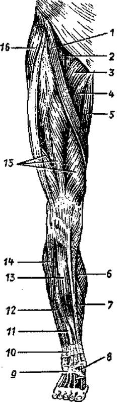
Fig. 35. Muscles of the right lower limb. Anterior view:
1 — sartorius muscle, 2 — iliopsoas muscle, 3 — pectineus muscle, 4 — adductor longus muscle, 5 — gracilis muscle, 6 — gastrocnemius muscle (medial head), 7 — soleus muscle, 8 — tendon of extensor hallucis longus, 9 — inferior extensor retinaculum, 10 — superior extensor retinaculum, 11 — extensor digitorum longus, 12 — peroneus brevis (fibularis brevis) muscle, 13 — tibialis anterior muscle, 14 — peroneus longus (fibularis longus) muscle, 15 — quadriceps femoris muscle, 16 — tensor fasciae latae

Fig. 36. Muscles of the right lower limb. Posterior view:
1 — gluteus maximus muscle, 2 — iliotibial tract, 3 — biceps femoris muscle, 4 — popliteal fossa, 5 — calcaneal (Achilles) tendon, 6 — gastrocnemius muscle, 7 — semitendinosus muscle, 8 — semimembranosus muscle
Internal pelvic muscles. The iliacus and psoas major muscles together form a large, powerful common iliopsoas muscle, which emerges onto the thigh beneath the inguinal ligament through the muscular lacuna and inserts via a common tendon onto the lesser trochanter of the femur.
The iliacus originates in the eponymous fossa of the ilium, whereas the psoas major arises from the lumbar vertebrae. The iliopsoas flexes the thigh at the hip joint and laterally rotates it. The psoas minor (inconstant) originates from the lumbar vertebrae, and its long, slender tendon blends into the iliac fascia, rendering it taut, and also attaches to the iliopubic eminence. The piriformis originates on the anterior surface of the sacrum, exits the pelvic cavity through the greater sciatic foramen, and inserts into the greater trochanter of the femur. This muscle laterally rotates the thigh. Within the greater sciatic foramen, the muscle leaves two clefts—the suprapiriform and infrapiriform foramina—through which vessels and nerves pass from the pelvic cavity into the gluteal region.
The obturator internus originates along the margins of the obturator foramen and on the obturator membrane, exits the pelvic cavity through the lesser obturator foramen, and inserts into the trochanteric fossa. The muscle laterally rotates the thigh.
External pelvic muscles. The gluteus maximus is a massive, powerful muscle originating from the external surface of the ala of the ilium, the sacrum, and the coccyx. The muscle inserts into the gluteal tuberosity of the femur, extending the thigh and rotating it laterally. It maintains the trunk in an upright posture and imparts a "military" posture to the body. The gluteus medius and minimus lie deep to the gluteus maximus; they originate on the external surface of the iliac ala and insert into the greater trochanter. These muscles abduct the thigh. The tensor fasciae latae originates from the anterior superior iliac spine, courses inferiorly, and blends into the fascia lata, keeping it taut. The obturator externus originates from the hip bone around the obturator foramen and on the obturator membrane, inserts into the greater trochanter, and laterally rotates the thigh. The quadratus femoris, originating on the lateral margin of the ischial tuberosity and inserting onto the intertrochanteric crest, rotates the thigh laterally. The thigh is likewise laterally rotated by the superior and inferior gemelli muscles, which originate from the ischial tuberosity (inferior) and the ischial spine (superior) and insert into the trochanteric fossa of the femur.
The Muscles of the Free Lower Limb comprise the muscles of the thigh, leg, and foot. Unlike the arm and forearm, the thigh and leg each feature three muscle groups rather than two. The foot also differs in its muscular structure and arrangement.
The muscles of the thigh are divided into anterior, posterior, and medial groups.
The anterior group includes the sartorius and quadriceps femoris muscles.
The sartorius muscle is long, narrow, and flattened, running obliquely across the thigh from top to bottom and medially. It originates at the anterior superior iliac spine and inserts into the tibial tuberosity. This muscle flexes the thigh and leg, and laterally rotates the thigh. The quadriceps femoris occupies the entire anterolateral surface of the thigh. The straight head of this muscle (rectus femoris) originates at the anterior inferior iliac spine. The other three heads (the vastus lateralis, vastus medialis, and vastus intermedius) originate on the anterior surface of the thigh. All four heads converge into a single, thick, broad tendon that inserts into the tibial tuberosity. Embedded within this tendon is a sesamoid bone (the Patella), which participates in the formation of the knee joint. The quadriceps femoris is the sole extensor of the leg at the knee joint, while its straight head (rectus femoris) also flexes the thigh at the hip joint.
The posterior group comprises the biceps femoris, semitendinosus, and semimembranosus muscles.
The long head of the biceps femoris originates at the ischial tuberosity, and the short head originates at the linea aspera of the femur. The muscle inserts into the head of the Fibula. It extends the thigh, flexes the leg, and laterally rotates it. The semitendinosus and semimembranosus muscles, which occupy the medial part of the posterior thigh, originate at the ischial tuberosity and insert into the Tibia. These muscles extend the thigh at the hip joint and flex the leg at the knee joint, while also laterally rotating the leg.
The Medial Group of the thigh consists of five muscles: the pectineus, gracilis, and the adductor magnus, adductor longus, and adductor brevis. These muscles originate on the pubic and ischial bones and insert (except for the gracilis) into the femur. All of these muscles adduct the thigh and slightly rotate it laterally. The gracilis muscle inserts into the tibia; it adducts the thigh, flexes the leg at the knee joint, and medially rotates it.
The muscles of the leg form three groups: anterior, posterior, and lateral. The anterior group consists of three muscles: the tibialis anterior, extensor digitorum longus, and extensor hallucis longus. All of these muscles originate on the anterior surface of the interosseous membrane of the leg, as well as on the tibia and fibula. The tibialis anterior inserts into the Base of the first metatarsal and the medial cuneiform bone, dorsiflexes the foot, and elevates its medial border. The extensor digitorum longus and extensor hallucis longus insert into the phalanges of the respective digits and extend the foot.
The posterior muscle group of the leg includes six muscles: the triceps surae, popliteus, tibialis posterior, plantaris, flexor digitorum longus, and flexor hallucis longus.
The triceps surae is located superficially, forming the contour of the posterior calf. Two of its heads (lateral and medial) originate on the respective epicondyles of the femur, forming the gastrocnemius muscle. The third head (deep) forms the soleus muscle, originating on the tibia and the tendinous arch. The soleus and gastrocnemius merge into a common tendon (the Achilles tendon), which inserts into the calcaneal tuberosity. This muscle is a powerful plantar flexor of the foot, and its medial and lateral heads also assist in flexing the leg at the knee joint. The plantaris is a thin, short muscle with a long tendon that originates on the lateral epicondyle of the femur, with its tendon blending into the calcaneal (Achilles) tendon. The popliteus is a short muscle lying posteriorly on the capsule of the knee joint, acting as its flexor. It also originates on the lateral epicondyle of the femur and inserts into the posterior surface of the tibia.
The tibialis posterior, flexor digitorum longus, and flexor hallucis longus lie beneath the triceps surae. They originate on the posterior surfaces of the tibia, fibula, and interosseous membrane. The tendons of these muscles pass into the foot posterior to the medial malleolus. All of these muscles are plantar flexors of the foot, with the latter two also acting as flexors of the corresponding digits. The tibialis posterior inserts into the navicular, cuneiform, and 4th Metatarsal Bones.
The Lateral Muscle Group of the leg consists of two muscles: the fibularis (peroneus) longus and fibularis (peroneus) brevis. They originate on the fibula, and their tendons course into the foot posterior to the lateral malleolus. The fibularis brevis inserts into the tuberosity of the 5th metatarsal bone. The insertion of the fibularis longus is complex: its tendon crosses the SOLE OF THE foot obliquely and inserts into the base of the medial cuneiform and the 1st and 2nd metatarsal bones. Consequently, the fibularis longus not only plantarflexes the foot and everts (pronates) its lateral border (along with the fibularis brevis), but also reinforces the transverse arch of the foot.
The foot contains dorsal and plantar muscles. The dorsum of the foot contains two muscles: the extensor digitorum brevis and extensor hallucis brevis. Both muscles originate on the dorsal surface of the calcaneus and insert into the dorsal phalanges of the respective digits.
The sole of the foot features three muscle groups: medial (the ball of the big toe), lateral (the ball of the little toe), and intermediate. The medial group comprises three muscles: the abductor hallucis, flexor hallucis brevis, and adductor hallucis. These muscles originate on the Tarsal Bones and insert into the proximal phalanx of the big toe.
The lateral group also includes three muscles: the abductor digiti minimi, flexor digiti minimi brevis, and opponens digiti minimi. These muscles originate on the PLANTAR ASPECT OF the calcaneus, the 5th metatarsal bone, and the long plantar ligament, inserting into the phalanges of the little toe.
The central elevation (intermediate group) comprises 13 muscles. The flexor digitorum brevis and quadratus plantae originate on the calcaneus. The former inserts into the middle phalanges of the lateral four digits, while the quadratus plantae attaches to the tendons of the flexor digitorum longus. Four lumbricals originate on the tendons of the flexor digitorum longus and insert into the proximal phalanges of digits 2–5. The interossei originate on the metatarsal bones (within the interosseous spaces) and insert into the phalanges. The plantar interossei adduct digits 3–5 toward the 2nd digit, while the dorsal interossei abduct digits 2–4 laterally (away from the midline).
Fasciae of the lower limb. The internal pelvic muscles, which originate on the pelvic bones and lumbar vertebrae, are covered by the iliac fascia. The external pelvic muscles, as well as the muscles of the free lower limb, are covered by superficial (subcutaneous) fascia, which is referred to as the fascia lata in the thigh, the crural fascia in the leg, and the gluteal fascia in the gluteal region, etc.
The fascia lata is a dense, thick, tendinous membrane. It encloses all the muscles of the thigh externally and forms three intermuscular septa that, together with the femur, delineate three muscular compartments in the thigh. This results in the formation of osteofascial sheaths for the anterior, posterior, and medial muscle groups of the thigh. In the upper medial thigh (inferior to the inguinal ligament), the fascia lata is pierced by numerous apertures transmitting blood vessels and nerves. This specialized area of the fascia is known as the cribriform fascia, which seals the exit of the femoral canal (its superficial, subcutaneous ring).
The crural fascia is a continuation of the fascia lata of the thigh. In the leg, two intermuscular septa extend from this fascia to the fibula, separating the lateral muscle group of the leg (peroneal muscles) from the anterior and posterior groups. At the level of the ankle joint, the crural fascia forms several transversely oriented thickenings known as retinacula. Anterior to the ankle joint are the superior and inferior extensor retinacula. These retinacula contribute to the formation of three osteofibrous tunnels through which the tendons of the tibialis anterior, extensor hallucis longus, and extensor digitorum longus pass onto the dorsum of the foot. Each tunnel contains a corresponding synovial sheath. Posterior to the medial malleolus, beneath the flexor retinaculum, there are likewise three synovial sheaths for the respective flexor tendons. Posterior to the lateral malleolus, beneath the peroneal retinaculum, there is initially a common synovial sheath for the fibular tendons, which subsequently bifurcates inferiorly into separate sheaths for the tendons of the fibularis longus and fibularis brevis.
The fasciae of the foot differ in structure between its dorsum and sole. On the dorsum of the foot, the fascia is thin and delicate. On the plantar aspect, the fascia is thick and reinforced by dense bundles of fibrous fibers; hence, the plantar fascia is designated as the plantar aponeurosis.
The fasciae and muscle tendons of the lower limb contribute to the formation of canals, fossae, and other structures of significant physiological and clinical importance. These include the femoral and adductor (subadductor) canals, the femoral triangle, and the popliteus (popliteal) fossa.
The femoral canal, which can serve as a site for femoral hernia formation, is located beneath the medial part of the inguinal ligament within the vascular lacuna. The walls of this canal, which is approximately 2 cm long, are formed by the inguinal ligament (anteriorly), the femoral vein (laterally), and the deep layer of the fascia lata (posteriorly). The internal ring (opening) of the femoral canal within the lesser pelvis is the region bounded by the attachment of the inguinal ligament to the pubic bone medially and the femoral vein laterally. The external opening of the canal is the cribriform fascia—a perforated plate within the fascia lata of the thigh.
The femoral canal is located in the upper medial region of the femoral triangle, which is bounded superiorly by the inguinal ligament, medially by the adductor longus, and laterally by the sartorius. The Clinical significance of this triangle lies in the passage of the femoral artery and vein from the midpoint of the inguinal ligament to the apex of the triangle.
From the apex of the femoral triangle originates the adductor (subadductor) canal, which transmits the femoral artery and vein and opens into the popliteal fossa. This fossa, covered by the popliteal fascia and shaped like a vertically oriented rhombus, contains adipose tissue, the popliteal artery and vein with their respective branches and tributaries, nerves, and lymph nodes.
REVIEW AND SELF-assessment questions:
1. How are the muscles of the upper limb categorized according to their anatomical location and function?
2. Name the muscles that adduct and abduct the arm at the shoulder joint.
3. Name the pronators and supinators of the forearm and hand.
4. Name the muscle groups of the hand. Which muscles are included in each group, and what functions do these muscles perform?
5. Name the canals, openings, fossae, and synovial sheaths of the upper limb. Where are they located, and what is their practical significance?
6. List the muscles of the lower limb girdle (pelvic girdle). Where are these muscles located, and what functions do they perform?
7. Name the muscle groups of the thigh and leg. Which muscles are included in each group, and what functions do these muscles perform?
8. Name the muscle groups of the foot. What functions do these muscles perform?
9. Name the canals, fossae, and synovial sheaths found in the lower limb. Where are they located, and what is their practical significance?
Last update: 10/08/2026
Editorial and Educational Adaptation: This material has been compiled based on the primary/original source text. The project team performed an editorial review, corrected technical inaccuracies, structured sections, and adapted the content for an educational format.
What was processed:
- elimination of formatting defects (OCR errors, structural breaks, corrupted characters);
- editorial organization of content;
- standardization of terminology in accordance with academic sources;
- verification of factual statements against the original source text.
All mentions of the author, publication year, and origin of the primary text have been preserved in accordance with the source.