Human Anatomy Part 1 - K. A. Dyubenko, A. K. Kolomiytsev, Yu. B. Chaykovsky 2002
Special Part
Axial Skeleton, sceleton axiale - Bones of the Neurocranium
Frontal bone
The BONES OF THE neurocranium include:
- frontal bone, os frontale,
- Sphenoid bone, os sphenoidale,
- Occipital bone, os occipitale,
- Temporal bone, os temporale,
- Parietal bone, os parietale,
- Ethmoid bone, os ethmoidale (partially).
The frontal bone, os frontale, is an unpaired bone (Fig. 36) that contributes to The formation of the cranial vault and base, as well as the orbits and Nasal cavity. It develops as a primary (membranous) bone from Connective Tissue. It is classified as a pneumatic bone because it contains a cavity, the frontal sinus, sinus frontalis. The frontal bone consists of four parts: the frontal squama, squama frontalis; two orbital parts, partes orbitales; and the nasal part, pars nasalis.
The frontal squama, squama frontalis, is convex anteriorly and presents the following surfaces: an external (frontal) surface, two lateral (temporal) surfaces, and an internal (endocranial) surface.
The external surface is smooth and features a midline suture in childhood that typically obliterates by the age of five, leaving a faint linear trace known as the metopic suture line, sutura frontalis metopica. Anteriorly, the frontal surface of the squama transitions into the orbital surface, forming the supraorbital margin, margo supraorbitalis, on each side. Below and parallel to the supraorbital margin lies an arched elevation, the superciliary arch, arcus superciliaris. Superior to each superciliary arch, approximately in the middle of the squama, is a rounded eminence termed the frontal eminence, eminentia frontalis, or frontal tuber, tuber frontale. Between the superciliary arches, slightly superiorly, is a smooth area known as the glabella, glabella. The medial part of the supraorbital margin exhibits a small supraorbital notch, incisura supraorbitalis, which varies considerably in anatomy and may form a complete foramen. Closer to the midline lies the frontal notch, incisura frontalis, which occasionally converts into a foramen transmitting Blood Vessels and nerves. Further laterally, the supraorbital margin continues into the zygomatic process, processus zygomaticus. Posteriorly and superiorly from the zygomatic process runs the temporal line, linea temporalis.
The internal surface of the frontal squama is concave and displays impressiones digitatae (gyral impressions), granular foveolae (for arachnoid granulations), and arterial sulci. Along the midline of the superior part of the internal surface runs the sulcus for the superior sagittal sinus, sulcus sinus sagittalis superioris. The margins of this groove converge inferiorly to form the frontal crest, crista frontalis, ending below in the foramen caecum.
The superior or parietal border, margo parietalis, is arched and articulates with the frontal margin of the parietal bones to form the coronal suture, sutura coronalis.
Class="center">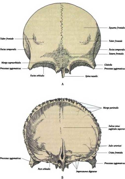
Fig. 36. Frontal bone, os frontale:
A — external surface; B — internal surface
The orbital part, pars orbitalis, of the frontal bone forms the roof of the Orbit. It presents inferior (orbital) and superior (cerebral) surfaces. Near the Base of the zygomatic process, the orbital surface bears a shallow lacrimal fossa, fossa glandulae lacrimalis. Medially, it features a small trochlear fossa, fovea trochlearis. The cerebral surface of the orbital part exhibits a series of impressiones digitatae and cerebral eminences, juga cerebralia (jugum meaning mountain ridge). Both orbital parts are separated from each other by the ethmoidal notch, incisura ethmoidalis, which is bridged by the cribriform plate of the ethmoid bone. This part of the bone contributes to the Formation of the anterior and posterior ethmoidal foramina.
The nasal part, pars nasalis, of the frontal bone is horseshoe-shaped and closes the anterior part of the ethmoidal notch, incisura ethmoidalis. Projecting anteriorly from the nasal part is the nasal spine, spina nasalis. It is flanked anteriorly and laterally by the nasal margin, margo nasalis, which articulates to form the frontonasal and frontomaxillary sutures. On either side of the nasal spine lies the aperture of the frontal sinus, apertura sinus frontalis, which leads into the cavity of the frontal sinus, sinus frontalis.
The frontal sinus is a paired air-filled cavity located within the diploë of the frontal squama, corresponding in position to the glabella and superciliary arches. In shape, the frontal sinus resembles a triangular pyramid with its base directed inferiorly and its apex superiorly. The right and left frontal sinuses are separated by a perpendicular intersinus septum, septum intersinuale frontale. The dimensions of the sinuses vary widely, but on average do not exceed 5 cm3 (Fig. 37). Infection of the sinus cavity leads to inflammation of its mucous membrane, known as frontal sinusitis (frontitis). Accessory frontal sinuses occasionally occur (Jovanović, 1961).
Ossification. Ossification centers appear at the end of the second month of intrauterine development in the region of the supraorbital margin. At birth, the frontal bone consists of two separate halves that fuse during the second year of life. The metopic suture may persist up to 5 years of age.
Last update: 08/08/2026
Editorial and Educational Adaptation: This material has been compiled based on the primary/original source text. The project team performed an editorial review, corrected technical inaccuracies, structured sections, and adapted the content for an educational format.
What was processed:
- elimination of formatting defects (OCR errors, structural breaks, corrupted characters);
- editorial organization of content;
- standardization of terminology in accordance with academic sources;
- verification of factual statements against the original source text.
All mentions of the author, publication year, and origin of the primary text have been preserved in accordance with the source.