MEDICAL BIOLOGY, HUMAN ANATOMY, PHYSIOLOGY AND PATHOLOGY - Ya.I. Fedonyuk 2010
ANATOMY, PHYSIOLOGY, PATHOLOGY
SECTION 2. THE LOCOMOTOR SYSTEM (THE MECHANISM OF MOVEMENT)
MUSCLES AND FASCIAE OF BODY REGIONS
MUSCLES AND FASCIAE OF THE TRUNK
3. MUSCLES AND FASCIAE OF THE ABDOMEN
The Abdominal Muscles are subdivided into anterior, lateral, and posterior groups.
The anterior group includes the rectus abdominis and pyramidalis muscles.
The rectus abdominis Muscle originates from the xiphoid process of the Sternum and the cartilages of the V and VI Ribs. It inserts onto the pubic bone (Fig. 2.86).
Class="center">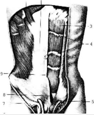
Fig. 2.86. Muscles of the abdomen; the anterior lamina of the rectus sheath has been removed on the left side.
Function: flexion of the trunk forward.
The pyramidalis muscle is a vestigial Structure that is well developed in marsupials, whereas in humans it acts to tense the linea alba.
The lateral group comprises the external and internal oblique muscles and the transversus abdominis muscle.
The external oblique muscle of the abdomen originates from the lower eight ribs. Its posterior fibers attach to the iliac crest, while the anterior fibers continue into a broad, flat aponeurosis. The lower margin of this aponeurosis is rolled inward to form the inguinal (Poupart's) ligament, which extends from the anterior superior iliac spine to the pubic tubercle. Function: Rotation of the trunk to the sides.
The internal oblique muscle of the abdomen originates from the thoracolumbar fascia, the iliac crest, and the inguinal ligament. Its posterior fibers attach to the lower ribs, while the anterior fibers transition into an aponeurosis. Function: rotation of the trunk to the sides.
The transversus abdominis muscle originates from the inner surface of the lower six ribs, the thoracolumbar fascia, the iliac crest, and the inguinal ligament.
Its muscle fibers run transversely and continue into an aponeurosis (Fig. 2.87). Function: rotation of the trunk to the sides.

Fig. 2.87. Muscles of the abdomen; lateral view.
The posterior group is represented by the quadratus lumborum muscle.
The quadratus lumborum muscle originates from the iliac crest and inserts onto the XII rib and the transverse processes of the lumbar vertebrae.
Function: lateral flexion of the trunk.
All abdominal muscles contribute to The formation of the walls of the Abdominal cavity. When they contract, intra-abdominal pressure increases, forming the abdominal press that maintains the viscera in a proper position. Furthermore, the muscles of the abdominal press assist in the evacuation of the bowels and bladder, and in women, they aid in childbirth.
By attaching to the ribs, these muscles also participate in Respiration.
Abdominal fascia
Beneath the Skin lies the external Fascia of the abdomen, which is a continuation of the external pectoral fascia. The walls of the abdominal cavity are lined on the inside with the Peritoneum (a serous membrane). Superficial to the peritoneum lies the transversalis fascia.
Last update: 08/08/2026
Editorial and Educational Adaptation: This material has been compiled based on the primary/original source text. The project team performed an editorial review, corrected technical inaccuracies, structured sections, and adapted the content for an educational format.
What was processed:
- elimination of formatting defects (OCR errors, structural breaks, corrupted characters);
- editorial organization of content;
- standardization of terminology in accordance with academic sources;
- verification of factual statements against the original source text.
All mentions of the author, publication year, and origin of the primary text have been preserved in accordance with the source.